Cardiac resynchronisation therapy (CRT-P and CRT-D): a Cath Lab 101 guide
Reviewed by Rodnie Oro, cath lab charge nurse · Last reviewed: November 2026 · Education only, not medical advice
Cardiac resynchronisation therapy in one minute
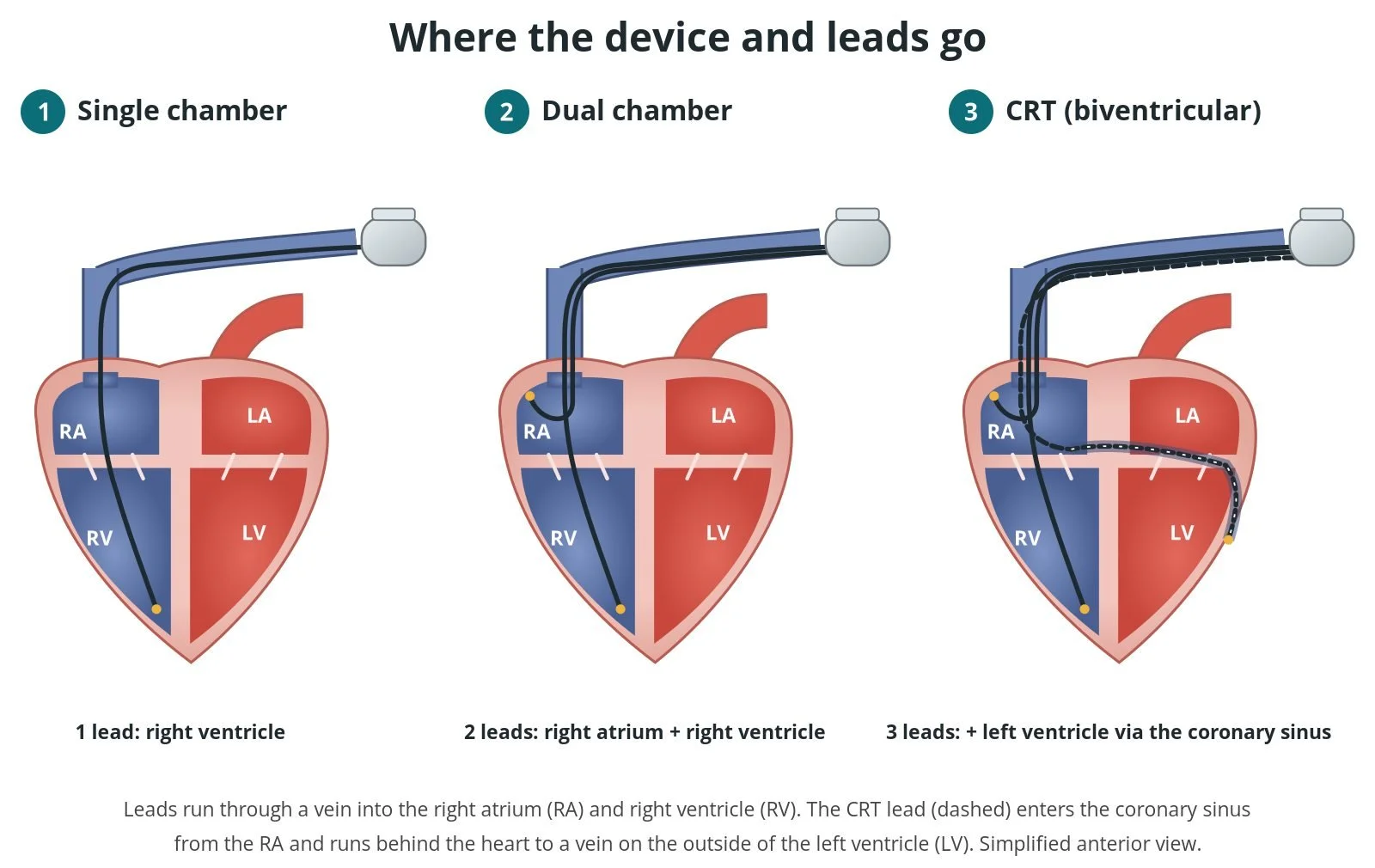
What it is: Cardiac resynchronisation therapy (CRT) uses a special pacemaker with three leads: one in the right atrium, one in the right ventricle and one in a vein on the outside of the left ventricle. Pacing both ventricles together helps them squeeze in step. CRT-P is a pacemaker only; CRT-D also has a defibrillator (ICD) to treat dangerous fast rhythms.
Who it is for: People with heart failure and a weak left ventricle, still breathless despite good medicines, whose ECG shows a wide QRS, especially left bundle branch block. It is also used when someone with a weak heart needs a lot of pacing.
Main risks: Bleeding or a bruise (haematoma) at the wound, infection, a lead moving out of place, a collapsed lung (pneumothorax), damage to the coronary sinus, and pacing of the diaphragm (phrenic nerve). Your team will explain your own risk.
Usually: Under local anaesthetic with sedation, about 1.5 to 3 hours, often home the same or next day.
On this page: What is cardiac resynchronisation therapy? · Who is it for? · When is it done? · Step by step · Recovery · Risks · Kit list · First time in the lab? · FAQ
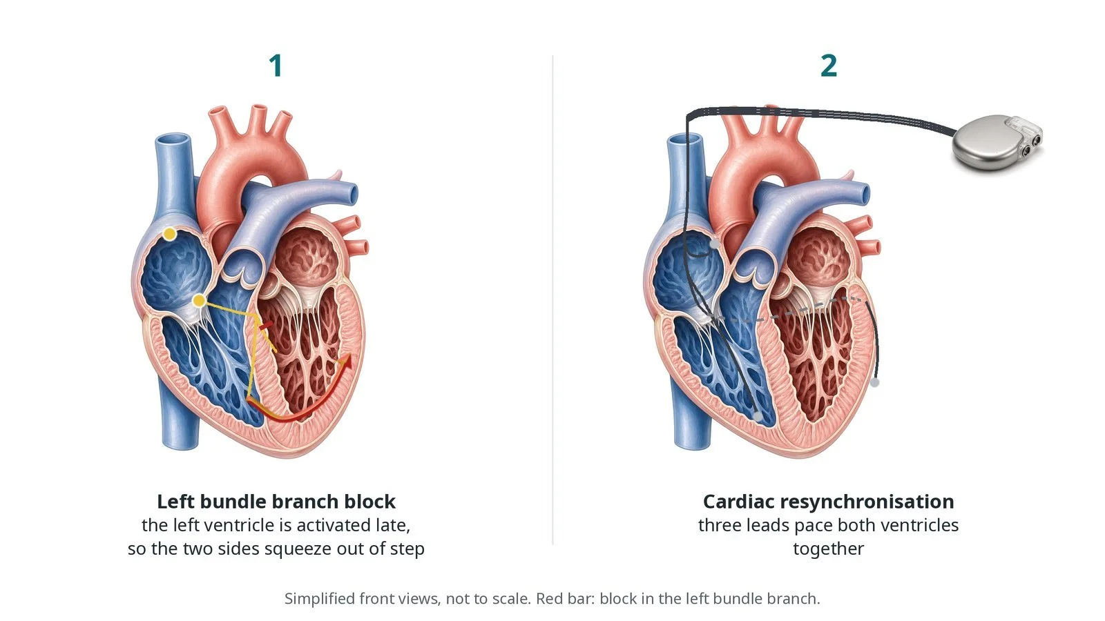
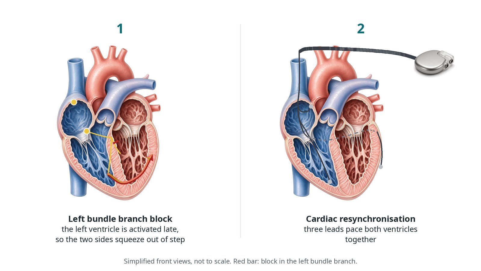
(1) Left bundle branch block: the left ventricle is activated late, so the two sides squeeze out of step. (2) CRT: three leads pace both ventricles together.
What is cardiac resynchronisation therapy?
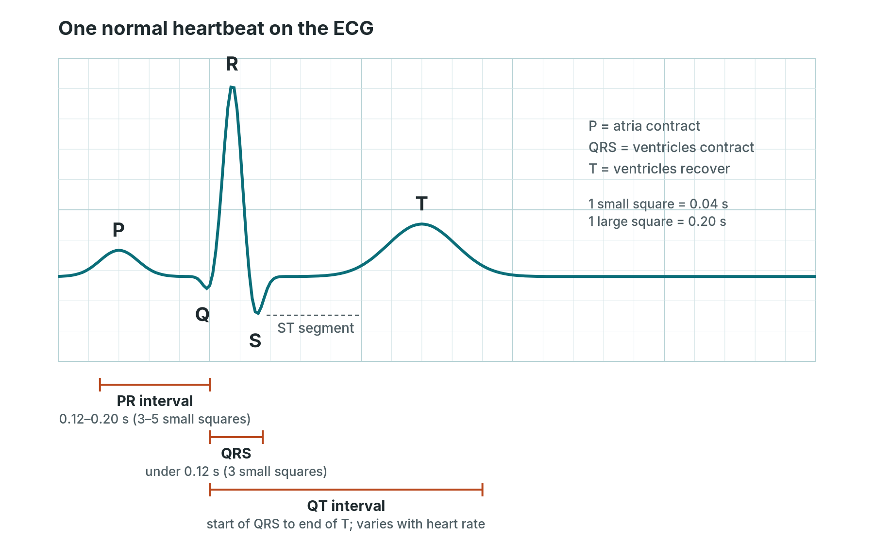
In a healthy heart the electrical signal travels down the right and left bundle branches at the same time, so both ventricles squeeze together. In left bundle branch block (LBBB) the left side is activated late. The ECG shows a wide QRS, and the two ventricles and the septum move out of step, which wastes energy in a heart that is already weak.
CRT paces the right ventricle and the left ventricle together, which brings them back into step (resynchronisation). In the right people this can improve symptoms, heart pumping and survival, alongside heart failure medicines.
The left ventricular lead does not go inside the left ventricle. It goes through the right atrium into the coronary sinus, the large vein that drains the heart muscle and runs in the groove at the back of the heart. From there it is passed into one of its side branches on the outside wall of the left ventricle, ideally a lateral or posterolateral vein.
The phrenic nerve, which works the diaphragm, runs close to the left side of the heart. If the LV lead is too near it, pacing can make the diaphragm twitch, so this is always tested.
New to the acronyms? See the Cath Lab Abbreviations A–Z.
Who is it for?
Decided by a heart failure and device team, using ESC and NICE guidance. Typical reasons:
heart failure symptoms despite good medicines
a weak left ventricle on echo (low ejection fraction)
a wide QRS on the ECG (usually 130 ms or more), most clearly with left bundle branch block
or a weak heart in someone who needs a pacemaker and will be paced a lot
CRT-D is chosen when there is also a high risk of dangerous fast rhythms and a defibrillator is right for the person.
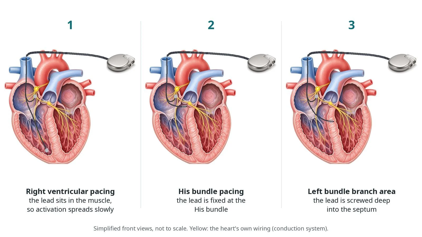
Who it may not suit: People with a narrow QRS, or a QRS without left bundle branch block, are less likely to benefit. An active infection must be treated first. Conduction system pacing may be an alternative in some people.
The decision is made by your heart team, who will talk through the reasons, the alternatives and the risks with you.
When is it done?
It is a planned procedure.
Before it, you usually have:
an ECG and echo, and blood tests including kidney function and blood count
a review of blood thinners, diabetes medicines and any infection
a talk about CRT-P versus CRT-D, driving, and what happens at the end of life with a defibrillator
gives you a chance to ask questions and give consent
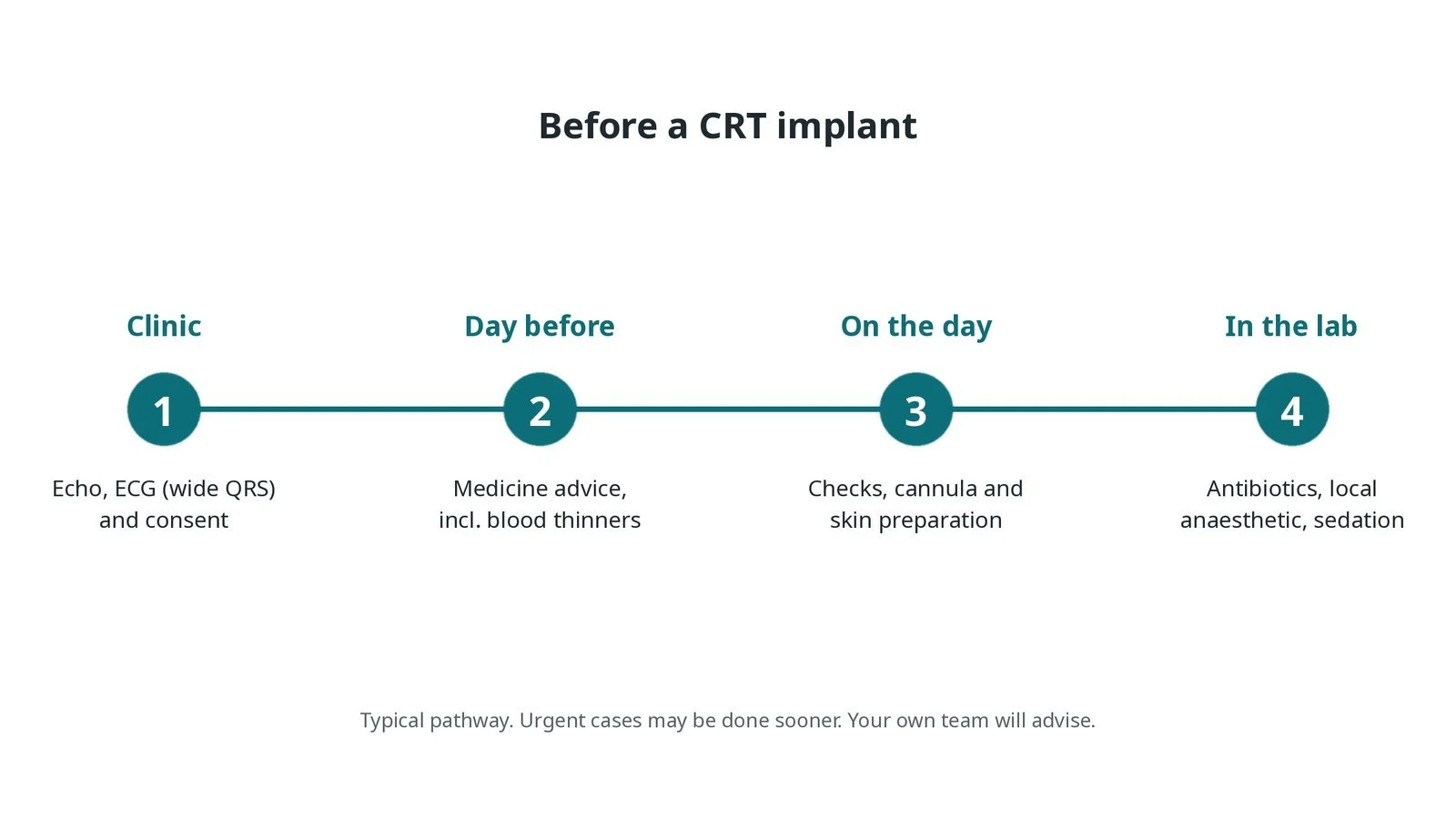
Before a CRT implant: echo, ECG and consent; medicine advice; checks and skin preparation on the day; antibiotics, local anaesthetic and sedation in the lab.
Step by step
1. Getting ready
You lie on the X-ray table with ECG, blood pressure and oxygen monitoring. Defibrillator pads are put on. Antibiotics are given before the skin cut, as per centre protocol. The skin below the left collarbone is cleaned and draped, and local anaesthetic is given, with sedation as needed.
2. Access and pocket
Three veins are entered below the collarbone (cephalic, axillary or subclavian vein, per operator), often with ultrasound or a contrast venogram to guide. A pocket is made under the skin, or under the muscle, for the generator.
Watch: Sudden breathlessness, falling oxygen or chest pain after subclavian access can mean a pneumothorax. Tell the operator and check the screen and observations.
3. The left ventricular lead
A. Coronary sinus. A long, shaped delivery sheath is passed into the right atrium and its tip is guided into the opening of the coronary sinus.
B. Venogram. A balloon catheter blocks the coronary sinus briefly while contrast is injected, to map the side veins on the outside of the left ventricle.
C. LV lead. A thin wire is passed into a suitable lateral or posterolateral vein and the LV lead is advanced over it until it is stable.
D. Test. Thresholds and sensing are measured, and high-output pacing checks for phrenic nerve (diaphragm) stimulation. The sheath is then cut away (slit) without moving the lead.
Watch: Contrast staining or a falling blood pressure during the venogram can mean a coronary sinus dissection or perforation. Tell the operator; have echo and a pericardiocentesis kit ready.
4. The right-sided leads
The right ventricular lead is fixed low in the right ventricle (an ICD lead with shock coils for CRT-D) and the right atrial lead in the right atrium. Each is tested with the pacing system analyser.
5. Connect and close
The leads are connected to the generator and the screws tightened. The pocket is washed out and an antibiotic envelope may be used, as per centre protocol. The wound is closed in layers. For a CRT-D, defibrillation testing is not routine and is per centre protocol.
6. Checks before home
A chest X-ray checks the lead positions and the lungs, and a device check confirms that both ventricles are being paced. An ECG shows the new paced QRS.
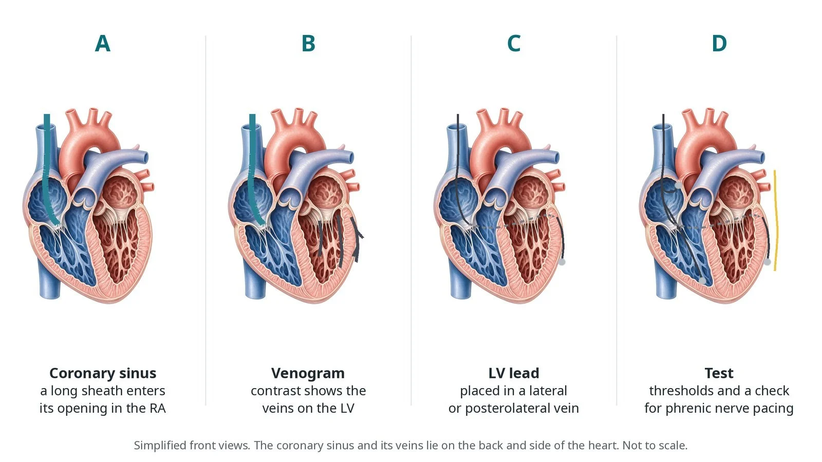
A: a long sheath enters the coronary sinus. B: a venogram shows the veins. C: the LV lead is placed in a lateral or posterolateral vein. D: thresholds and a phrenic nerve check.
Recovery and aftercare
First hours: rest, with checks of the wound, blood pressure and heart rhythm, as per centre protocol.
Going home: often the same or next day, after a chest X-ray and device check.
Arm and wound: keep the arm below shoulder height and avoid heavy lifting for about 4 to 6 weeks, or as advised; keep the wound dry until it has healed.
Medicines: heart failure medicines continue; blood thinners restart as advised.
Driving: DVLA rules depend on the device and why it was fitted. You usually cannot drive for at least a week after a CRT-P, and for longer after a CRT-D. Different rules apply for bus and lorry licences.
Follow-up: wound check, then device and heart failure clinics, with remote monitoring.
Call 999 if you have chest pain, sudden breathlessness or fainting, or more than one shock from a CRT-D.
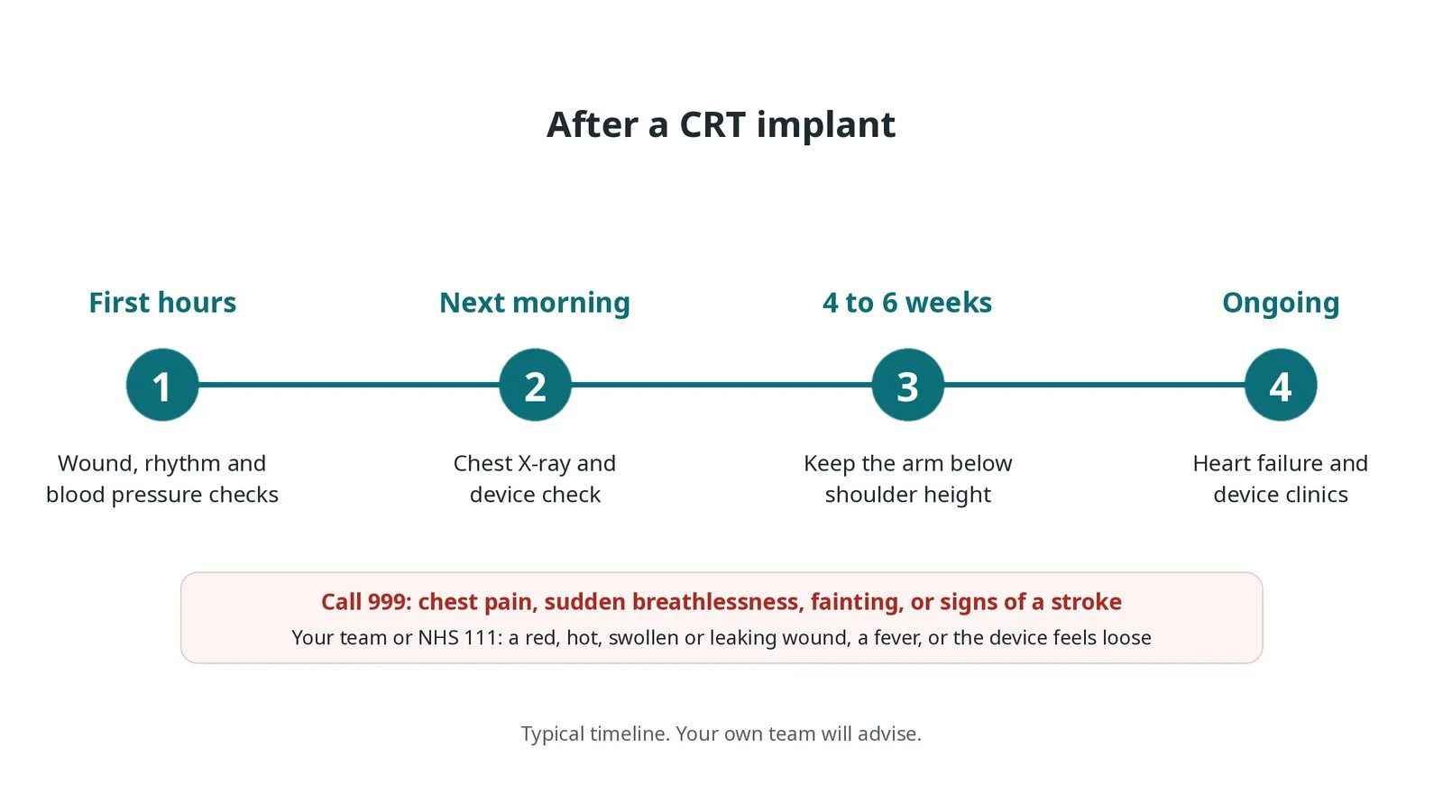
After a CRT implant: (1) wound, rhythm and blood pressure checks; (2) chest X-ray and device check; (3) arm below shoulder height for 4 to 6 weeks; (4) heart failure and device clinics.
Risks and complications
Your team will explain the risks that apply to you. Possible problems include:
Bleeding or a bruise (haematoma) at the wound
Infection of the pocket or leads (uncommon): Can mean removing the whole system.
A lead moving out of place, most often the LV lead (uncommon)
Diaphragm twitching from phrenic nerve pacing: Often fixed by reprogramming.
Collapsed lung (pneumothorax) (uncommon)
Damage to the coronary sinus or bleeding around the heart (rare)
Not responding to CRT as hoped: Some people feel little change.
Cath lab kit list (printable)
Barts example kit list
An example from one UK centre, written generically. Every lab has its own protocol, and sizes and doses vary.
pacemaker or ICD implant pack, sterile drapes and skin prep
venous access needle, wires and peel-away sheaths
coronary sinus delivery sheath and inner catheter, per operator
venogram balloon catheter and contrast
left ventricular lead and guidewires, per operator
right atrial and right ventricular leads (ICD lead for CRT-D)
CRT-P or CRT-D generator
pacing system analyser and programmer
slitting tool for the delivery sheath
antibiotic envelope, if used, as per centre protocol
defibrillator pads, echo and a pericardiocentesis kit nearby
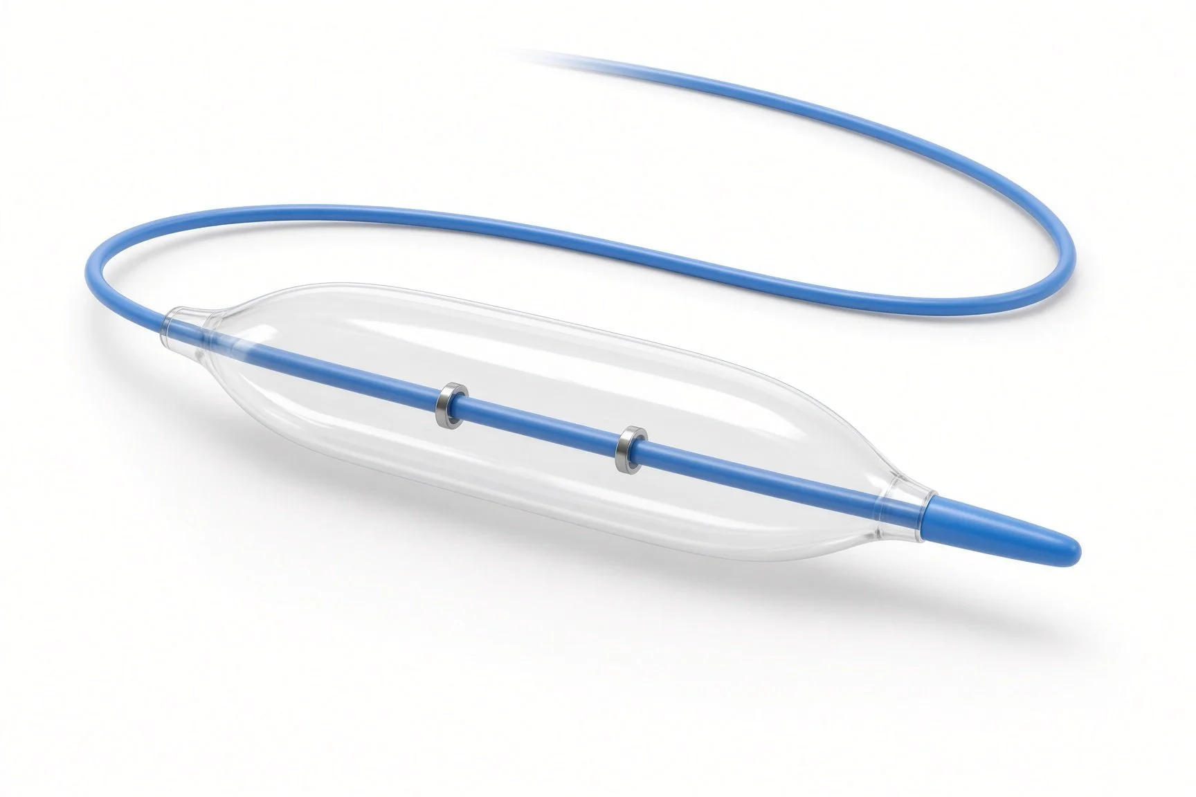
Venogram balloon catheter.
Venogram balloon catheter.
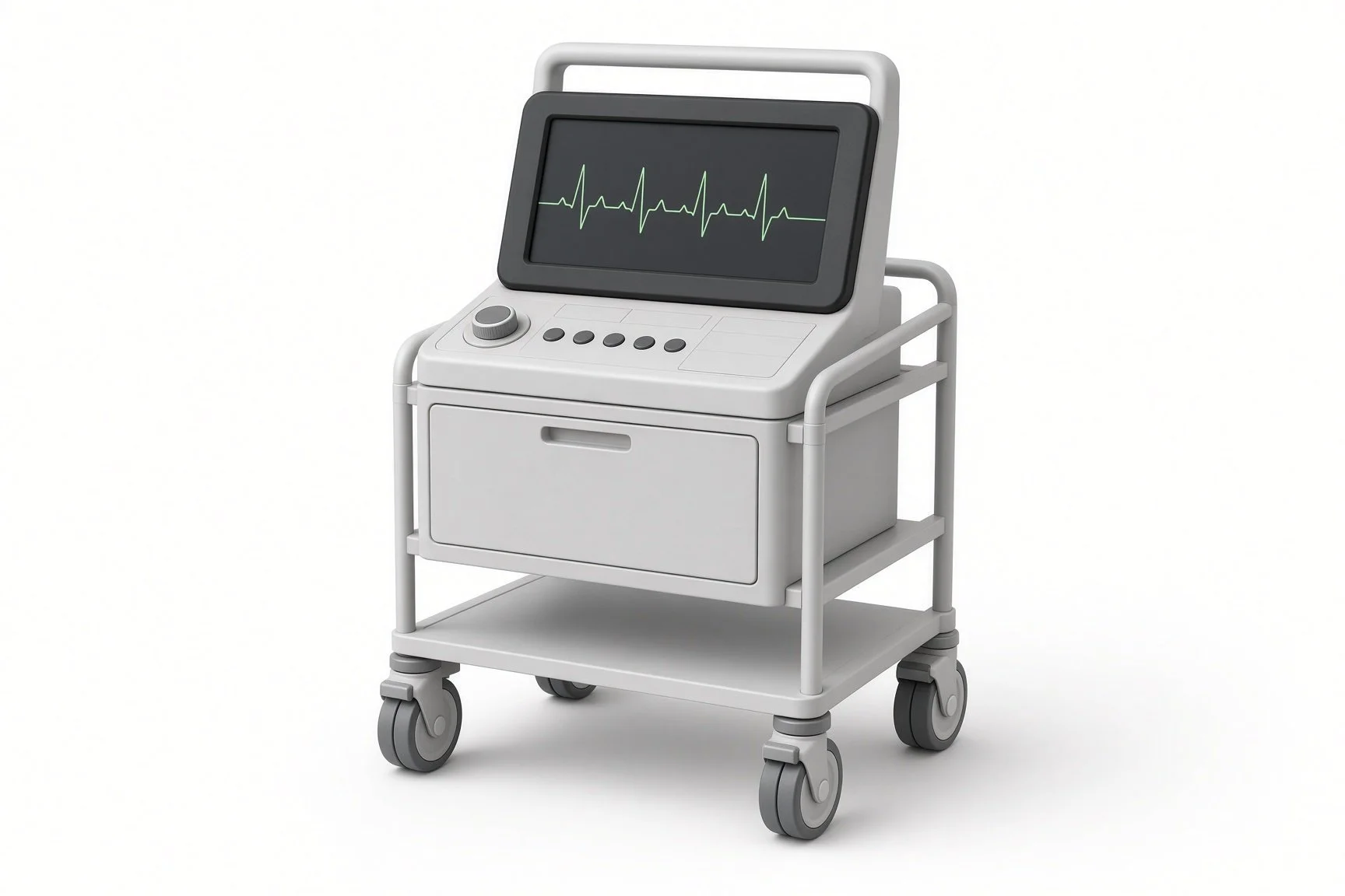
Pacing system analyser.
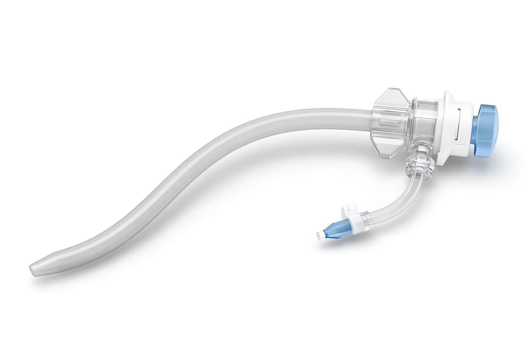
Coronary sinus delivery sheath.
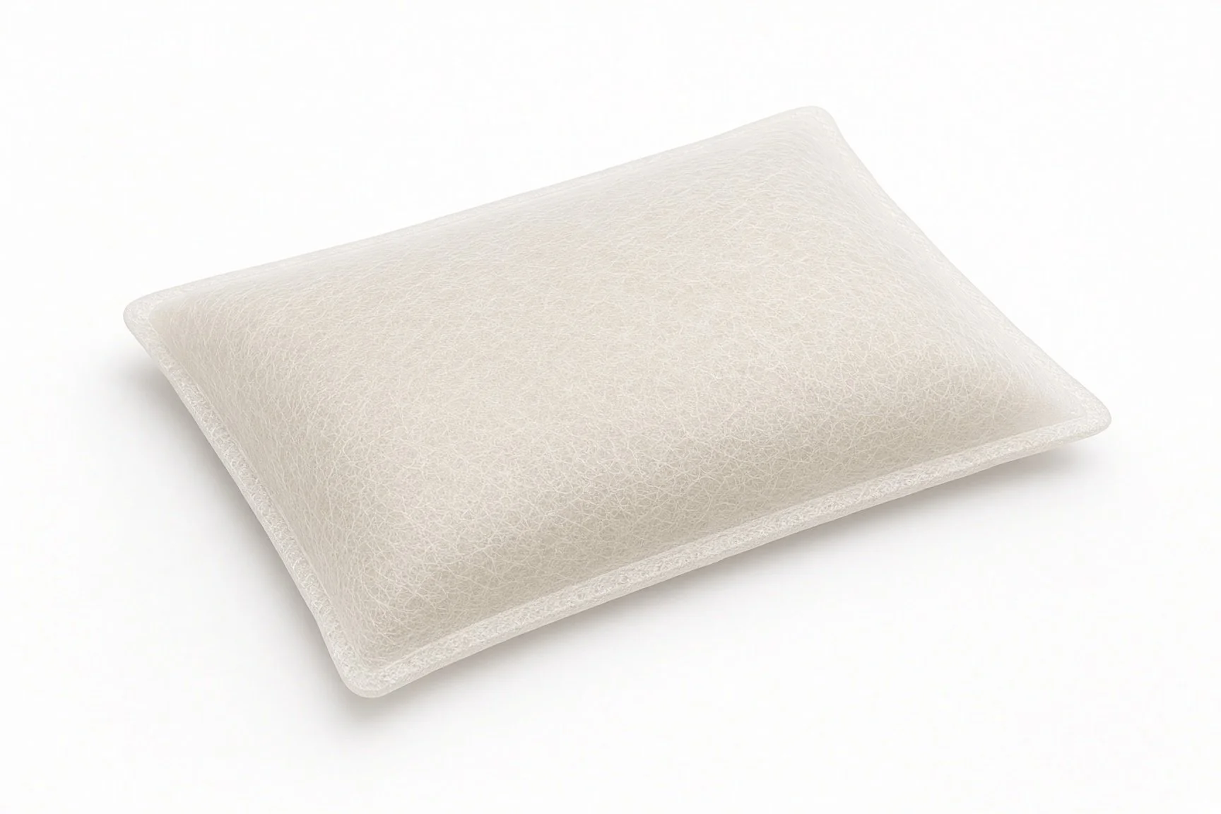
Antibiotic envelope.
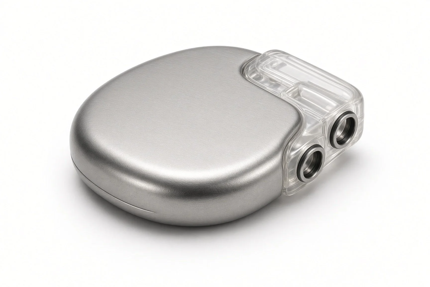
CRT generator.
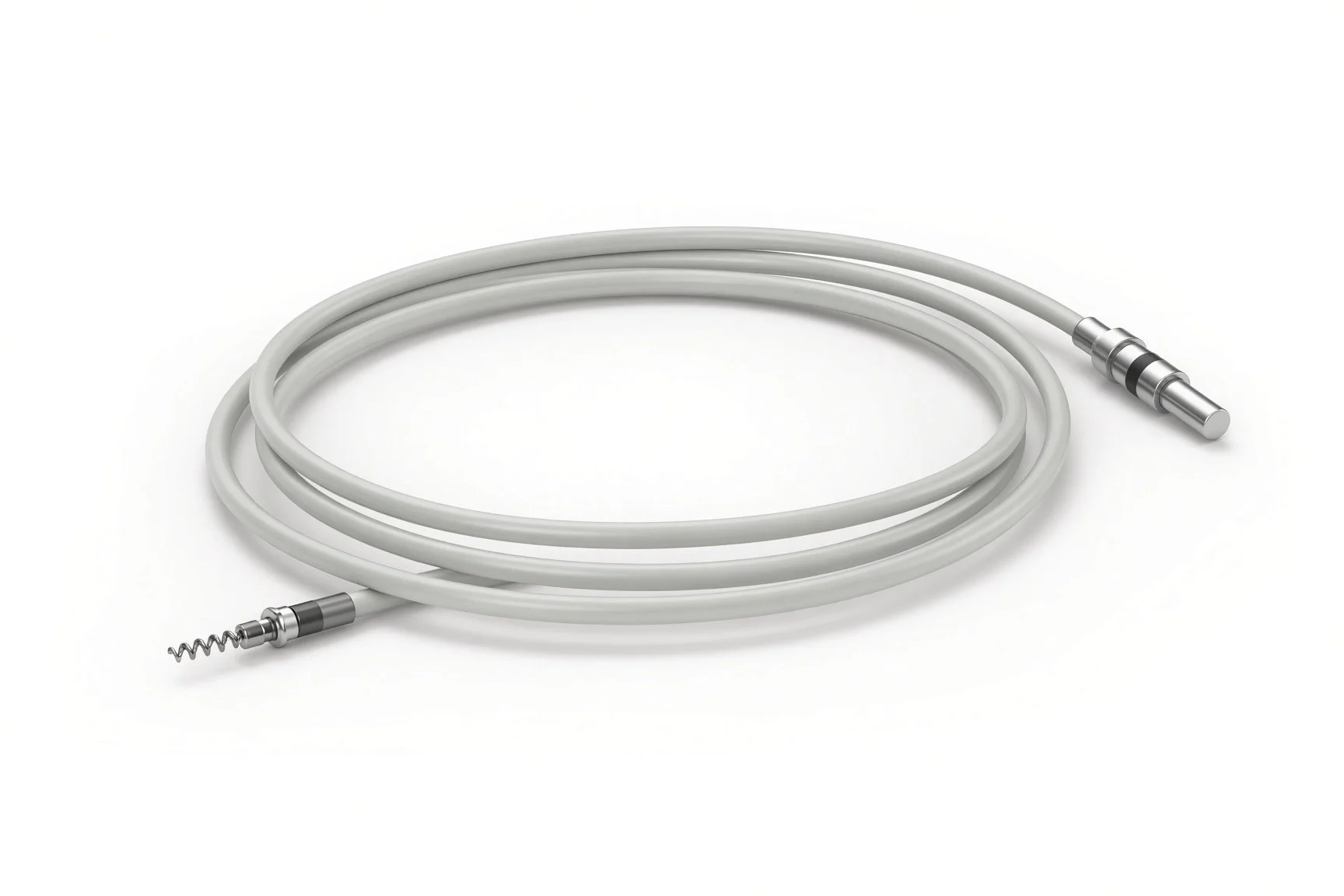
Right atrial and ventricular leads.
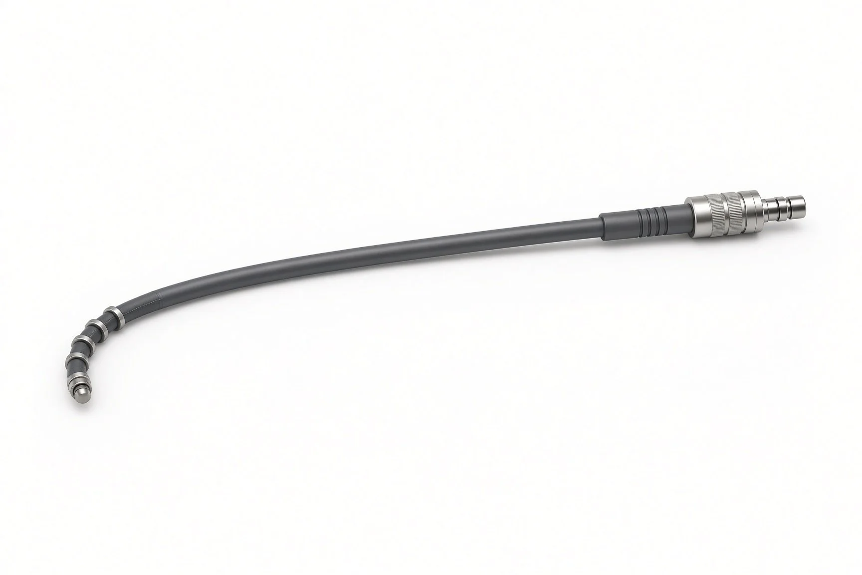
Left ventricular lead.
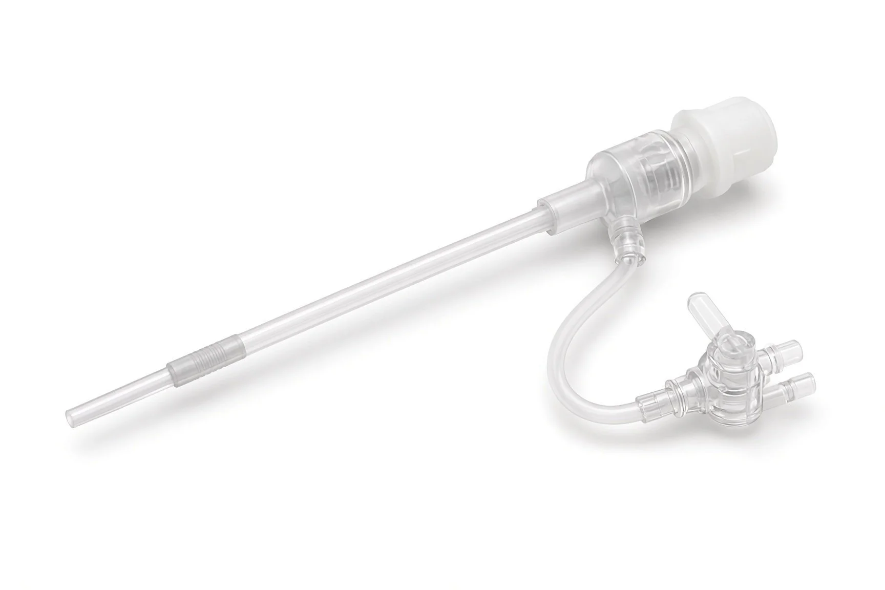
Peel-away sheaths.
Pacing system analyser.
Procedural steps (Barts example)
Getting ready
Access and pocket
The left ventricular lead
The right-sided leads
Connect and close
Checks before home
First time in the cath lab?
Expect a longer case. The LV lead can take time when the veins are small or twisted. Plan breaks and keep the sterile field tidy.
Learn the coronary sinus kit. Know which sheath, inner catheter, balloon and wires your centre uses, and have back-up shapes ready.
Watch for the diaphragm. Phrenic nerve pacing shows as a hiccup-like twitch. Say what you see during high-output tests.
Infection prevention matters. Keep the door shut, the field covered and handling of the generator to a minimum.
Infection prevention starts here: skin prep, timing of antibiotics and keeping door traffic low all matter for implants.
Watch the patient's abdomen and shoulder during high-output testing. Diaphragm twitching can be subtle under drapes.
Keep track of the slitting tool and the sheath hub. Slitting is a two-person job.
Note the paced QRS after the case. A much narrower QRS than before is reassuring.
FAQ
Will I feel the pacing?
Usually not. Tell your team if you notice hiccups or twitching in your tummy, as the settings can often be changed.
What is the difference between CRT-P and CRT-D?
Both pace the heart in step. A CRT-D can also give a shock to stop a dangerous fast rhythm.
How soon will I feel better?
Some people feel better within weeks; for others it takes months. Some feel little change.
Can I have an MRI scan?
Many systems are MRI conditional. Your device team will check your system and set it up before a scan.
Related Cath Lab 101 guides
Conduction system pacing (His bundle and left bundle branch area pacing) (coming soon)
Pacemaker and ICD generator change and upgrade (coming soon)
Sources
All checked on 12 November 2026. Each page's own date is shown where it gives one.
Glikson M, et al. 2021 ESC Guidelines on cardiac pacing and cardiac resynchronization therapy. Eur Heart J 2021;42(35):3427–3520
McDonagh TA, et al. 2021 ESC Guidelines for the diagnosis and treatment of acute and chronic heart failure. Eur Heart J 2021;42(36):3599–3726
NICE. Implantable cardioverter defibrillators and cardiac resynchronisation therapy for arrhythmias and heart failure (TA314). nice.org.uk/guidance/ta314
StatPearls (NCBI Bookshelf), search: cardiac resynchronization therapy. ncbi.nlm.nih.gov/books
Disclaimer
Education only. This page is for general education for healthcare students, cath lab staff and curious readers. It isn't medical advice and doesn't replace advice from your own doctor, heart team or local unit protocols. Equipment and medicines are described generically. No product or manufacturer is recommended. If you think someone is having a heart emergency, call 999.
Disclosure: I work for a company that makes a device used in this procedure. This page is independent education, isn't sponsored or reviewed by any company, and doesn't recommend any product.
All diagrams on this page are original illustrations created for rodnieoro.com.