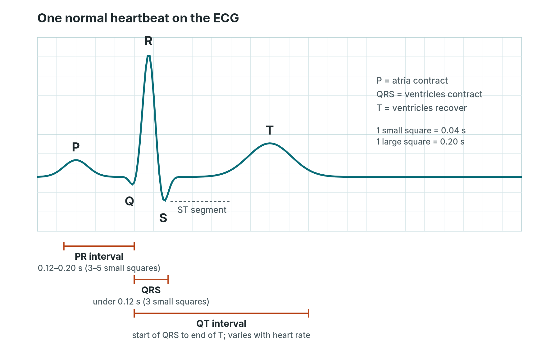
ECG basics: a Cath Lab 101 guide
Reviewed by Rodnie Oro, cath lab charge nurse · Last reviewed: October 2026 · Education only, not medical advice
ECG in one minute
What it is: an ECG (electrocardiogram) is a painless recording of the heart's electrical activity, picked up by sticky electrodes on the skin. It shows how fast the heart is beating, whether the rhythm is regular, and whether part of the heart muscle may be short of blood.
Who it's for: nearly every patient who comes through the cath lab. A 12-lead ECG is taken before most procedures, the monitor shows a live ECG throughout, and a repeat ECG is often taken afterwards.
Why it matters in the lab: the ECG is often the first sign that something is changing: a new rhythm, a slow heart rate, or ST changes during a balloon inflation.
Usually: a 12-lead recording takes under 5 minutes. Rhythm monitoring runs for the whole case.
On this page: What is an ECG? · 12-lead placement · The normal waveform · Reading a strip · Key rhythms · STEMI patterns · During procedures · Checklist · First time in the lab? · FAQ
What is an ECG?
Every heartbeat starts with a small electrical signal. In a normal heart it begins in the sinus node (the heart's natural pacemaker, high in the right atrium), spreads across both atria, pauses briefly at the AV node, then races down the bundle of His and the bundle branches into the ventricles.
Electrodes on the skin pick up this signal. The ECG machine draws it as a line on a grid: up when the signal travels towards an electrode, down when it travels away.
You will hear a few different ECGs in the lab:
- 12-lead ECG: 10 electrodes give 12 "views" of the heart. Used for diagnosis, before and after procedures, and when anything changes.
- 3- or 5-lead monitoring: the live trace on the lab monitor. Good for rate and rhythm, not for diagnosing a heart attack.
- Intracardiac signals: recorded from catheters inside the heart during an EP study. See Electrophysiology (EP) study.
New to the acronyms? See the Cath Lab Abbreviations A–Z.
12-lead electrode placement
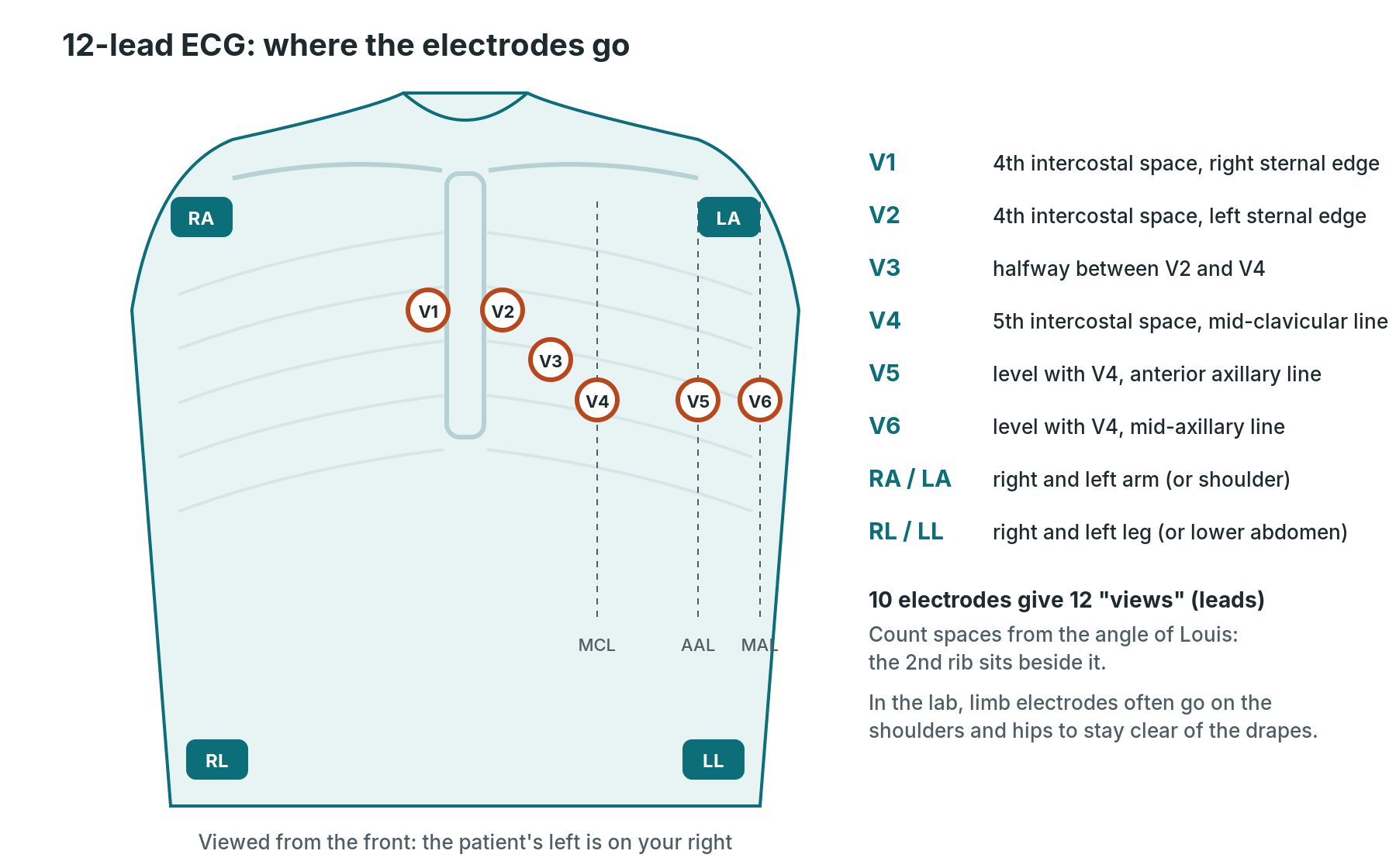
V1 to V6 sit across the left chest in a curve. The four limb electrodes (RA, LA, RL, LL) give the limb leads. In the lab they are often placed on the shoulders and hips to keep the chest clear for the sterile field.
| Electrode | Where it goes |
|---|---|
| V1 | 4th intercostal space, right edge of the sternum |
| V2 | 4th intercostal space, left edge of the sternum |
| V3 | halfway between V2 and V4 |
| V4 | 5th intercostal space, mid-clavicular line |
| V5 | same level as V4, anterior axillary line |
| V6 | same level as V4, mid-axillary line |
| RA / LA | right and left arm (or shoulder) |
| RL / LL | right and left leg (or lower abdomen) |
Finding the spaces: feel for the bony ridge near the top of the sternum (the angle of Louis). The 2nd rib joins beside it. The space below is the 2nd intercostal space. Count down to the 4th for V1 and V2.
Getting a clean trace: explain what you are doing and keep the patient covered as much as you can. Dry the skin, clip hair if needed, and make sure the patient is relaxed, warm and still. Use fresh electrodes. Most poor traces are caused by muscle tremor, a loose electrode or swapped limb leads.
The 12 leads
- Limb leads (I, II, III, aVR, aVL, aVF) look at the heart from the front, in an up-down and side-to-side plane.
- Chest leads (V1–V6) look across the heart from the right side of the sternum round to the left side of the chest.
- Extra leads: right-sided leads (V4R) and posterior leads (V7–V9) are added when a right ventricle or posterior heart attack is suspected, as per centre protocol.
The normal waveform
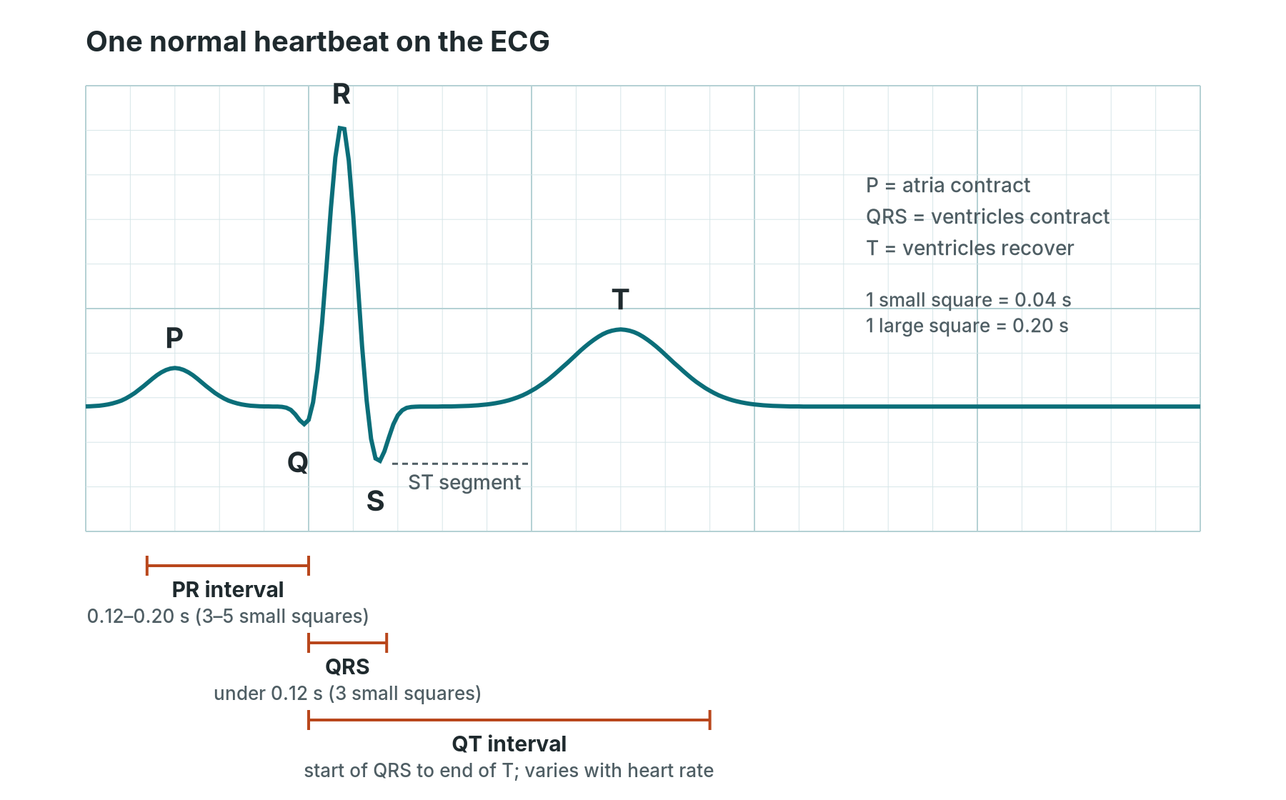
One normal heartbeat. P: atria contract. QRS: ventricles contract. T: ventricles recover, ready for the next beat.
The ECG paper is a grid. At the standard speed (25 mm per second):
- 1 small square = 0.04 seconds
- 1 large square (5 small squares) = 0.20 seconds
- 5 large squares = 1 second
| Part | What it shows | Normal (adult) |
|---|---|---|
| P wave | the atria contracting | small, rounded, upright in lead II |
| PR interval | start of P to start of QRS: the pause at the AV node | 0.12–0.20 s (3–5 small squares) |
| QRS complex | the ventricles contracting | under 0.12 s (under 3 small squares) |
| ST segment | end of QRS to start of T | flat, level with the baseline |
| T wave | the ventricles recovering | usually upright in most leads |
| QT interval | start of QRS to end of T | changes with heart rate; corrected as QTc |
The point where the QRS ends and the ST segment begins is called the J point. You measure ST elevation or depression just after it.
Reading a rhythm strip, step by step
Always look at the patient first. A flat line with a talking patient is a lead off. Then use the same six steps every time:
- Rate. Count the QRS complexes in a 6-second strip (30 large squares) and multiply by 10. For a regular rhythm you can also divide 300 by the number of large squares between two R waves. Normal is 60–100 beats per minute.
- Rhythm. Are the R waves evenly spaced? Mark two R waves on a piece of paper and walk it along the strip. Regular, regularly irregular, or irregularly irregular?
- P waves. Is there a P wave? Is there one before every QRS? Is there a QRS after every P?
- PR interval. Is it normal (3–5 small squares), long, or changing from beat to beat?
- QRS width. Narrow (under 3 small squares) means the beat is using the normal pathway. Broad means it starts in the ventricles or the conduction is blocked.
- ST segment and T waves. On a 12-lead, look for ST elevation, ST depression or new T wave inversion.
Then put it together and name the rhythm. If you are not sure, describe what you see ("broad, regular, about 180") and get help. A clear description is safer than a wrong label.
Key rhythms the cath lab sees
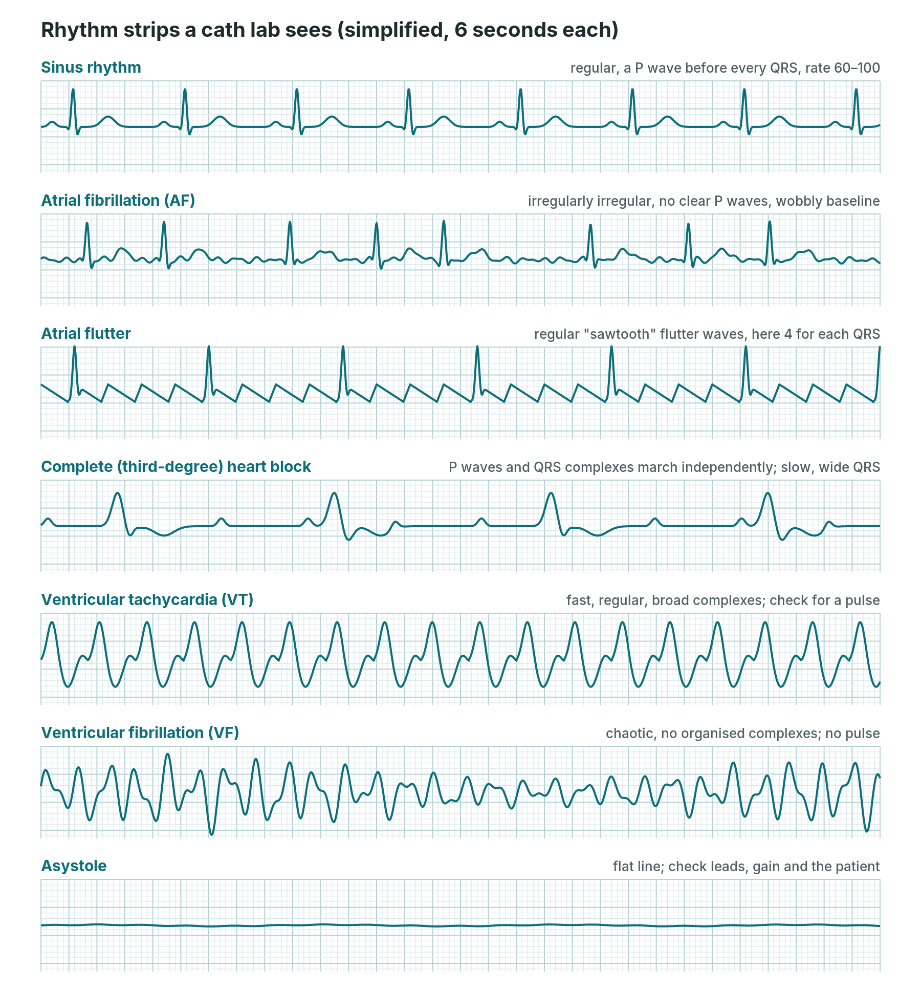
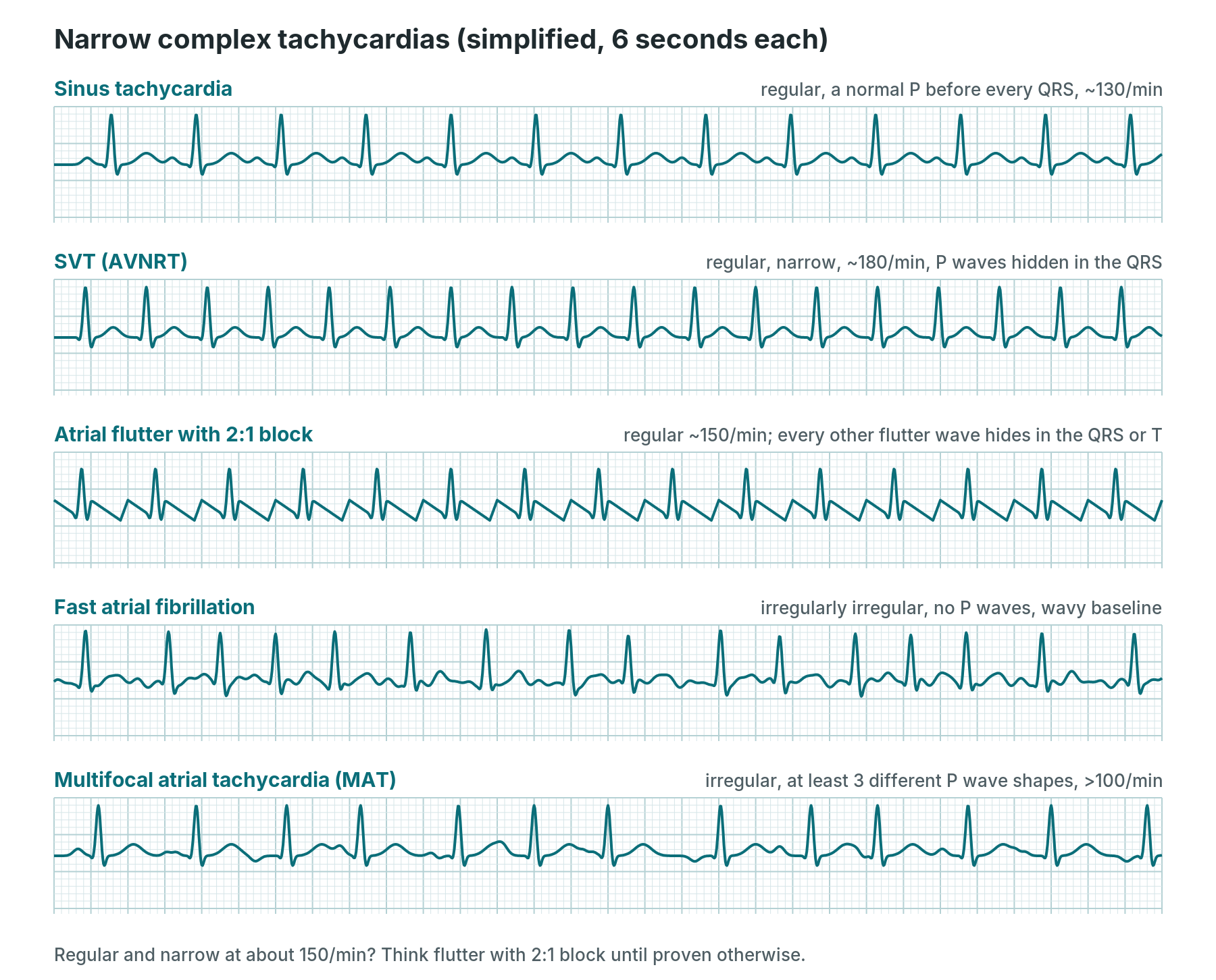
Simplified drawings to show the pattern of each rhythm. Real traces are messier.
Sinus rhythm. Regular, a P wave before every QRS, rate 60–100. Under 60 is sinus bradycardia; over 100 is sinus tachycardia. Both are common in the lab: slow with vagal reactions or right coronary work, fast with pain, anxiety, bleeding or dehydration.
Atrial fibrillation (AF). Irregularly irregular, with no clear P waves and a wobbly baseline. The atria quiver instead of contracting. Common in older patients and in TAVI and mitral work. Tell the operator if it is new.
Atrial flutter. A regular "sawtooth" baseline, often best seen in II, III and aVF. The QRS is usually regular, with a fixed ratio such as 2:1 or 4:1. A regular narrow rhythm at about 150 may be flutter with 2:1 block.
Heart blocks. The signal is delayed or stopped at the AV node.
- First-degree: every P is followed by a QRS, but the PR interval is long (over 5 small squares).
- Second-degree, Mobitz I (Wenckebach): the PR gets longer each beat until one P has no QRS. Then the cycle repeats.
- Second-degree, Mobitz II: the PR stays the same, but some P waves suddenly have no QRS. This can progress to complete block.
- Third-degree (complete) heart block: P waves and QRS complexes march independently. The QRS is often slow and broad.
New heart block can happen during TAVI, alcohol septal ablation, VSD closure and right coronary work. It may need a temporary pacing wire or a permanent pacemaker.
Ventricular tachycardia (VT). Fast, regular and broad. It can occur with or without a pulse. Check the patient and the pulse straight away and call for help.
Ventricular fibrillation (VF). Chaotic and disorganised, with no pulse. This is a cardiac arrest. Start the arrest protocol and defibrillate as per centre protocol. In the lab it can happen with a blocked or reopened coronary artery, or with contrast or a catheter in a coronary.
Asystole and PEA. Asystole is a near-flat line. First check it is real: leads on, gain turned up, a second lead viewed. PEA (pulseless electrical activity) is the dangerous one: the monitor can show a fairly normal-looking rhythm while the patient has no pulse. In the lab, think of causes such as tamponade, major bleeding or a blocked artery. Treat the patient, not the monitor.
Arrest drugs, shock energies and pacing settings: always as per centre protocol and current resuscitation guidelines.
STEMI patterns and territories
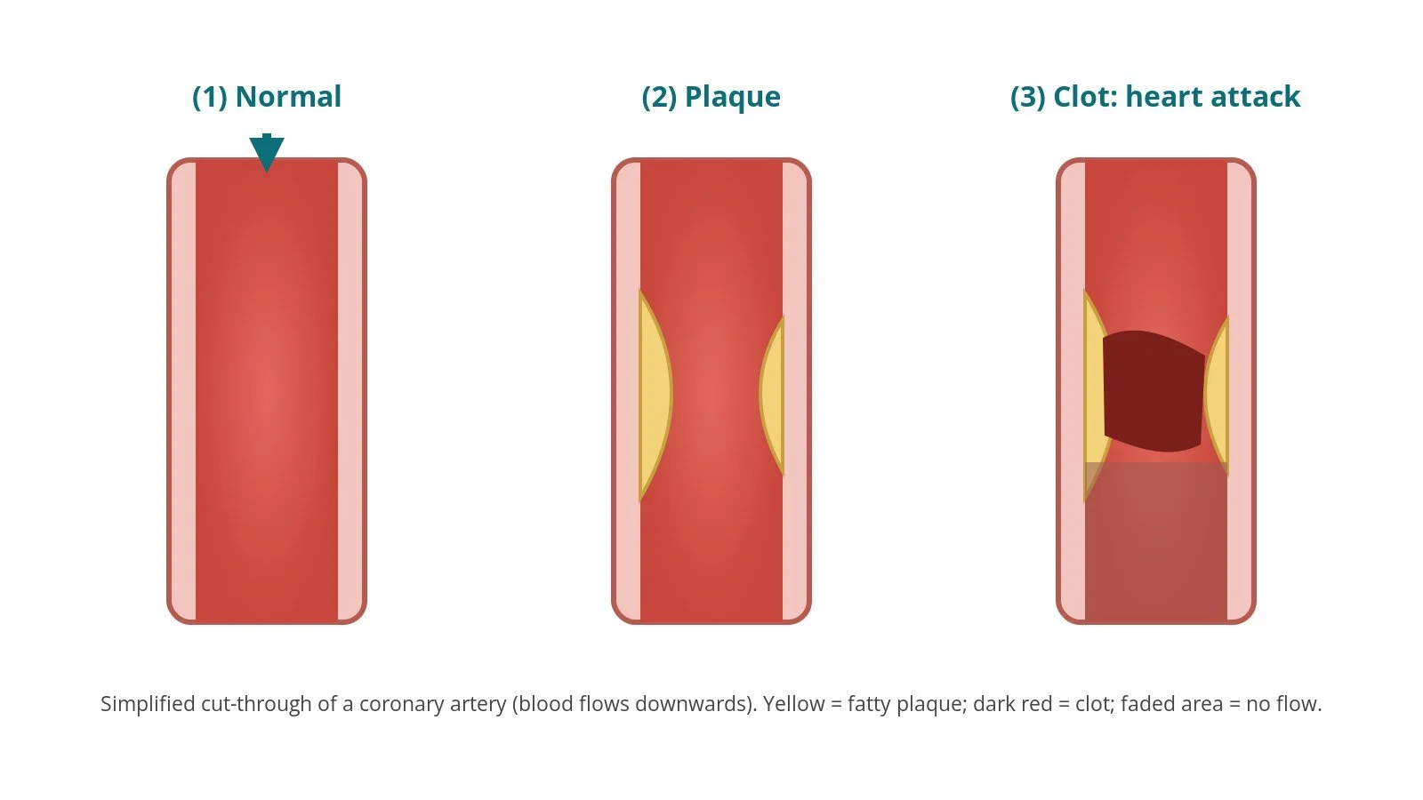
A STEMI (ST-elevation myocardial infarction) is a heart attack where an artery is usually completely blocked. It shows up on the 12-lead ECG as ST elevation in leads that look at the same area of the heart. It is an emergency, usually treated with primary PCI.
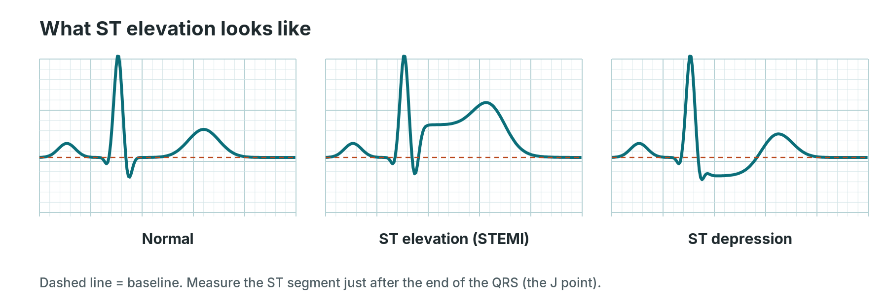
ST elevation: the segment after the QRS sits above the baseline. ST depression: it sits below.
What counts as ST elevation? New ST elevation at the J point in at least two neighbouring leads, of at least 1 mm (1 small square). V2 and V3 have higher thresholds, which vary with age and sex. A new left bundle branch block, or ST depression in V1–V3 (a possible posterior STEMI), can also need urgent review. The decision belongs to the cardiology team, following the 2023 ESC guideline and local pathways.
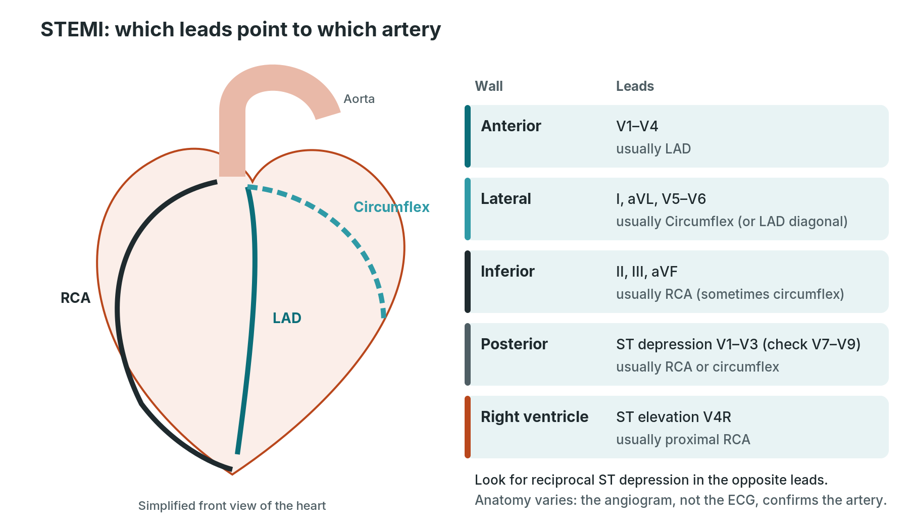
Each group of leads looks at one wall of the heart, and each wall is usually supplied by one artery.
| Wall | Leads with ST elevation | Usual artery |
|---|---|---|
| Anterior | V1–V4 (septal V1–V2) | LAD (left anterior descending) |
| Lateral | I, aVL, V5–V6 | circumflex or an LAD diagonal |
| Inferior | II, III, aVF | RCA (right coronary), sometimes circumflex |
| Posterior | ST depression V1–V3; elevation in V7–V9 | RCA or circumflex |
| Right ventricle | ST elevation in V4R (with inferior STEMI) | proximal RCA |
Reciprocal changes: ST depression in leads on the opposite side of the heart. For example, an inferior STEMI often shows ST depression in I and aVL. Reciprocal changes support the diagnosis.
Why this helps in the lab: it tells the team which artery to look at first, and what to expect. Inferior STEMIs often come with slow rhythms and heart block, and right ventricle involvement can make the blood pressure drop. Any STEMI can be complicated by VT or VF, so keep the pads and defibrillator ready.
The ECG points to the artery. The coronary angiogram confirms it, because anatomy varies.
What to watch for during procedures
The monitor is your early-warning system. Know your baseline, watch the trend, and say what you see out loud.
Before the case
- Record or check a baseline 12-lead ECG. Note the rhythm, rate, PR, QRS width and any existing ST changes, so you can tell what is new.
- Put on defibrillator pads before draping when your centre protocol says so (for example primary PCI, EP and TAVI). Check the defibrillator works.
- Choose monitor leads that show the P wave clearly (often II) and, for coronary work, a lead that looks at the target artery.
- Make sure electrodes and cables are clear of the X-ray field and the sterile area.
During the case: what is expected and what to call out
| Moment | Often seen | Tell the operator if |
|---|---|---|
| Catheter in the heart (angiogram, right heart catheter) | extra beats (ectopics) as the catheter touches the heart wall | runs of VT, or beats that don't stop when the catheter moves |
| Contrast in the right coronary | brief slowing of the heart rate | a long pause, heart block or low blood pressure |
| Balloon inflation (PCI) | brief ST changes and chest pain in that territory | ST changes or pain that don't settle after the balloon is down; VT or VF |
| Reopening a blocked artery (primary PCI) | reperfusion rhythms, such as a slow broad rhythm, or slowing of the heart rate | VF, VT, or a drop in blood pressure |
| Valve, septal and structural work (TAVI, alcohol septal ablation, VSD closure) | rapid pacing runs on purpose; brief rhythm changes | new heart block, especially if it doesn't go away |
| EP and ablation | rhythms triggered on purpose | anything the patient can't tolerate, or a blood pressure drop |
| Groin or wrist access, sheath removal | slow heart rate and low blood pressure (vagal reaction) | it doesn't recover quickly |
Always check the patient as well as the screen: colour, breathing, conscious level, pain, nausea and blood pressure. A sudden fall in blood pressure with a slowing heart rate in a patient who has had a puncture into the heart (for example transseptal work or pacing) may be tamponade. Say it early. See Pericardiocentesis.
After the case: record a repeat 12-lead ECG if your centre protocol asks for one, and compare it with the baseline. This is routine after PCI, TAVI, pacing and structural work in many labs.
Cath lab ECG checklist (printable)
Equipment, brands and settings vary by unit. Everything below is generic. Follow your own centre protocol.
For a 12-lead ECG
- ECG machine, charged, with paper
- fresh, in-date electrodes (radiolucent electrodes are useful in the lab)
- skin prep: gauze or wipes, and a clipper for chest hair
- check the patient's name and ID details on the machine before you record
- standard settings: 25 mm/s and 10 mm/mV, unless asked otherwise
For the case
- lab monitor with a 3- or 5-lead ECG, invasive pressure and oxygen saturation
- defibrillator pads on, defibrillator checked, as per centre protocol
- temporary pacing kit available, as per centre protocol
- arrest trolley and drugs checked, with doses as per centre protocol
Write down or annotate
- baseline rhythm and ST changes
- the time of any rhythm change and what was happening at that moment
- any shocks, pacing and drugs given, as per centre protocol
First time in the cath lab?
💡 First-time tip
Look at the patient before the monitor. A flat line with a patient who is talking to you is a lead off. Know the baseline. Glance at the pre-procedure ECG so you know what is old and what is new. Watch the trend, not just one beat. Rate slowly falling, ST slowly rising and blood pressure slowly dropping all matter. Say what you see. "Heart rate's dropping, 45" is a perfect sentence. In a good lab, calling it out is always welcome. Learn where the pads and the defibrillator are before the case starts, not during it.
FAQ
Does an ECG hurt?
No. It just records the heart's own electrical signals. Nothing is sent into the body. Peeling off the electrodes can sting a little.
What's the difference between the monitor and a 12-lead?
The monitor shows a few leads all the time and is best for rate and rhythm. A 12-lead takes a snapshot from 12 angles and is needed to look for a heart attack or other changes.
Why are the limb electrodes on the shoulders and hips in the lab?
To keep them out of the X-ray picture and the sterile field. It changes the trace slightly, so note it on the ECG if your centre asks you to.
Why does the heart rate drop when contrast goes into the right coronary?
The right coronary often supplies the sinus and AV nodes, so contrast or a blocked artery there can slow the heart. It usually recovers within seconds, but tell the operator.
Can I diagnose a heart attack from the monitor?
No. Monitor leads can hint at ST changes, but a full 12-lead ECG is needed, and the diagnosis is made by the medical team.
Related Cath Lab 101 guides
- Heart attack (acute MI) and primary PCI
- PCI (coronary angioplasty and stents)
- Coronary angiogram
- Electrophysiology (EP) study
- Radiofrequency (RF) ablation · AF cryoablation · Pulsed field ablation
- Permanent pacemaker (PPM) and ICD · Subcutaneous ICD (S-ICD)
- Temporary pacing wire
- Ventricular septal defect (VSD) closure
- Alcohol septal ablation (ASA)
- TAVI procedure
- Pericardiocentesis
- Cath Lab Abbreviations A–Z
- ECG advanced: a Cath Lab 101 guide
Sources
All checked on 22 October 2026. Each source's own date is shown where it gives one.
- NHS. Electrocardiogram (ECG). nhs.uk/tests-and-treatments/electrocardiogram
- British Heart Foundation. Electrocardiogram (ECG). bhf.org.uk
- Campbell B, Richley D, Ross C, Eggett CJ. Clinical Guidelines by Consensus: Recording a standard 12-lead electrocardiogram. SCST, 2024 (issued September 2024, review September 2028). scst.org.uk
- Byrne RA, et al. 2023 ESC Guidelines for the management of acute coronary syndromes. Eur Heart J 2023;44(38):3720–3826. PubMed 37622654
- Thygesen K, et al. Fourth universal definition of myocardial infarction (2018). Eur Heart J 2019;40(3):237–269. PubMed 30165617
- Resuscitation Council UK. 2021 Resuscitation Guidelines: Adult advanced life support. resus.org.uk
Disclaimer
Education only. This page is for general education for healthcare students, cath lab staff and curious readers. It isn't medical advice and doesn't replace advice from your own doctor, cardiology team or local unit protocols. ECG interpretation in clinical practice must be confirmed by a qualified clinician. Equipment and medicines are described generically. No product or manufacturer is recommended. If you have chest pain now, call 999.
Disclosure: I work in the medical device industry. This page is independent education and isn't sponsored or reviewed by any company.
All diagrams on this page are original illustrations created for rodnieoro.com. The rhythm strips are simplified drawings, not recordings from patients.