Temporary pacing wire insertion: a Cath Lab 101 guide
Reviewed by Rodnie Oro, cath lab charge nurse · Last reviewed: October 2026 · Education only, not medical advice
Temporary pacing in one minute
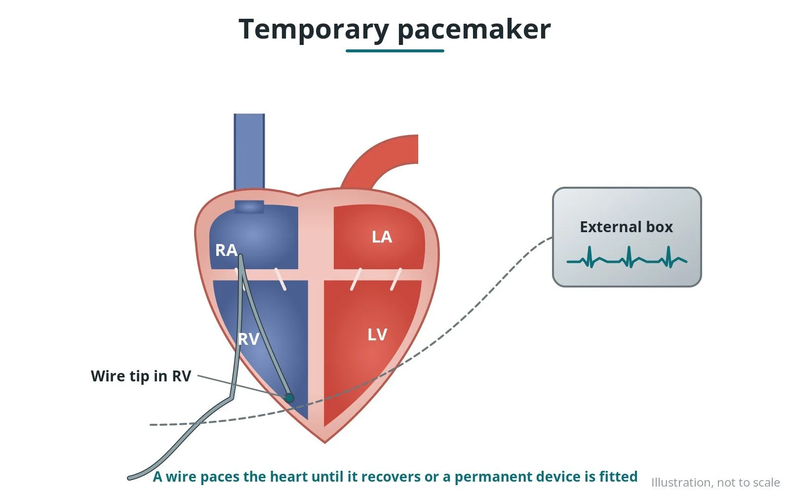
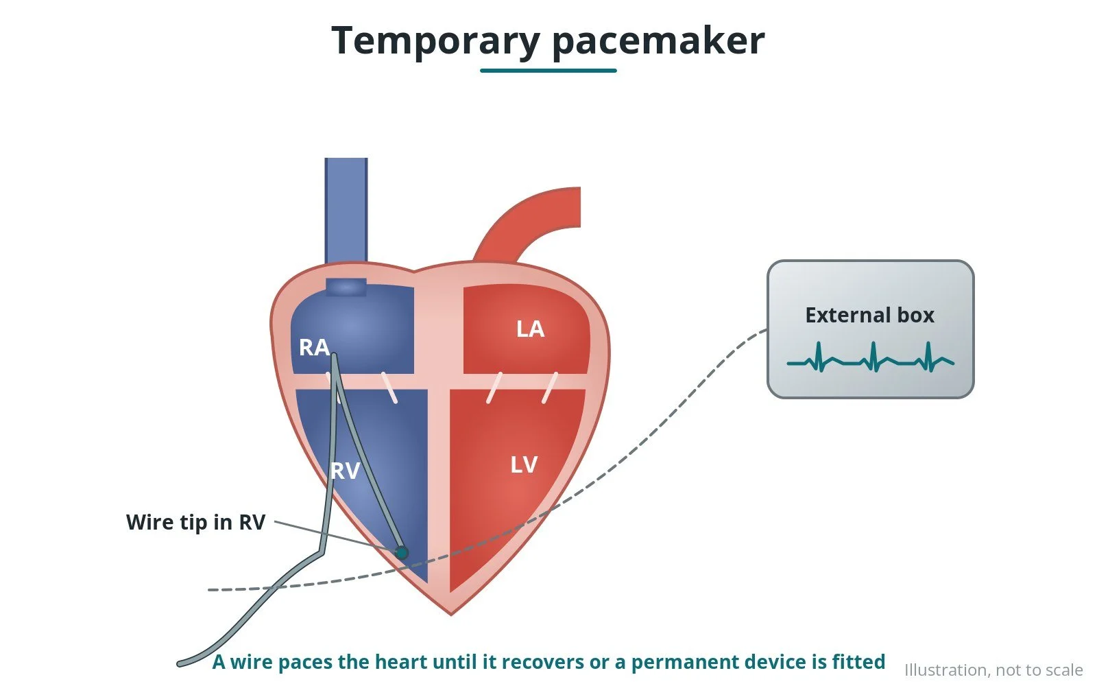
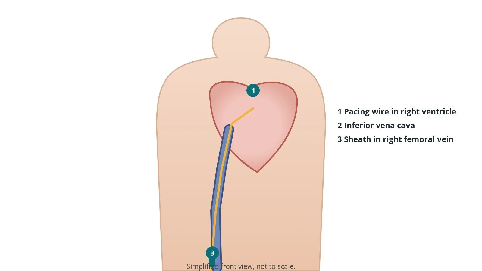
What it is: passing a thin pacing wire through a vein into the right ventricle and connecting it to an external pacing box, to keep the heart beating at a safe rate for a short time.
Who it is for: people with a dangerously slow heart rate (for example heart block) until it recovers or a permanent pacemaker is fitted, and during some procedures.
Main risks: the wire moving out of place, infection, bleeding, rhythm problems and rarely a hole in the heart.
Usually: under local anaesthetic, 20–40 minutes; the wire stays in for hours to a few days.
On this page: What is temporary pacing? · Who is it for? · When is it done? · Step by step · Recovery · Risks · Kit list · First time in the lab? · FAQ
What is temporary pacing?
The heart has its own electrical system. If it fails, the heart can beat too slowly.
A temporary pacing wire:
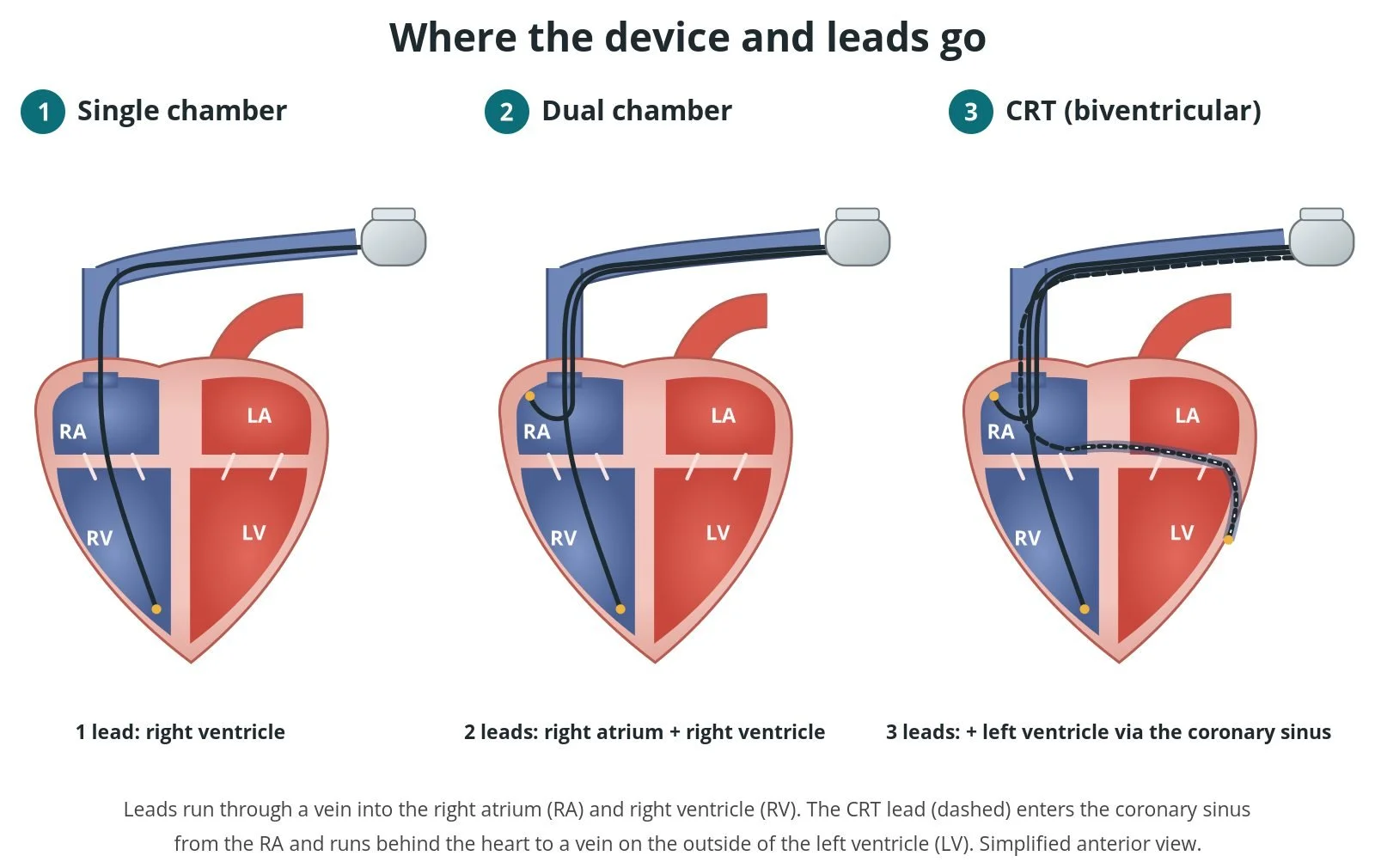
- is passed through a vein (neck, arm or groin) into the right ventricle
- is connected to a pacing box outside the body
- sends small electrical pulses so the heart beats at a set rate
It is a short-term fix. A permanent pacemaker may follow if the problem does not recover.
New to the acronyms? See the Cath Lab Abbreviations A–Z.
Who is it for?
It may be considered for people with:
- complete heart block or very slow heart rates causing symptoms
- slow rhythms after a heart attack or heart surgery
- during procedures such as TAVI or some atherectomy
- drug-related slow rhythms until they wear off
The decision is made by your heart team, who will talk through the reasons, the alternatives and the risks with you.
When is it done?
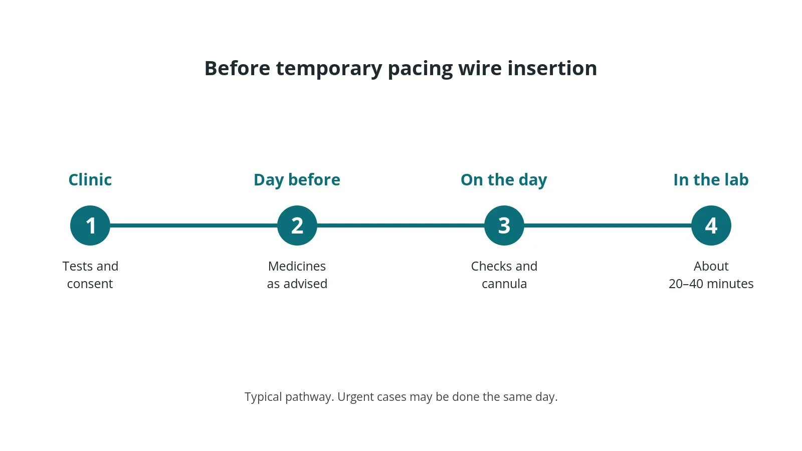
It is often urgent. Before it, the team will:
- check the ECG and blood tests
- explain and ask for consent if possible
- give medicines or external pacing if needed, as per centre protocol
Step by step
1. Getting ready
You lie on the X-ray table. ECG, blood pressure and oxygen levels are monitored. The skin over the access site is cleaned and numbed with local anaesthetic; some people also have light sedation, as per centre protocol.
2. Access
A sheath is placed in a vein: the internal jugular in the neck, the femoral vein in the groin, or an arm vein. Ultrasound guides the needle.
3. Placing the wire
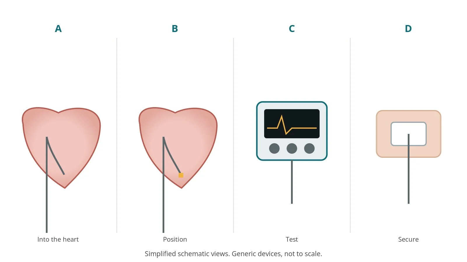
- A. Into the heart. The wire is passed under X-ray into the right ventricle.
- B. Position. The tip is placed against the ventricle wall.
- C. Test. The threshold (the lowest output that works) is checked.
- D. Secure. The wire is secured and the box set.
4. Finishing
The wire and sheath are stitched and dressed. The pacing box is secured and settings recorded.
Recovery and aftercare
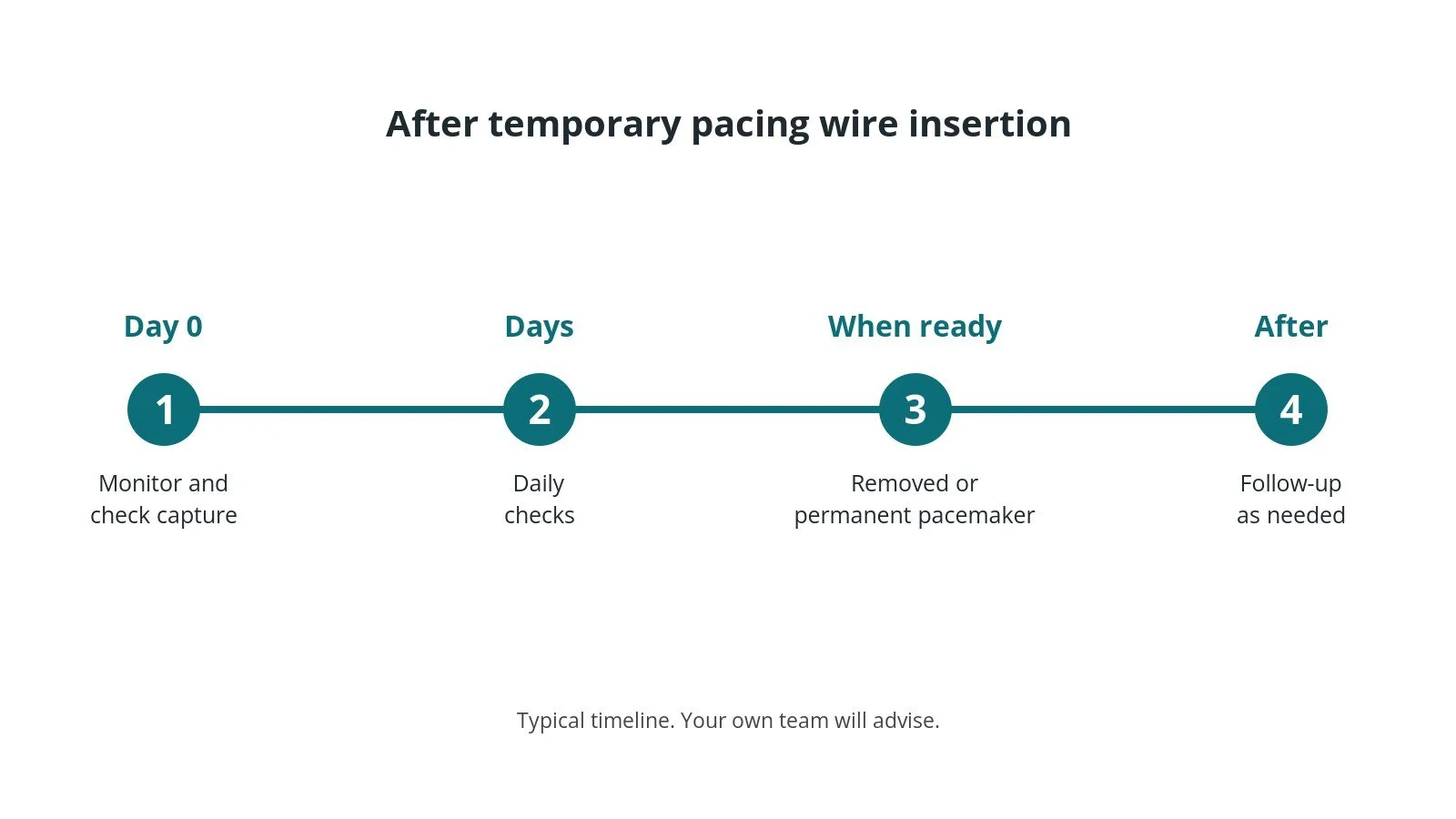
- Monitoring: continuous ECG on a cardiac ward or unit.
- Lying still: limit movement of the arm or leg with the wire, as advised.
- Daily checks: threshold and site checks, as per centre protocol.
- Next step: the wire is removed when the rhythm recovers or a permanent pacemaker is fitted.
Call 999 if you have chest pain, breathlessness, fainting, signs of a stroke, or heavy bleeding or swelling at the access site.
Risks and complications
Your team will explain the risks that apply to you. Possible problems include:
- the wire moving and stopping pacing
- infection or bleeding at the site
- extra heartbeats or other rhythm problems
- punctured lung (neck approach)
- hole in the heart (rare)
Cath lab kit list (printable)
Barts example kit list
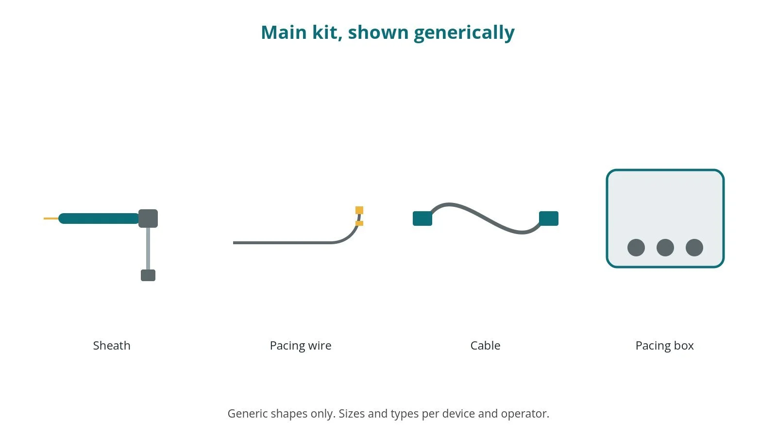
An example from one UK centre, written generically. Every lab has its own protocol, and sizes and doses vary.
- venous sheath, size per device and operator
- temporary pacing wire, per device and operator
- external pacing box with fresh battery
- connecting cable
- ultrasound for vein access
- dressings and suture
Procedural steps (Barts example)
- Ultrasound-guided venous access
- Wire to RV apex or septum under X-ray
- Threshold and sensing checked
- Rate and output set, as per centre protocol
- Secure, dress and document settings
First time in the cath lab?
💡 First-time tip
- Fresh battery. Check the pacing box battery.
- Write it down. Record threshold, rate and output.
- Secure the box. Don't let it hang on the cable.
- Watch capture. Every spike should be followed by a beat.
FAQ
Is it permanent?
No. It is a short-term solution.
Can I get up?
Usually limited; the team will advise.
Will I need a pacemaker?
Sometimes; it depends on the cause.
Related Cath Lab 101 guides
- Permanent pacemaker (PPM), ICD and CRT
- TAVI (transcatheter aortic valve implantation)
- Cath Lab Abbreviations A–Z
Sources
All checked on 10 October 2026. Each page's own date is shown where it gives one.
- Glikson M, et al. 2021 ESC Guidelines on cardiac pacing and cardiac resynchronization therapy. Eur Heart J 2021;42(35):3427–3520. escardio.org
- StatPearls (NCBI Bookshelf), search: temporary transvenous pacing. ncbi.nlm.nih.gov/books
Disclaimer
Education only. This page is for general education for healthcare students, cath lab staff and curious readers. It isn't medical advice and doesn't replace advice from your own doctor, heart team or local unit protocols. Equipment and medicines are described generically. No product or manufacturer is recommended. If you think someone is having a heart emergency, call 999.
Disclosure: I work in the medical device industry. This page is independent education and isn't sponsored or reviewed by any company.
All diagrams on this page are original illustrations created for rodnieoro.com.