ECG advanced: a Cath Lab 101 guide
Reviewed by Rodnie Oro, cath lab charge nurse · Last reviewed: October 2026 · Education only, not medical advice
Advanced ECG in one minute
What it is: a deeper guide to abnormal ECGs. For each rhythm or pattern it covers what you see, common causes, symptoms, how it is usually treated, and what to do or call out in the cath lab.
Who it's for: student nurses, new cath lab and coronary care staff, and anyone who has read ECG basics and wants the next step.
Why it matters in the lab: rhythms and ST changes can change within seconds during a case. Spotting the pattern early, and saying it out loud, gives the team time to act.
The golden rule: treat the patient, not the monitor. Check the patient, the pulse and the arterial pressure trace before you name any rhythm.
On this page: Before you start · Quick approach · Fast rhythms · Slow rhythms and heart block · Conduction and inherited patterns · Ischaemia and infarction · Other patterns · Pacing, ectopics and artefact · Arrest rhythms · Summary tables · First time in the lab? · FAQ
Before you start
This guide builds on ECG basics: a Cath Lab 101 guide, which covers electrode placement, the normal waveform and the six-step way to read a strip. If terms like PR interval, QRS width or J point are new, start there.
A few things hold true for every abnormal ECG in this guide:
- Look at the patient first. Are they awake, talking, pale, sweaty, in pain? Is there a pulse and a good arterial trace?
- Compare with the baseline. Most labs record a 12-lead ECG before the case. A pattern that was there yesterday is less worrying than one that appeared five minutes ago.
- Describe before you diagnose. "Broad, regular, about 180, patient is clammy" gets the right help faster than a confident wrong label.
- Know the danger signs. The Resuscitation Council UK lists four life-threatening features in any patient with an abnormal rhythm: shock, syncope (fainting), myocardial ischaemia and severe heart failure. Any of these means the patient is unstable and needs urgent senior help.
- Medicines, shock energies and pacing settings in this guide are always as per centre protocol and current resuscitation guidelines. Drug names are given only so you recognise them.
Quick approach
Fast rhythms (over 100 beats per minute). Ask two questions: is the QRS narrow or broad (under or over 3 small squares), and is it regular or irregular?
| Regular | Irregular | |
|---|---|---|
| Narrow QRS | sinus tachycardia, SVT (AVNRT, AVRT), atrial flutter, atrial tachycardia | atrial fibrillation, flutter with variable block, multifocal atrial tachycardia |
| Broad QRS | VT (treat as VT until proven otherwise), SVT with bundle branch block, antidromic AVRT | AF with bundle branch block, pre-excited AF (WPW), polymorphic VT and torsades |
Slow rhythms (under 60 beats per minute). Ask: is there a P wave before every QRS, and a QRS after every P? Then decide whether the patient is unstable, and whether there is a risk of asystole. The Resuscitation Council UK lists this risk as recent asystole, Mobitz II block, complete heart block with a broad QRS, or a pause of more than 3 seconds.
ST and T wave changes. Ask: is it in one coronary territory with reciprocal change (think heart attack), or everywhere (think pericarditis, electrolytes, drugs or a normal variant)? Is there anything that changes the baseline, such as bundle branch block, pacing or LVH?
Fast rhythms (tachyarrhythmias)
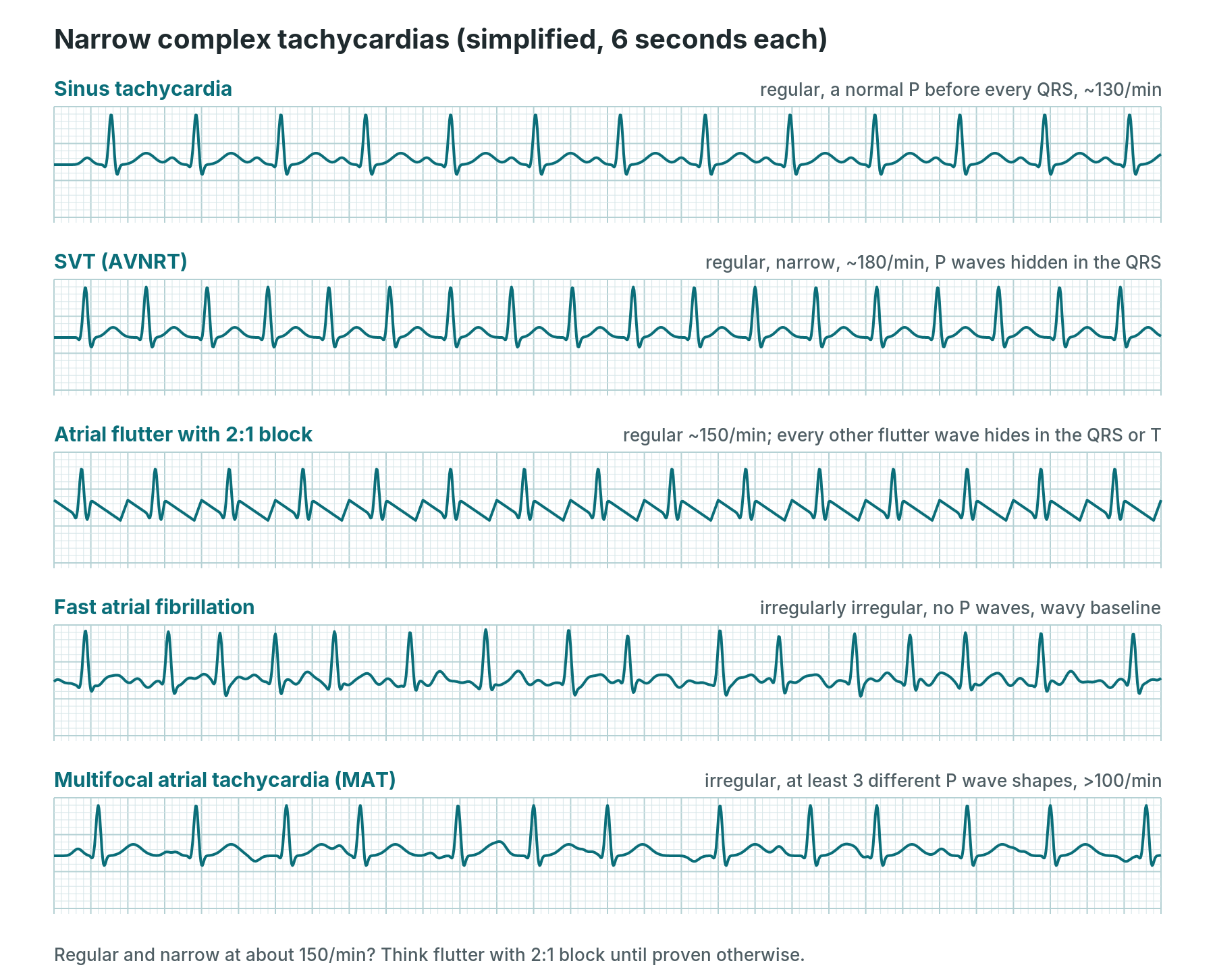
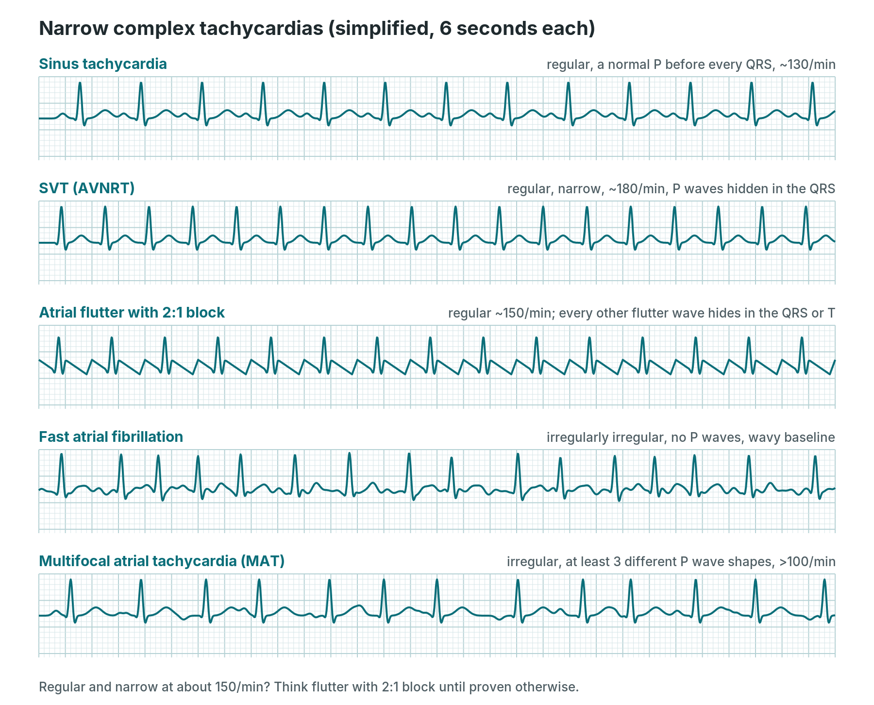
Simplified drawings, 6 seconds each. Real traces are messier.
Sinus tachycardia
- What you see: regular rhythm over 100 per minute, a normal P wave before every QRS, normal PR. It speeds up and slows down gradually.
- Causes: pain, anxiety, fever, bleeding or dehydration, sepsis, low oxygen, anaemia, pulmonary embolism, heart failure, an overactive thyroid, caffeine and drugs such as salbutamol or atropine.
- Symptoms: often none. Sometimes palpitations, breathlessness or the symptoms of the cause.
- Treatment: treat the cause, not the rate. Pain relief, fluids or blood, oxygen, treating infection. Slowing the heart with drugs can be dangerous when the fast rate is compensating for something.
- In the cath lab: a rising heart rate with a falling blood pressure can be the first sign of bleeding (for example a groin or retroperitoneal bleed) or tamponade. Tell the operator and check the access site.
Supraventricular tachycardia (AVNRT and AVRT)
- What you see: regular, narrow and fast, usually 140–250 per minute. In AVNRT the P waves are hidden in or just after the QRS (sometimes a small "pseudo r′" in V1). In AVRT (via an extra pathway) the P wave often sits in the ST segment. It starts and stops suddenly.
- Causes: a re-entry circuit. In AVNRT the circuit is in or around the AV node. In AVRT it uses an accessory pathway, as in Wolff–Parkinson–White. Triggers include caffeine, alcohol, stress, tiredness and sudden movement.
- Symptoms: sudden pounding palpitations, light-headedness, chest tightness, breathlessness, a need to pass urine afterwards. Rarely fainting.
- Treatment: if stable, vagal manoeuvres (such as a modified Valsalva), then adenosine with continuous ECG recording, then a beta-blocker or verapamil, all as per centre protocol. If unstable, synchronised DC cardioversion under sedation or anaesthesia. For recurrent episodes, catheter ablation is a long-term option. Adenosine is avoided when pre-excitation is known.
- In the cath lab: SVT is often triggered on purpose during an EP study. It can also start when a catheter or wire touches the heart. Record a strip or 12-lead when it starts and stops: the onset and offset help the diagnosis.
Atrial fibrillation (AF)
- What you see: irregularly irregular QRS, no clear P waves, a wavy (fibrillatory) baseline. Fast AF usually runs at 110–180 per minute.
- Causes: age, high blood pressure, heart valve disease (especially mitral), heart failure, coronary disease, an overactive thyroid, alcohol, sleep apnoea, infection, after heart surgery, and catheters or wires in the atria.
- Symptoms: palpitations, breathlessness, tiredness, dizziness, chest discomfort, or none at all. The big risk is stroke, from clots forming in the left atrium.
- Treatment: stroke prevention with anticoagulants, based on a stroke risk score. Then rate control (beta-blockers, diltiazem or verapamil, digoxin) or rhythm control (electrical or drug cardioversion, antiarrhythmic drugs, AF cryoablation, radiofrequency ablation or pulsed field ablation). Unstable fast AF needs synchronised DC cardioversion, as per centre protocol.
- In the cath lab: tell the operator about new AF, note the time, and watch the blood pressure. New AF is common during transseptal, mitral and TAVI work. It can affect the anticoagulation plan, so it should be documented.
Atrial flutter
- What you see: regular "sawtooth" flutter waves at about 300 per minute, best seen in II, III, aVF and V1. The QRS is usually regular with a fixed ratio: 2:1 gives a rate of about 150, 4:1 about 75. Variable block makes it irregular.
- Causes: similar to AF. Typical flutter uses a circuit around the tricuspid valve in the right atrium. Also seen after heart surgery and in congenital heart disease.
- Symptoms: palpitations, breathlessness, tiredness, dizziness. Stroke risk similar to AF.
- Treatment: anticoagulation as for AF. Rate control is often harder than in AF. Cardioversion (electrical or drug) as per centre protocol. Catheter ablation of the cavotricuspid isthmus is a common first-line long-term option for typical flutter.
- In the cath lab: a regular narrow rhythm at about 150 is flutter with 2:1 block until proven otherwise. Adenosine, given by the medical team, can briefly slow the ventricles and show the flutter waves.
Atrial tachycardia
- What you see: regular narrow rhythm, usually 100–250 per minute, with P waves that look different from sinus P waves and a flat baseline between them. It may come with AV block.
- Causes: a focus in the atria firing fast. Seen with structural heart disease, scars from previous surgery or ablation, lung disease, and digoxin toxicity (classically atrial tachycardia with block).
- Symptoms: palpitations, tiredness, breathlessness. Long-lasting atrial tachycardia can weaken the heart muscle.
- Treatment: treat the cause (check digoxin levels and potassium). Beta-blockers or calcium channel blockers for rate. Catheter ablation, often with electroanatomical mapping, for recurrent cases.
- In the cath lab: often mapped and ablated in the EP lab. A 12-lead of the tachycardia helps the team find where it comes from.
Multifocal atrial tachycardia (MAT)
- What you see: irregular rhythm over 100 per minute, with at least three different P wave shapes and varying PR intervals. Easily mistaken for AF, but P waves are visible.
- Causes: severe lung disease (such as COPD flare-ups), low oxygen, low potassium or magnesium, sepsis, theophylline.
- Symptoms: usually those of the underlying illness: breathlessness, tiredness.
- Treatment: treat the cause: oxygen, treat the chest infection, correct potassium and magnesium. Some rate-slowing drugs may be used. Cardioversion does not usually help.
- In the cath lab: uncommon. More likely to be seen in a very unwell patient coming from critical care. Tell the team, and check the latest blood gas and electrolytes.
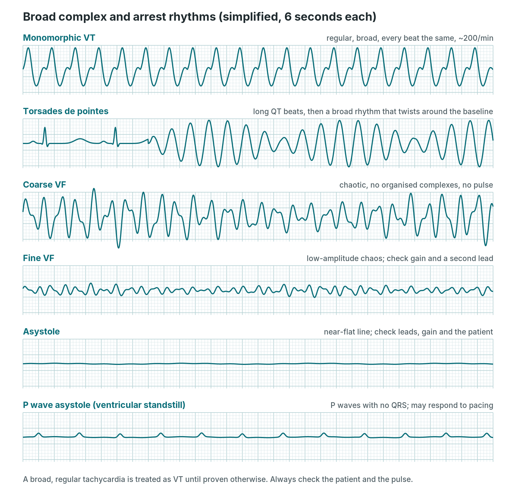
Simplified drawings. A broad regular tachycardia is treated as VT until proven otherwise.
Ventricular tachycardia (VT): monomorphic
- What you see: fast (usually 120–250), regular, broad QRS (over 3 small squares), every complex looking the same. Clues that it is VT rather than SVT with a bundle branch block: P waves marching through independently (AV dissociation), capture or fusion beats, all chest leads pointing the same way (concordance), and a known history of heart attack or heart failure.
- Causes: scar from an old heart attack, heart muscle disease (cardiomyopathy), acute ischaemia, low potassium or magnesium, some drugs, and catheters touching the ventricle.
- Symptoms: palpitations, dizziness, chest pain, breathlessness, fainting. It can be pulseless, which is a cardiac arrest.
- Treatment: no pulse: treat as cardiac arrest (shockable rhythm). Pulse with life-threatening features: synchronised DC cardioversion under sedation or anaesthesia. Pulse and stable: expert help, 12-lead ECG, correct electrolytes; drug treatment (for example amiodarone) or electrical cardioversion as per centre protocol. Long term: treat the cause, consider an ICD or VT ablation.
- In the cath lab: short runs are common when a catheter crosses the aortic valve or sits in the ventricle. Call it if it does not stop when the catheter moves. Look at the arterial pressure trace straight away: no trace and no pulse means arrest.
Polymorphic VT and torsades de pointes
- What you see: fast, broad, chaotic-looking complexes that change shape beat to beat. In torsades de pointes the complexes twist around the baseline, getting bigger and smaller, and the beats before it show a long QT. It often starts after a short–long–short sequence of beats.
- Causes: torsades: QT-prolonging drugs (some antiarrhythmics, antibiotics, antipsychotics, antiemetics and others), low potassium, magnesium or calcium, slow heart rates, inherited long QT syndrome. Polymorphic VT with a normal QT: think acute ischaemia first.
- Symptoms: dizziness, fainting, seizures, or cardiac arrest. It may stop by itself or turn into VF.
- Treatment: pulseless or sustained: defibrillate (an unsynchronised shock, because there is no regular R wave to sync to) as per centre protocol. For torsades: intravenous magnesium, correct potassium, stop QT-prolonging drugs, and speed up the heart (isoprenaline or temporary pacing) as per centre protocol. Polymorphic VT with ischaemia needs urgent revascularisation.
- In the cath lab: check the QTc on the baseline ECG and tell the team about QT-prolonging drugs on the chart. Polymorphic VT during primary PCI or after an artery reopens is treated as an emergency.
Ventricular fibrillation (VF)
- What you see: chaotic, irregular waves of varying size with no recognisable QRS. Coarse VF has large waves. Fine VF can look almost flat: check the gain and a second lead.
- Causes: acute heart attack and blocked arteries, reperfusion, scar, severe electrolyte disturbance, an R-on-T ectopic, electric shock, drugs, inherited conditions such as Brugada or long QT.
- Symptoms: sudden collapse. No pulse, not breathing normally. This is a cardiac arrest.
- Treatment: start the arrest protocol: call for help, CPR, defibrillate as per centre protocol and the Resuscitation Council UK algorithm (see Arrest rhythms).
- In the cath lab: most likely during STEMI work, with contrast or a catheter in a coronary artery, or as an artery reopens. If it is witnessed and monitored with the defibrillator ready, current UK guidance allows up to three quick successive shocks before starting compressions, as per centre protocol.
Slow rhythms and heart block (bradyarrhythmias)
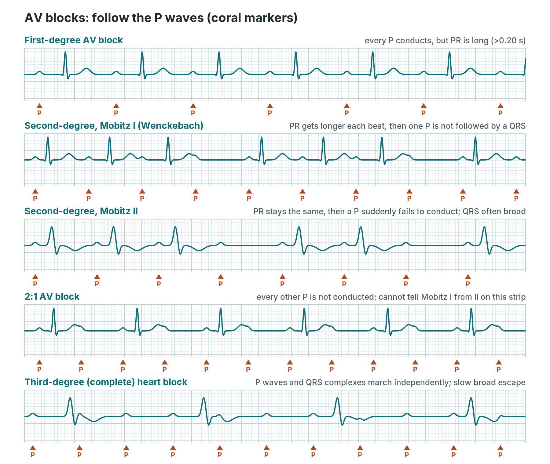
Follow the P waves (coral markers) and ask: does each one produce a QRS, and is the PR the same each time?
Sinus bradycardia
- What you see: regular rhythm under 60 per minute, a normal P wave before every QRS, normal PR.
- Causes: fitness and sleep (normal), vagal reactions (pain, sheath removal, vomiting, fainting), inferior heart attack or right coronary work, drugs (beta-blockers, diltiazem, verapamil, digoxin, amiodarone), an underactive thyroid, hypothermia, raised pressure in the skull, high potassium.
- Symptoms: often none. Dizziness, tiredness, fainting, low blood pressure.
- Treatment: none if well. If there are life-threatening features: atropine, then other drugs (isoprenaline or adrenaline) or pacing, as per centre protocol and the Resuscitation Council UK bradycardia algorithm. Stop or reduce slowing drugs.
- In the cath lab: expected briefly with contrast in the right coronary and with vagal reactions during access or sheath removal. Call it if it does not recover quickly, or if the blood pressure falls.
Sick sinus syndrome (sinus node dysfunction)
- What you see: inappropriate sinus bradycardia, sinus pauses or sinus arrest (no P wave at all for a while), and often tachy-brady syndrome: bursts of AF or flutter followed by long pauses when they stop.
- Causes: age-related scarring of the sinus node, some drugs, heart surgery, infiltrative disease.
- Symptoms: dizziness, fainting, tiredness, palpitations, breathlessness.
- Treatment: stop slowing drugs if possible. A permanent pacemaker for symptoms. Once paced, drugs can safely be used to control the fast rhythms.
- In the cath lab: these patients often come for pacemaker implants. Pauses can follow the end of an AF episode, so have pacing support ready as per centre protocol.
First-degree AV block
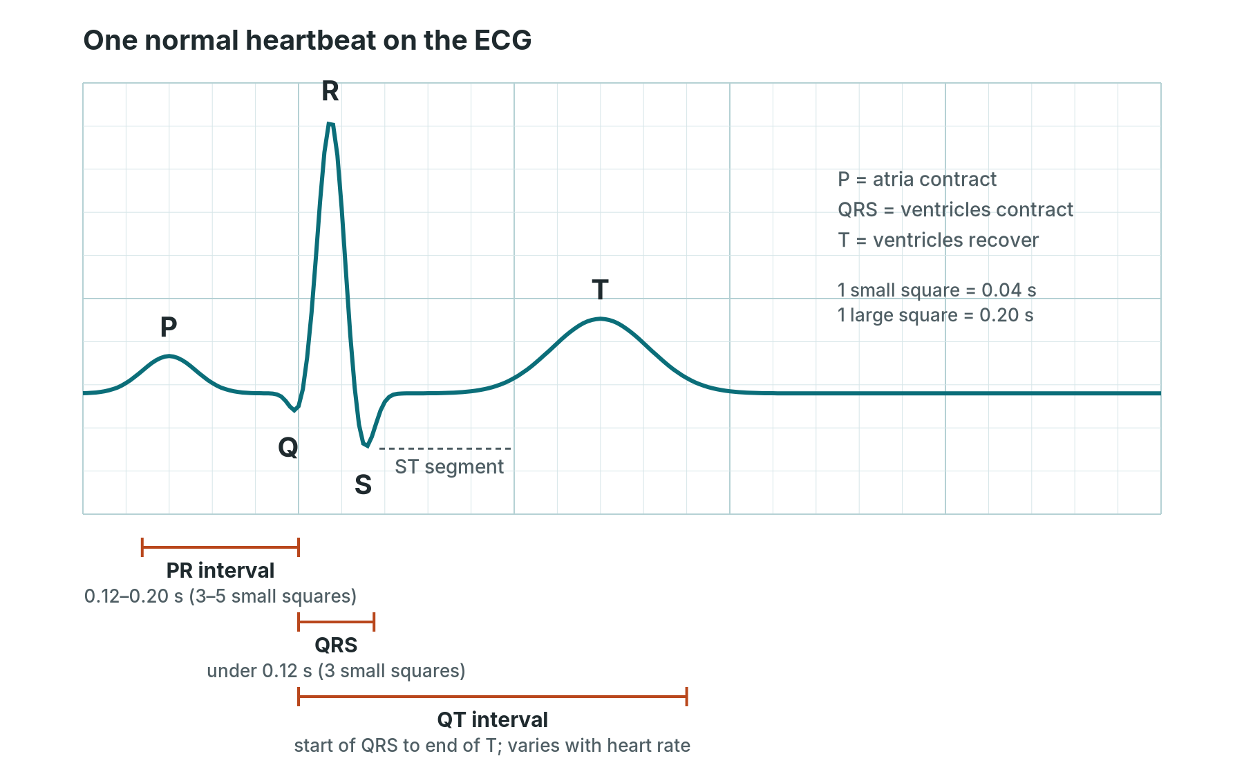
- What you see: every P wave is followed by a QRS, but the PR interval is longer than 0.20 seconds (5 small squares). It stays the same length.
- Causes: athletes and high vagal tone, ageing of the conduction system, drugs (beta-blockers, calcium channel blockers, digoxin), inferior heart attack, low or high potassium, some infections and inflammatory conditions.
- Symptoms: usually none.
- Treatment: usually none. Review drugs and monitor.
- In the cath lab: note it on the baseline. A long PR before TAVI, or a PR that gets longer during or after a valve, septal ablation or VSD case, can be an early warning of higher-degree block.
Second-degree AV block, Mobitz I (Wenckebach)
- What you see: the PR interval gets longer with each beat until one P wave is not followed by a QRS. Then the cycle starts again. The QRS is usually narrow and the rhythm looks "grouped".
- Causes: the block is usually in the AV node. Vagal tone (in sleep and athletes), inferior heart attack, drugs that slow the AV node, myocarditis, after heart surgery.
- Symptoms: usually none. Occasionally dizziness.
- Treatment: usually none, apart from treating the cause. Atropine or pacing only if the patient is unstable, as per centre protocol.
- In the cath lab: often seen with inferior STEMI and usually temporary. Watch for progression and keep pacing support available.
Second-degree AV block, Mobitz II
- What you see: the PR stays the same, then a P wave suddenly fails to conduct, with no warning lengthening. The QRS is often broad because the block is usually lower down, in the His–Purkinje system.
- Causes: scarring of the conduction system, anterior heart attack, after TAVI, alcohol septal ablation, VSD closure or heart surgery, infiltrative disease.
- Symptoms: dizziness, fainting, breathlessness, tiredness.
- Treatment: it can suddenly progress to complete heart block or asystole. It is a "risk of asystole" rhythm. Temporary pacing may be needed, and usually a permanent pacemaker.
- In the cath lab: call it straight away. Make sure the defibrillator with external pacing and temporary pacing wire kit are ready, as per centre protocol.
2:1 AV block
- What you see: every second P wave is not followed by a QRS. Because there are never two conducted beats in a row, you cannot see whether the PR lengthens, so you cannot tell Mobitz I from Mobitz II on this strip alone.
- Causes: as for Mobitz I and II.
- Symptoms: depend on the ventricular rate: often tiredness, dizziness, breathlessness.
- Treatment: a narrow QRS suggests the block is in the AV node (more like Mobitz I). A broad QRS suggests it is lower down (more like Mobitz II), which is higher risk. Look at a longer strip and get expert review.
- In the cath lab: record a long rhythm strip. Treat as higher risk until a senior has reviewed it.
Third-degree (complete) heart block
- What you see: P waves and QRS complexes march independently. The P rate is faster than the QRS rate, and the PR varies at random. The escape rhythm is either junctional (narrow, 40–60) or ventricular (broad, 20–40).
- Causes: ageing and scarring of the conduction system, inferior heart attack (often temporary), anterior heart attack (usually more serious), after TAVI, alcohol septal ablation, VSD closure or heart surgery, drugs, high potassium, and some infections and infiltrative diseases.
- Symptoms: tiredness, dizziness, breathlessness, fainting (sometimes called Stokes–Adams attacks), low blood pressure, heart failure.
- Treatment: if unstable: atropine (more likely to help with a narrow escape), isoprenaline or adrenaline, external pacing, then temporary transvenous pacing, as per centre protocol. Most patients need a permanent pacemaker unless there is a reversible cause.
- In the cath lab: one of the most important rhythms to spot. Say it out loud, check the blood pressure, and get the pacing kit ready. During TAVI, the team may watch the rhythm for longer, or keep a temporary wire in place, as per centre protocol.
Junctional and escape rhythms
- What you see: junctional rhythm: narrow QRS at 40–60 with no P wave, or an inverted P wave just before or after the QRS. Accelerated junctional rhythm: the same at 60–100. Ventricular escape (idioventricular) rhythm: broad QRS at 20–40. Accelerated idioventricular rhythm (AIVR): broad QRS at roughly 50–110, often taking over gently from sinus rhythm.
- Causes: escape rhythms are the heart's back-up when higher pacemakers fail (sinus arrest, heart block). Accelerated junctional rhythm: digoxin toxicity, after heart surgery, inferior heart attack, myocarditis. AIVR: classically a reperfusion rhythm after an artery is reopened.
- Symptoms: depend on the rate. Slow escape rhythms can cause dizziness, low blood pressure or fainting.
- Treatment: treat the cause. Never try to suppress an escape rhythm with antiarrhythmic drugs: it may be the only thing keeping the heart going. AIVR is usually short-lived and not treated, unless the patient is unstable.
- In the cath lab: AIVR during primary PCI as the artery opens is common. Tell the operator, check the blood pressure, and watch for VT or VF.
Conduction problems and inherited patterns
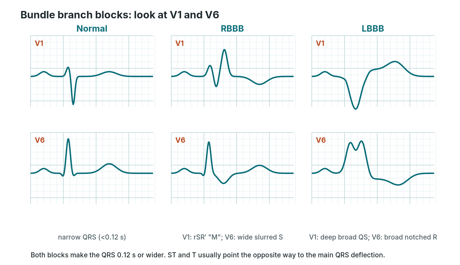
In bundle branch block, one side of the ventricles is activated late, so the QRS widens. V1 and V6 show which side.
Right bundle branch block (RBBB)
- What you see: QRS 0.12 s or wider; an rSR′ ("M") pattern in V1; a wide, slurred S wave in I and V6. T waves in V1–V3 are often inverted. Incomplete RBBB has the same shape with a QRS of 0.11–0.12 s.
- Causes: can be normal, especially in younger people. Also right heart strain (pulmonary embolism, lung disease), coronary disease, atrial septal defect, after heart surgery, and catheters in the right ventricle.
- Symptoms: none from the block itself.
- Treatment: none on its own. Look for the cause if it is new.
- In the cath lab: a right heart catheter can cause a temporary RBBB. In a patient who already has LBBB, that can cause complete heart block. Check the baseline ECG before right heart work and keep pacing support ready, as per centre protocol.
Left bundle branch block (LBBB)
- What you see: QRS 0.12 s or wider; a broad, notched or slurred R wave in I, aVL, V5 and V6, with no small septal q waves; a deep QS or rS wave in V1. The ST segment and T wave point the opposite way to the QRS (discordant).
- Causes: coronary disease, high blood pressure, aortic stenosis, cardiomyopathy, ageing of the conduction system, and after TAVI (a common new finding).
- Symptoms: none from the block itself. It often signals underlying heart disease.
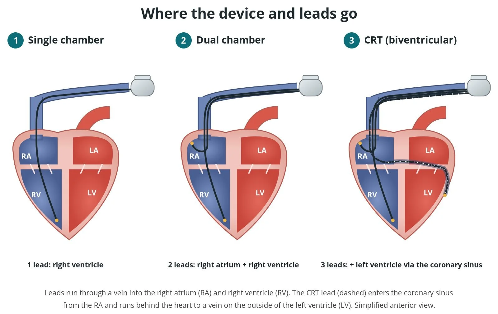
- Treatment: treat the cause. In patients with heart failure and LBBB, cardiac resynchronisation therapy (biventricular pacing) may be considered.
- In the cath lab: LBBB hides the usual signs of a heart attack. A patient with LBBB and symptoms of an ongoing heart attack is managed along a STEMI pathway, and the Sgarbossa criteria help (see Ischaemia). Remember the risk of complete heart block with right heart catheters.
Fascicular blocks (hemiblocks)
- What you see: left anterior fascicular block: left axis deviation (about −45° or more), small q and tall R in I and aVL, small r and deep S in II, III and aVF, QRS under 0.12 s. Left posterior fascicular block: right axis deviation, small r and deep S in I and aVL, small q and tall R in II, III and aVF, once other causes of right axis deviation are excluded.
- Causes: coronary disease, high blood pressure, aortic valve disease, cardiomyopathy, ageing of the conduction system. Posterior block is rarer and usually means more extensive disease.
- Symptoms: none on their own.
- Treatment: none on their own.
- In the cath lab: note them on the baseline. They matter more in combination with other blocks.
Bifascicular and "trifascicular" block
- What you see: bifascicular block: RBBB plus left anterior or left posterior fascicular block. "Trifascicular" block is used loosely, usually for bifascicular block plus first-degree AV block. True trifascicular block means alternating bundle branch block, or bifascicular block with complete heart block.
- Causes: widespread disease of the conduction system, coronary disease, valve disease, after valve procedures.
- Symptoms: none, or dizziness and fainting if intermittent complete block occurs.
- Treatment: monitoring. Unexplained fainting needs prompt specialist review, as a pacemaker may be needed. Alternating bundle branch block usually needs pacing.
- In the cath lab: these patients have little conduction reserve. During TAVI, right heart work or septal procedures, have pacing ready as per centre protocol.
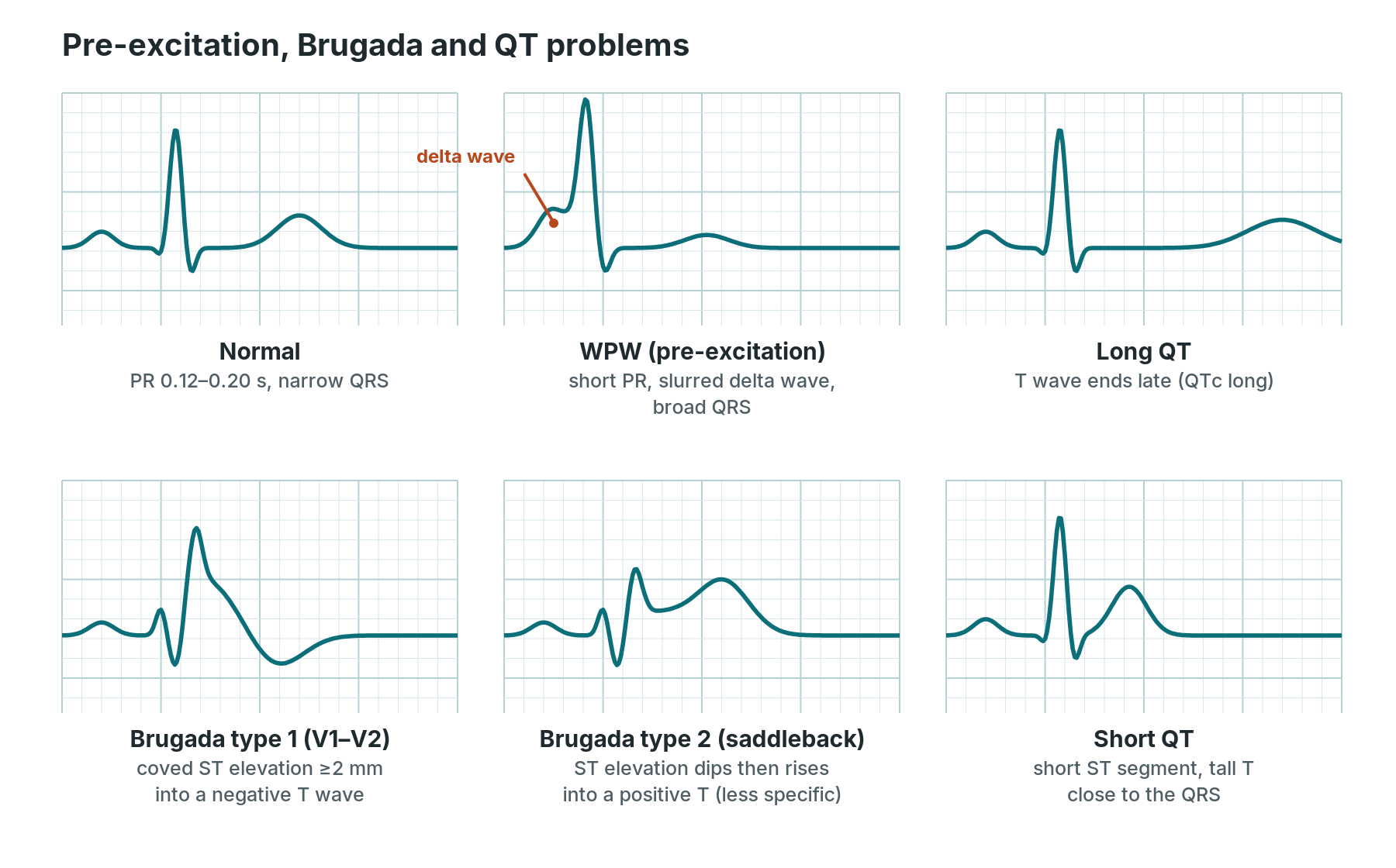
Simplified single beats. Brugada patterns are looked for in V1 and V2.
Wolff–Parkinson–White (WPW) and pre-excitation
- What you see: in sinus rhythm: a short PR (under 0.12 s), a delta wave (a slurred start to the QRS), a broad QRS and secondary ST–T changes. During AVRT, usually a regular narrow tachycardia. Pre-excited AF: an irregular, very fast, broad and bizarre rhythm with QRS shapes that vary.
- Causes: an extra electrical connection (accessory pathway) between the atria and ventricles, present from birth.
- Symptoms: often none (the "WPW pattern"). With arrhythmias ("WPW syndrome"): palpitations, dizziness, fainting. Pre-excited AF can turn into VF.
- Treatment: AVRT: as for SVT. Pre-excited AF: avoid drugs that block the AV node (adenosine, verapamil, diltiazem, digoxin and beta-blockers), because they can speed up conduction down the pathway. Unstable: synchronised DC cardioversion. Stable: specialist drugs as per centre protocol. Long term: catheter ablation of the pathway.
- In the cath lab: accessory pathways are mapped and ablated in the EP lab. If you see an irregular broad fast rhythm in a young patient, ask "could this be pre-excited AF?" before anyone reaches for AV-nodal drugs.
Long QT
- What you see: a QT interval that is long for the heart rate. The QTc (corrected QT) is roughly over 450 ms in men and over 460–470 ms in women; over 500 ms is high risk. The T wave may be broad, notched or have a U wave on it.
- Causes: drugs (some antiarrhythmics, antibiotics, antifungals, antipsychotics, antidepressants, antiemetics and methadone), low potassium, magnesium or calcium, slow heart rates, hypothermia, ischaemia, brain injury, and inherited long QT syndromes.
- Symptoms: often none. Fainting, seizures or palpitations, often triggered by exercise, emotion or sudden noise in inherited types. Risk of torsades and sudden death.
- Treatment: stop QT-prolonging drugs, correct electrolytes, and treat slow rates. Inherited types: beta-blockers, lifestyle advice, sometimes an ICD.
- In the cath lab: check the QTc before giving QT-prolonging drugs. Some antiemetics and antiarrhythmics used around procedures prolong the QT. Flag a long QTc to the team.
Short QT
- What you see: a short QT (QTc roughly under 340–360 ms), often with tall, peaked T waves and very little ST segment.
- Causes: rare inherited short QT syndrome, high calcium, digoxin, high body temperature, acidosis.
- Symptoms: palpitations, fainting, AF, or sudden cardiac arrest in inherited types. Often none.
- Treatment: treat acquired causes. Inherited type: specialist care, sometimes an ICD.
- In the cath lab: rare. Note it on the baseline and check the calcium and drug chart.
Brugada pattern and syndrome
- What you see: type 1 (the diagnostic pattern): coved ST elevation of 2 mm or more in V1 and/or V2, going down into an inverted T wave. Type 2 ("saddleback"): ST elevation that dips and then rises into a positive T wave; it is less specific. The pattern can come and go, and is easier to see with V1–V2 placed one or two rib spaces higher.
- Causes: an inherited sodium channel problem. The pattern can be brought out by fever, some drugs (sodium channel blockers, some antidepressants and anaesthetic drugs), cocaine, alcohol and large meals.
- Symptoms: often none. Fainting, palpitations, abnormal breathing at night, or sudden cardiac arrest, typically at rest or during sleep.
- Treatment: treat fever promptly, avoid trigger drugs, family screening. An ICD for people at high risk. Specialist drugs or ablation in some cases.
- In the cath lab: drug challenge tests to unmask Brugada are done in EP settings with full resuscitation equipment ready, as per centre protocol. Fever in a patient with Brugada is treated quickly.
Ischaemia and infarction
The ECG basics guide shows the five STEMI territories and their arteries. This section goes deeper.
STEMI by territory
- What you see: new ST elevation at the J point in at least two neighbouring leads. ESC thresholds are 1 mm or more in most leads; in V2–V3 it is 2.5 mm in men under 40, 2 mm in men 40 and over, and 1.5 mm in women. Reciprocal ST depression supports the diagnosis. Convex ("tombstone") ST elevation and new Q waves may follow.
- Anterior (V1–V4): LAD. Septal (V1–V2). Lateral (I, aVL, V5–V6): circumflex or an LAD diagonal. High lateral (I, aVL): first diagonal or circumflex.
- Inferior (II, III, aVF): usually RCA, sometimes circumflex. ST elevation bigger in III than II, with ST depression in I and aVL, points to the RCA.
- ST elevation in aVR (and often V1) with widespread ST depression in six or more leads: suggests left main or severe multi-vessel disease, especially if the patient is unstable.
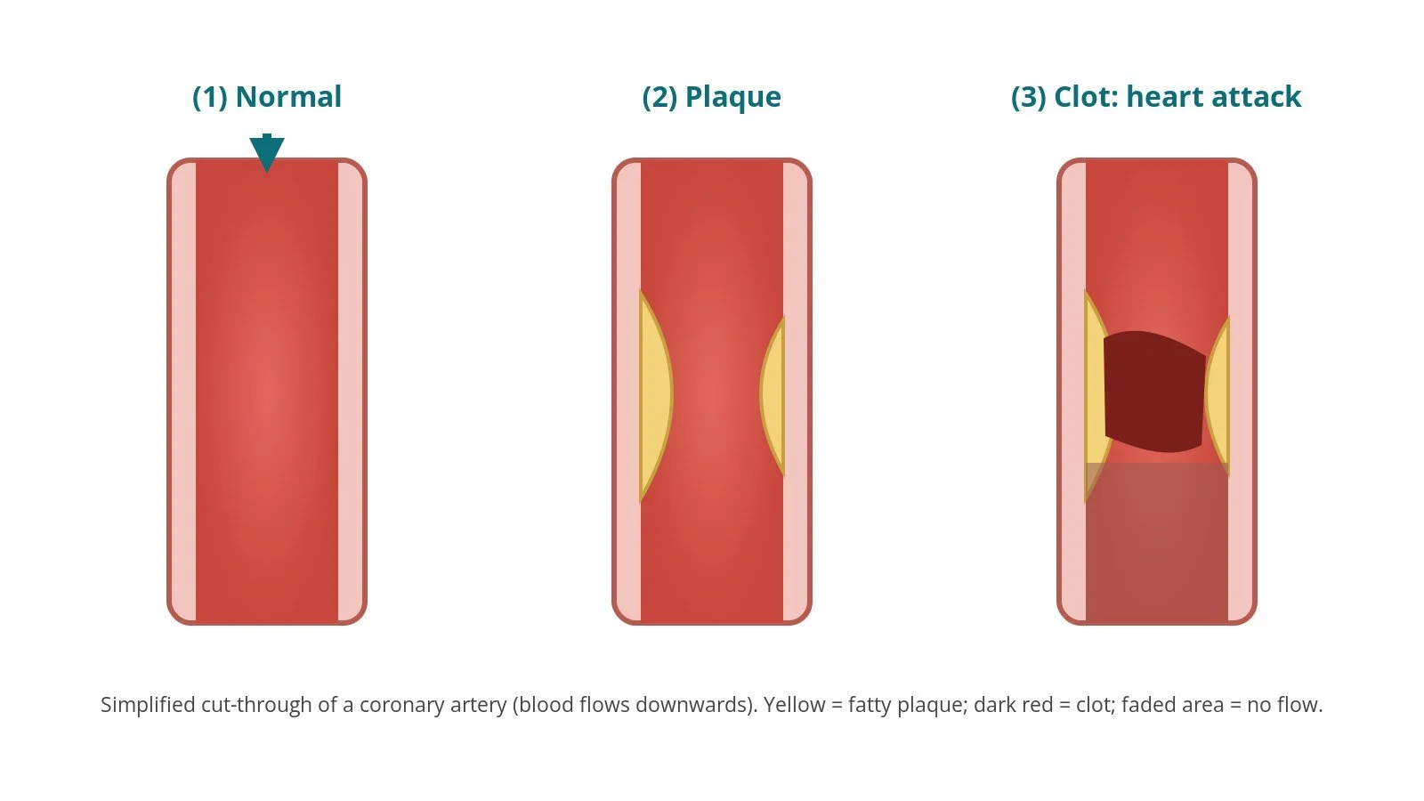
- Causes: a coronary artery is suddenly blocked, usually by a clot on a ruptured plaque. Less often by spontaneous coronary artery dissection (SCAD), embolism or spasm.
- Symptoms: central chest pain or pressure, often spreading to the arm, jaw or back, with sweating, nausea, breathlessness. Older people, women and people with diabetes may have atypical or no pain.
- Treatment: emergency reperfusion, usually primary PCI, with antiplatelet and anticoagulant drugs as per centre protocol. See Heart attack (AMI) and primary PCI and PCI.
- In the cath lab: pads on, defibrillator ready, and expect VT, VF, AIVR and heart block, especially as the artery opens. Inferior STEMIs often bring slow rhythms and low blood pressure.
Posterior STEMI
- What you see: ST depression in V1–V3, often with tall R waves and upright T waves there (a "mirror image" of ST elevation). Posterior leads V7–V9 show ST elevation of 0.5 mm or more (1 mm in men under 40). Often comes with an inferior or lateral STEMI.
- Causes: a blocked circumflex or RCA.
- Symptoms: as for any heart attack.
- Treatment: a STEMI equivalent: managed along the primary PCI pathway when confirmed, as per centre protocol.
- In the cath lab: if you see ST depression in V1–V3 in a patient with chest pain, suggest posterior leads. It is easily missed.
Right ventricular infarction
- What you see: an inferior STEMI with ST elevation in V4R (a right-sided chest lead). ST elevation in V1, and in III more than II, are clues.
- Causes: a blockage high up in the RCA.
- Symptoms: a heart attack with low blood pressure, raised neck veins and clear lungs. Slow rhythms and heart block are common.
- Treatment: primary PCI. The right ventricle depends on filling, so the team avoids drugs that lower preload (such as nitrates) and may give fluids, as per centre protocol. Pacing may be needed for heart block.
- In the cath lab: record V4R when an inferior STEMI is suspected. Watch the blood pressure closely, and be ready for bradycardia and heart block as the RCA is opened.
NSTEMI, unstable angina and ST depression
- What you see: new horizontal or downsloping ST depression of 0.5 mm or more in two neighbouring leads, and/or new T wave inversion. The ECG can also be normal. The diagnosis of NSTEMI needs a rise in high-sensitivity troponin.
- Causes: a partly blocked coronary artery, or a mismatch between oxygen supply and demand (fast AF, anaemia, low blood pressure, severe aortic stenosis). Other causes of ST depression: reciprocal change from a STEMI elsewhere, digoxin, LVH strain, bundle branch block, low potassium and fast rates.
- Symptoms: chest pain at rest or on minimal effort, breathlessness, sweating.
- Treatment: antithrombotic drugs and a coronary angiogram. Timing depends on risk: immediate if the patient is unstable, has ongoing pain, dangerous arrhythmias, heart failure or dynamic ST changes; usually early (within 24 hours) for other high-risk patients, as per the 2023 ESC guideline and local pathways.
- In the cath lab: watch for ST changes during balloon inflations in the culprit territory, and say if they don't settle.
T wave inversion
- What you see: T waves pointing down. Normal in aVR and V1, and sometimes in III. Abnormal when new or in two or more neighbouring leads.
- Causes: ischaemia, Wellens syndrome, after a heart attack, pulmonary embolism (V1–V4 and III), LVH or RVH strain, bundle branch block and pacing (secondary changes), hypertrophic cardiomyopathy (deep inversion, especially apical), brain bleeds (deep, broad "cerebral" T waves), and "T wave memory" after pacing or tachycardia.
- Symptoms: depend on the cause.
- Treatment: treat the cause. New inversion with chest pain is treated as possible acute coronary syndrome until proven otherwise.
- In the cath lab: compare with old ECGs, and note whether the inversion is in one territory or everywhere.
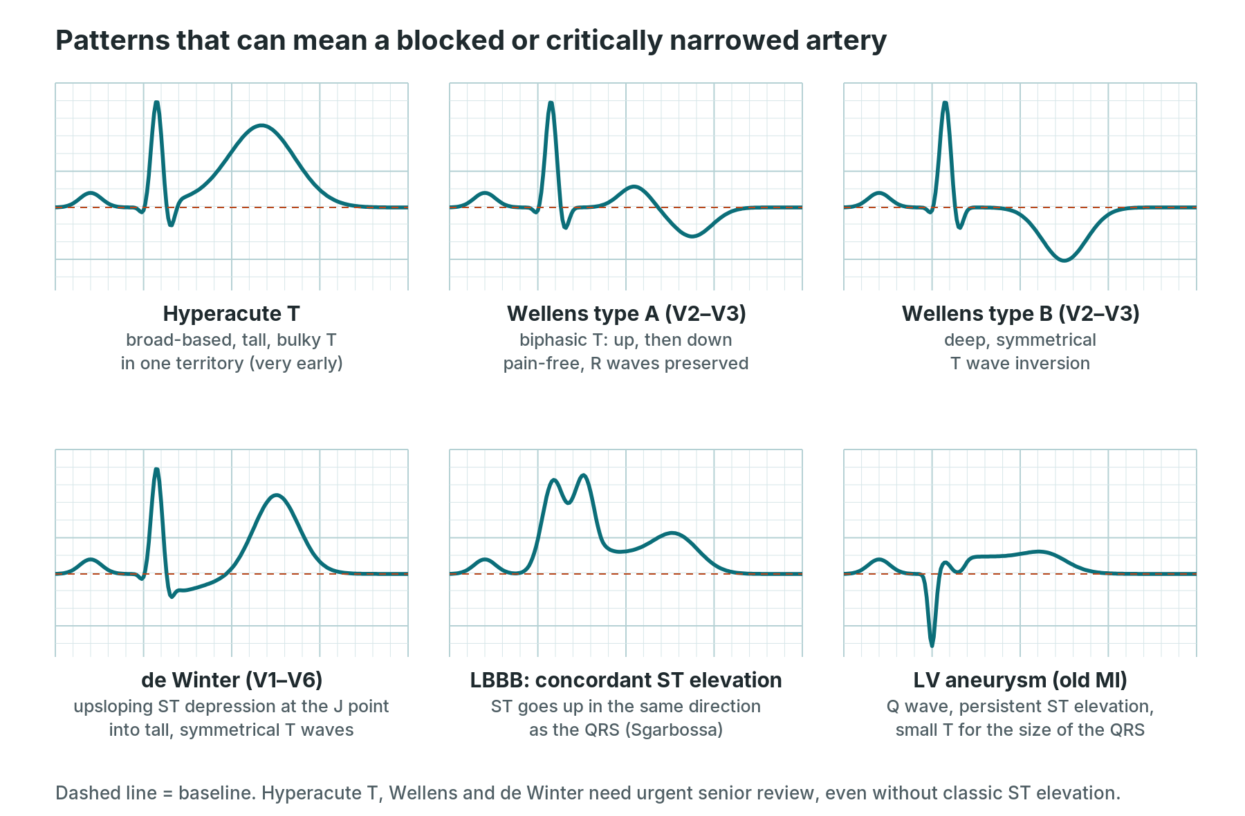
Some blocked or critically narrowed arteries do not show classic ST elevation. These patterns need urgent senior review.
Wellens syndrome
- What you see: in a patient whose chest pain has settled: type A, biphasic T waves (up then down) in V2–V3; type B, deep symmetrical T wave inversion in V2–V3, sometimes V1–V6. Little or no ST elevation, no Q waves, R waves preserved.
- Causes: a critical narrowing of the proximal LAD that has temporarily reopened.
- Symptoms: recent chest pain, now pain-free. The patient may look well.
- Treatment: urgent coronary angiography. Exercise stress testing is avoided, because it can trigger a large anterior heart attack.
- In the cath lab: these patients may arrive looking well but have a very tight LAD. Expect the ECG to change if the pain comes back.
de Winter pattern
- What you see: 1–3 mm upsloping ST depression at the J point in V1–V6, running into tall, symmetrical T waves, often with slight ST elevation in aVR. There is no classic ST elevation.
- Causes: usually a blocked proximal LAD.
- Symptoms: heart attack symptoms.
- Treatment: recognised as a STEMI equivalent: urgent reperfusion along the primary PCI pathway, as per centre protocol.
- In the cath lab: treat as an anterior STEMI. Same preparation, same urgency.
Sgarbossa criteria (heart attack with LBBB or a paced rhythm)
- What you see: in LBBB, ST and T normally point opposite to the QRS. Suspect a heart attack if there is: concordant ST elevation of 1 mm or more in any lead where the QRS is positive; concordant ST depression of 1 mm or more in V1–V3; or excessively discordant ST elevation (the original rule used 5 mm or more; the Smith-modified rule uses an ST/S ratio of −0.25 or more extreme).
- Causes: a blocked coronary artery in a patient whose baseline ECG is already abnormal.
- Symptoms: heart attack symptoms.
- Treatment: a positive result with symptoms supports the STEMI pathway, as per centre protocol. The criteria are also used with right ventricular paced rhythms.
- In the cath lab: have the old ECG to hand. A patient with LBBB and ongoing chest pain is treated seriously even if the criteria are not met.
Hyperacute T waves
- What you see: broad-based, tall, bulky T waves in one coronary territory, sometimes with slight ST elevation. They appear in the first minutes of an artery blocking.
- Causes: very early heart attack. The main look-alike is high potassium, where the T waves are tall but narrow-based and pointed.
- Symptoms: heart attack symptoms, often of recent onset.
- Treatment: repeat ECGs every 15–30 minutes, or sooner with ongoing pain. Senior review. Treat as a possible occlusion.
- In the cath lab: may be seen if a vessel closes suddenly during a case. Watch the ST trend in the monitor lead for that territory.
Pathological Q waves
- What you see: Q waves that are wide (0.03–0.04 s or more, about 1 small square) or deep (25% or more of the R wave height) in two neighbouring leads. Normal small Q waves are seen in I, aVL, V5–V6 and sometimes III.
- Causes: a previous (or evolving) heart attack, where scar no longer conducts. Look-alikes: hypertrophic cardiomyopathy, LBBB, WPW, poor lead placement.
- Symptoms: none from the Q waves themselves. They show the heart has been damaged.
- Treatment: secondary prevention and treatment of heart failure, as per the medical team.
- In the cath lab: Q waves in a territory help the team interpret the angiogram (for example, an old blocked vessel with collateral supply).
Left ventricular aneurysm
- What you see: ST elevation that persists weeks after a heart attack, usually with deep Q waves in the same leads (typically anterior). The T waves are small compared with the QRS, which helps tell it apart from an acute STEMI.
- Causes: a thinned, scarred area of the left ventricle after a large heart attack.
- Symptoms: may be none. Heart failure, VT, or stroke from a clot forming in the aneurysm.
- Treatment: heart failure medicines, anticoagulation if there is a clot, and treatment of VT. Rarely surgery.
- In the cath lab: compare with old ECGs. Persistent ST elevation with old Q waves and no new symptoms is reviewed, but may not mean a new blockage. The echo helps confirm.
Other patterns
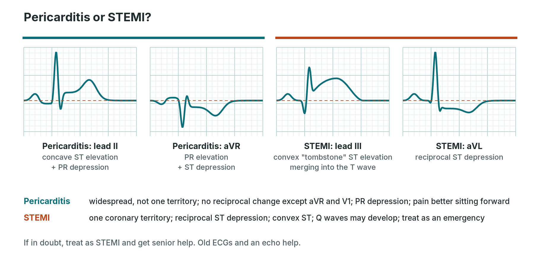
Pericarditis is widespread. STEMI follows a coronary territory and has reciprocal change.
Pericarditis (and pericardial effusion)
- What you see: widespread concave ST elevation across many leads, not one territory; PR depression (best in II); PR elevation and ST depression in aVR. No reciprocal ST depression other than in aVR and V1. The changes evolve over days to weeks. A large effusion gives small (low-voltage) complexes and electrical alternans (QRS height alternating beat to beat).
- Causes: viral infections, after a heart attack, after heart surgery or procedures, autoimmune disease, kidney failure, cancer, radiotherapy, tuberculosis.
- Symptoms: sharp chest pain that is worse lying flat and breathing in, and better sitting forward; fever; a scratchy "rub" heard with a stethoscope.
- Treatment: usually anti-inflammatory medicines such as aspirin or another NSAID, with colchicine, and rest from strenuous exercise, as per the 2025 ESC guideline and the medical team. Large effusions or tamponade may need drainage.
- In the cath lab: after a procedure, new chest pain with widespread ST changes, rising heart rate and falling blood pressure could mean pericardial bleeding or tamponade. Say so early. See Pericardiocentesis and Percutaneous balloon pericardiotomy.
Left ventricular hypertrophy (LVH)
- What you see: tall voltages: for example the Sokolow–Lyon criterion (S in V1 plus R in V5 or V6 of 35 mm or more) or the Cornell criterion (R in aVL plus S in V3 over 28 mm in men or 20 mm in women). Often a "strain" pattern: ST depression and asymmetric T wave inversion in I, aVL, V5–V6. Left axis deviation and left atrial enlargement may be present.
- Causes: high blood pressure, aortic stenosis, hypertrophic cardiomyopathy, athletic training (voltage only).
- Symptoms: none from the ECG; symptoms relate to the cause (breathlessness, chest pain, fainting in severe aortic stenosis).
- Treatment: treat the cause: blood pressure control, valve treatment such as TAVI or surgical AVR.
- In the cath lab: common in patients with aortic stenosis. The strain pattern makes ischaemia harder to read, so compare with the baseline.
Right ventricular hypertrophy (RVH)
- What you see: right axis deviation, a dominant R wave in V1 (R bigger than S), deep S waves in V5–V6, T wave inversion in V1–V3 (RV strain), and often P pulmonale.
- Causes: pulmonary hypertension, chronic lung disease, chronic pulmonary emboli, pulmonary valve stenosis and other congenital heart disease, mitral stenosis.
- Symptoms: breathlessness, tiredness, ankle swelling, fainting on exertion.
- Treatment: treat the cause, with specialist pulmonary hypertension care where needed.
- In the cath lab: these patients may come for right heart catheterisation. They can tolerate low blood pressure and slow rhythms poorly.
Atrial enlargement
- What you see: left atrial enlargement (P mitrale): a broad (over 0.12 s), notched P wave in II, and a deep, broad negative part to the P wave in V1. Right atrial enlargement (P pulmonale): a tall, peaked P wave over 2.5 mm in II (over 1.5 mm in V1).
- Causes: left: mitral valve disease, high blood pressure, LVH, heart failure. Right: pulmonary hypertension, lung disease, tricuspid valve disease, congenital heart disease.
- Symptoms: those of the cause. Left atrial enlargement raises the chance of AF.
- Treatment: treat the cause.
- In the cath lab: a clue to the underlying valve or lung disease, for example before balloon mitral valvuloplasty.
Pulmonary embolism (PE) pattern
- What you see: most often just sinus tachycardia. Other signs of right heart strain: T wave inversion in V1–V4 and III, new RBBB, right axis deviation, P pulmonale, AF. The "S1Q3T3" pattern (S wave in I, Q wave and inverted T in III) is classic but uncommon. The ECG can be normal.
- Causes: a clot travelling to the lungs, usually from the leg veins. Risk factors include surgery, immobility, cancer, pregnancy and previous clots.
- Symptoms: sudden breathlessness, chest pain on breathing, coughing up blood, fainting, collapse.
- Treatment: anticoagulation; for high-risk PE with shock, clot-busting (thrombolysis), catheter-directed treatment or surgery, as per centre protocol and the ESC guideline.
- In the cath lab: some labs perform pulmonary angiography and catheter-directed treatment. PE is also a reversible cause of arrest (the "T" for thrombosis).
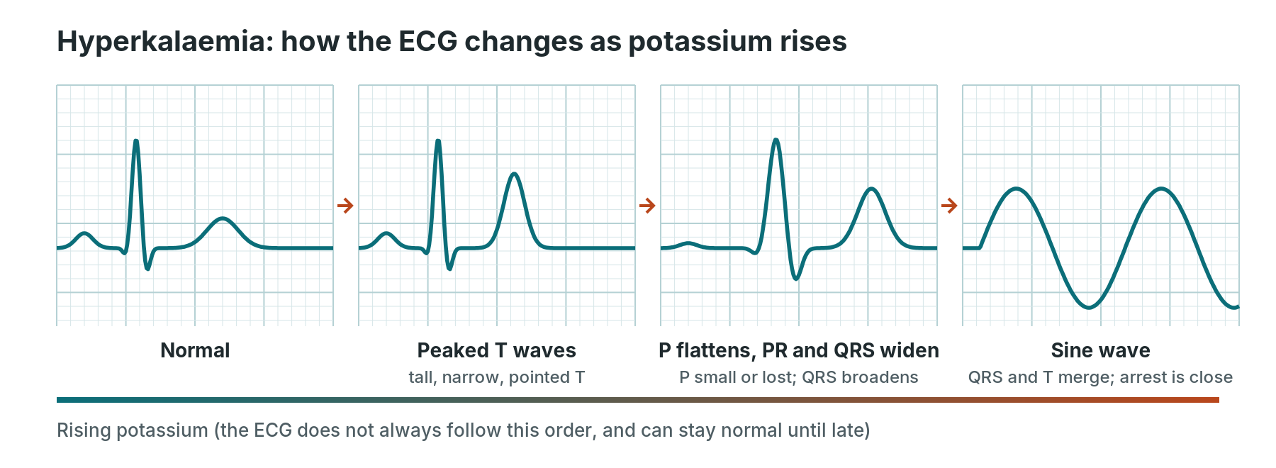
As potassium rises the ECG can change roughly in this order, but it can also jump straight to a dangerous rhythm.
Hyperkalaemia (high potassium)
- What you see: tall, narrow, peaked T waves, then flattening or loss of P waves, a longer PR, a widening QRS, and finally a sine wave pattern. It can cause slow rhythms, heart block, VT, VF or asystole. The ECG does not reliably match the blood level.
- Causes: kidney failure, drugs (ACE inhibitors, ARBs, spironolactone and similar drugs, potassium supplements, trimethoprim), muscle breakdown, diabetic ketoacidosis, tissue injury. A haemolysed blood sample can give a falsely high result.
- Symptoms: often none. Weakness, tiredness, palpitations, or muscle paralysis when severe.
- Treatment: follow the UK Kidney Association steps, as per centre protocol: protect the heart with IV calcium if there are ECG changes, shift potassium into cells with insulin and glucose (plus salbutamol), remove potassium (potassium binders or dialysis), then monitor potassium and glucose. Stop drugs that raise potassium.
- In the cath lab: check potassium in renal patients before the case. High potassium raises pacing thresholds, so a pacing wire may stop capturing. It is one of the "Hs" in cardiac arrest.
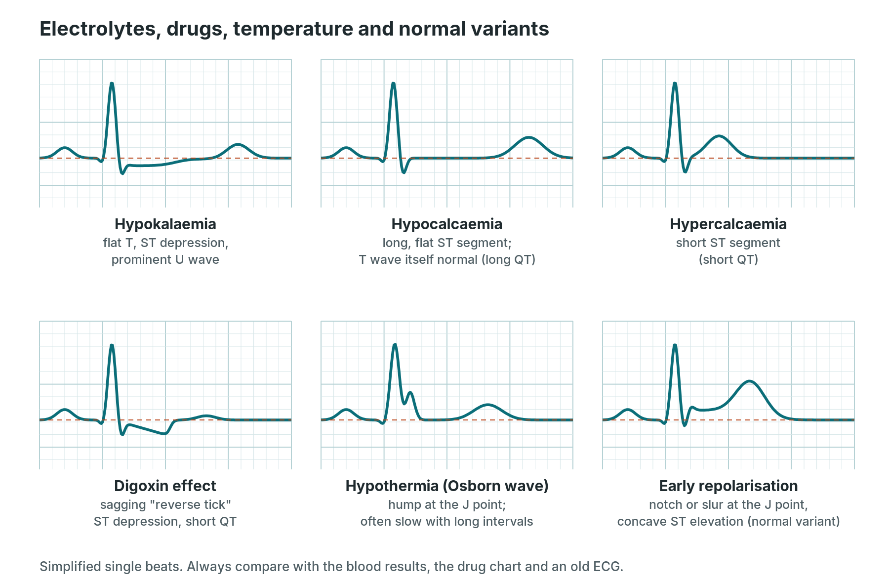
Simplified single beats. Always check the bloods, the drug chart and an old ECG.
Hypokalaemia (low potassium)
- What you see: flattened or inverted T waves, ST depression, prominent U waves (a small wave after the T), and an apparently long QT (really a QU interval). Ectopic beats are common.
- Causes: diuretics, vomiting and diarrhoea, insulin, salbutamol, steroids, high aldosterone, low magnesium.
- Symptoms: weakness, cramps, tiredness, palpitations, constipation.
- Treatment: oral or IV potassium as per centre protocol, and check and correct magnesium.
- In the cath lab: low potassium increases the risk of VT, VF and torsades, and makes digoxin toxicity more likely. Many labs check potassium before EP studies and ablation.
Hypocalcaemia and hypercalcaemia
- What you see: low calcium: a long QT caused by a long, flat ST segment, with a normal-looking T wave. High calcium: a short QT with a short ST segment; very high levels can cause J (Osborn-like) waves, slow rhythms and heart block.
- Causes: low calcium: after thyroid or parathyroid surgery, vitamin D deficiency, kidney disease, pancreatitis, massive blood transfusion. High calcium: overactive parathyroid glands, cancer, too much vitamin D, sarcoidosis.
- Symptoms: low calcium: tingling around the mouth and fingers, muscle cramps and spasms, seizures. High calcium: thirst, passing a lot of urine, constipation, tummy pain, confusion, kidney stones.
- Treatment: low: calcium replacement (IV if severe or symptomatic), as per centre protocol. High: IV fluids and specific medicines, as per centre protocol, and treat the cause.
- In the cath lab: low calcium is seen after massive transfusion and during ECMO or large blood loss. See ECMO in the cath lab.
Digoxin effect and digoxin toxicity
- What you see: digoxin effect (a sign of the drug, not toxicity): a sagging "reverse tick" ST depression, best in the lateral leads, a short QT and flattened T waves. Digoxin toxicity: almost any arrhythmia, classically frequent ectopics and bigeminy, atrial tachycardia with block, accelerated junctional rhythm, AV block, a slow "regularised" AF, and bidirectional VT.
- Causes: toxicity is more likely with kidney impairment, low potassium or magnesium, high calcium, older age, and drug interactions (such as amiodarone and verapamil).
- Symptoms of toxicity: nausea, vomiting, loss of appetite, confusion, blurred or yellow-tinted vision, palpitations, dizziness.
- Treatment: stop digoxin, correct potassium, and for life-threatening toxicity give digoxin-specific antibody fragments, as per centre protocol and poisons advice.
- In the cath lab: tell the team before any cardioversion if the patient is on digoxin. Digoxin effect makes ST depression harder to interpret.
Hypothermia (Osborn or J waves)
- What you see: Osborn (J) waves: a hump at the end of the QRS, getting bigger as the temperature falls. Also slow rhythms, longer PR, QRS and QT, shivering artefact, AF, and VF or asystole when very cold.
- Causes: exposure, near-drowning, older or frail patients, sedation and anaesthesia, long procedures, and therapeutic temperature control after cardiac arrest.
- Symptoms: shivering (which stops when severe), confusion, drowsiness, slow breathing.
- Treatment: rewarming, as per centre protocol. Resuscitation is modified when the core temperature is low: follow the Resuscitation Council UK special circumstances guidance.
- In the cath lab: keep patients warm on long cases, particularly after cardiac arrest or during ECMO. Shivering artefact can mimic arrhythmias.
Early repolarisation
- What you see: concave ST elevation, mostly in V2–V5 (and sometimes inferior leads), with a notch or slur at the J point and prominent T waves. It stays the same over time, with no reciprocal change and no PR depression.
- Causes: a common normal variant, especially in young, fit men and people of African or Afro-Caribbean heritage. A rare "early repolarisation syndrome" with inferolateral J waves is linked to VF.
- Symptoms: none.
- Treatment: none for the benign pattern. Specialist review if there is a history of unexplained fainting or cardiac arrest.
- In the cath lab: the main risk is confusing it with STEMI or pericarditis. Old ECGs, symptoms and serial ECGs help. If in doubt, a senior reviews it.
Pacing, ectopics and artefact
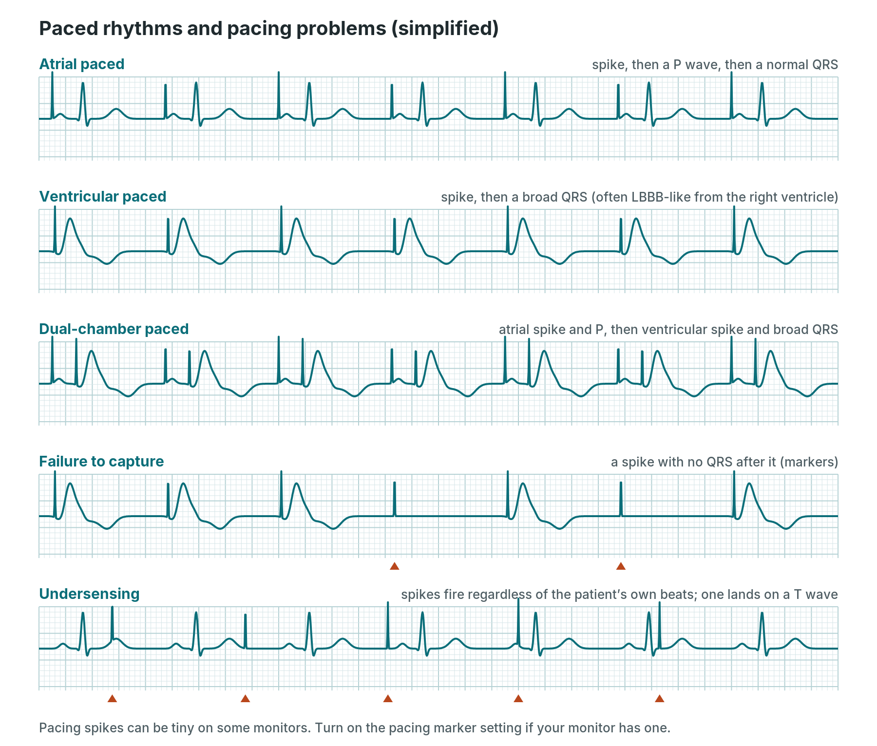
Pacing spikes can be tiny on some monitors. Use the pacing marker setting if your monitor has one.
Paced rhythms
- What you see: a sharp vertical pacing spike. Atrial pacing: spike, then a P wave, then the patient's own (usually narrow) QRS. Right ventricular pacing: spike, then a broad QRS, often LBBB-like, with discordant ST–T. Dual-chamber pacing: both. Biventricular pacing and conduction system pacing usually give a narrower paced QRS.
- Causes: a permanent pacemaker, an ICD with pacing, a temporary transvenous wire or external pacing.
- Symptoms: none when working well.
- Treatment: none needed for normal pacing. Device checks are done by the cardiac physiologist or device team.
- In the cath lab: ask about devices before the case. Ventricular pacing hides ischaemia in the same way as LBBB (the Sgarbossa criteria can help). Diathermy can interfere with devices: follow your centre protocol. See Permanent pacemaker and ICD, Subcutaneous ICD and Temporary pacing wire.
Pacing failure
- What you see: failure to capture: a spike with no P wave or QRS after it. Failure to pace (output failure): no spike when one is expected, with a pause. Undersensing: spikes appear when they should not, because the device has missed the patient's own beats; a spike landing on a T wave can trigger VT or VF. Oversensing: the device mistakes noise or muscle activity for heartbeats and stops pacing, causing pauses.
- Causes: lead displacement (especially in the first days after implant, or a temporary wire that has moved), a raised threshold (high potassium, ischaemia, acidosis, some antiarrhythmic drugs), a loose or disconnected cable on a temporary system, battery depletion, lead fracture, settings that need adjusting, and electrical interference.
- Symptoms: dizziness, fainting, breathlessness, tiredness, or collapse if the patient depends on pacing.
- Treatment: check the patient, then check connections and the battery on temporary systems. Output and sensitivity are adjusted only by trained staff, as per centre protocol. Have external pacing and drugs such as atropine or isoprenaline ready. Use of a magnet on a device is as per centre protocol. Get the device team.
- In the cath lab: if a temporary wire stops capturing, tell the operator immediately: it may need repositioning. Check the cables are secure before moving or transferring the patient.
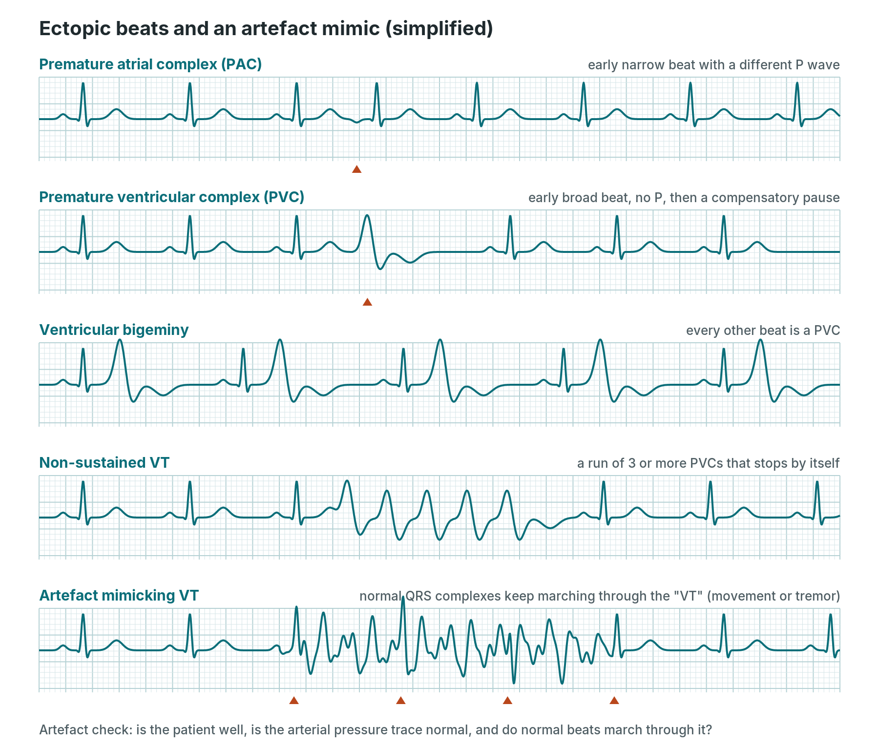
Markers show the ectopic beat, or (bottom strip) the normal QRS complexes marching through the artefact.
Ectopic beats: PACs and PVCs
- What you see: premature atrial complex (PAC): an early beat with a different-shaped P wave and usually a narrow QRS, followed by a short pause. Premature ventricular complex (PVC): an early broad QRS with no P wave before it, the T wave pointing the opposite way, and usually a full compensatory pause. Patterns include bigeminy (every other beat is ectopic), trigeminy (every third), couplets (two in a row) and non-sustained VT (three or more in a row, stopping by itself within 30 seconds).
- Causes: often none. Caffeine, alcohol, stress, lack of sleep, low potassium or magnesium, an overactive thyroid, ischaemia, heart failure, cardiomyopathy, digoxin toxicity, and catheters or wires touching the heart wall.
- Symptoms: none, or a "missed beat", "thump" or fluttering. Very frequent PVCs can cause tiredness and, rarely, weaken the heart over time.
- Treatment: reassurance, reducing triggers, correcting electrolytes. Beta-blockers or other drugs for troublesome symptoms. Catheter ablation for very frequent or problematic ectopics, as per the specialist team.
- In the cath lab: ectopics are expected when a catheter crosses a valve or sits in a chamber. Say so if they come in runs, if they don't stop when the catheter is moved, or if they appear with ST changes.
Artefact that mimics arrhythmias
- What you see: irregular, chaotic or fast-looking traces that do not behave like a real rhythm: the patient's normal QRS complexes keep marching through the "arrhythmia", the arterial pressure trace stays normal, and the "rhythm" may be seen in only some leads. Flat lines from a lead falling off.
- Causes: patient movement, shivering and tremor, brushing teeth or scratching, CPR, loose electrodes or cables, electrical interference (mains, diathermy, ablation energy), and swapped limb leads (for example arm leads reversed, giving an upside-down lead I and an upright aVR).
- Symptoms: none from the artefact: the patient is well.
- Treatment: check the patient first, then the electrodes and cables. Never treat a rhythm on the monitor without checking the patient.
- In the cath lab: artefact is common during ablation, diathermy and patient movement. The arterial pressure trace is your best check: a normal, pulsatile trace means the heart is pumping.
Arrest rhythms
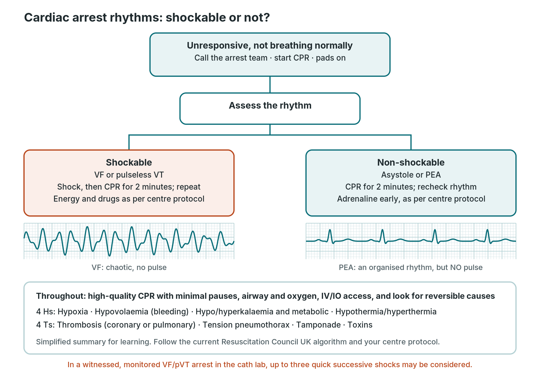
A simplified learning summary. Always follow the current Resuscitation Council UK algorithm and your centre protocol.
In cardiac arrest, the rhythm decides one thing: shock or no shock. The Resuscitation Council UK adult advanced life support algorithm (2021, updated in October 2025 with no major changes to the core algorithm) divides arrest rhythms into two groups.
Shockable: VF and pulseless VT
- What you see: VF (chaotic, no QRS) or VT with no pulse.
- Causes: acute heart attack, ischaemia and reperfusion, scar, electrolyte disturbance, drugs, inherited conditions, and the reversible causes below.
- Symptoms: collapse, unresponsive, not breathing normally, no pulse.
- Treatment: call for help, start high-quality CPR, attach the defibrillator, and defibrillate, then continue CPR for 2 minutes before reassessing. Adrenaline and amiodarone are given after the third shock. Energy, drugs and doses as per centre protocol and the current algorithm.
- In the cath lab: for a witnessed, monitored VF or pulseless VT arrest with the defibrillator ready, up to three quick successive shocks may be considered before compressions start; these count as the first shock in the algorithm. The team may continue the procedure (for example PCI) using mechanical CPR, and some centres use ECMO for selected patients.
Non-shockable: asystole and PEA
- What you see: asystole: a near-flat line (check the leads, gain and a second lead; look for P waves). PEA (pulseless electrical activity): an organised rhythm on the monitor, sometimes looking almost normal, but no pulse and no arterial trace.
- Causes: the reversible causes below. In the cath lab think especially of tamponade, major bleeding, acute vessel closure or stent thrombosis, and massive PE.
- Symptoms: collapse, unresponsive, no pulse.
- Treatment: high-quality CPR, adrenaline as soon as possible, and find and treat the cause. Reassess the rhythm every 2 minutes. If it is unclear whether a rhythm is asystole or very fine VF, the guidance is to continue CPR rather than shock.
- In the cath lab: P wave asystole (P waves with no QRS) may respond to pacing. PEA in the lab is a cue to look for tamponade (echo) and bleeding straight away. See Pericardiocentesis.
Reversible causes (4 Hs and 4 Ts):
| 4 Hs | 4 Ts |
|---|---|
| Hypoxia | Thrombosis (coronary or pulmonary) |
| Hypovolaemia (including bleeding) | Tension pneumothorax |
| Hypo/hyperkalaemia and other metabolic causes | Tamponade (cardiac) |
| Hypothermia / hyperthermia | Toxins (drugs and poisons) |
Summary tables
Fast rhythms
| Rhythm | Key ECG features | First thing to say or do in the lab |
|---|---|---|
| Sinus tachycardia | regular, P before every QRS, >100 | look for the cause: pain, bleeding, tamponade |
| SVT (AVNRT/AVRT) | regular, narrow, 140–250, P hidden or after the QRS | record a 12-lead; check BP; vagal manoeuvres or adenosine as per protocol |
| Atrial fibrillation | irregularly irregular, no P waves | note the time; tell the operator if new; check BP |
| Atrial flutter | sawtooth waves, often 2:1 at ~150 | "regular at 150, could be flutter" |
| Atrial tachycardia | abnormal P waves, regular, flat baseline between Ps | 12-lead; check digoxin and potassium |
| MAT | irregular, 3+ P wave shapes | check oxygen, gas and electrolytes |
| Monomorphic VT | regular, broad, uniform | check pulse and arterial trace; call for help |
| Torsades / polymorphic VT | twisting broad complexes; long QT before | pulse check; magnesium as per protocol; defibrillate if pulseless |
| VF | chaos, no QRS | arrest call; defibrillate as per protocol |
Slow rhythms and blocks
| Rhythm | Key ECG features | Risk | In the lab |
|---|---|---|---|
| Sinus bradycardia | regular, P before every QRS, <60 | usually low | expected with RCA contrast and vagal reactions; call if it lasts |
| Sick sinus syndrome | pauses, sinus arrest, tachy-brady | moderate | pacing support ready |
| First-degree AV block | PR >0.20 s, all conducted | low | note on baseline; watch after TAVI |
| Mobitz I | PR lengthens, then a dropped beat | usually low | common with inferior STEMI; watch |
| Mobitz II | fixed PR, sudden dropped beat | high | call it; pacing ready |
| 2:1 block | every other P not conducted | uncertain | long strip; senior review |
| Complete heart block | P and QRS independent | high | call it; check BP; pacing ready |
| Junctional / escape rhythm | narrow 40–60 or broad 20–40 | depends | never suppress an escape rhythm |
| AIVR | broad, 50–110, gentle onset | usually low | classic after reperfusion; watch for VT/VF |
Conduction and inherited patterns
| Pattern | Key ECG features | Why it matters in the lab |
|---|---|---|
| RBBB | rSR′ in V1, wide S in V6 | right heart catheter in a patient with LBBB can cause complete block |
| LBBB | broad notched R in V6, QS in V1 | hides STEMI; use Sgarbossa; common after TAVI |
| Fascicular block | left or right axis deviation | little conduction reserve when combined with RBBB |
| WPW | short PR, delta wave | avoid AV-nodal drugs in pre-excited AF |
| Long QT | QTc >450–470 ms (>500 high risk) | torsades risk; check drugs given in the lab |
| Short QT | QTc <340–360 ms | rare; check calcium and digoxin |
| Brugada type 1 | coved ST elevation V1–V2 into negative T | treat fever; avoid trigger drugs |
Ischaemia and infarction
| Pattern | Key ECG features | Usual artery or meaning |
|---|---|---|
| Anterior STEMI | ST elevation V1–V4 | LAD |
| Lateral STEMI | ST elevation I, aVL, V5–V6 | circumflex or diagonal |
| Inferior STEMI | ST elevation II, III, aVF | RCA (sometimes circumflex) |
| Posterior STEMI | ST depression V1–V3; elevation V7–V9 | circumflex or RCA |
| RV infarction | ST elevation V4R with inferior STEMI | proximal RCA; avoid preload-lowering drugs |
| aVR elevation + widespread ST depression | ST up in aVR, down in 6+ leads | left main or multi-vessel disease |
| Wellens | biphasic or deep T inversion V2–V3, pain-free | critical proximal LAD |
| de Winter | upsloping ST depression into tall T, V1–V6 | proximal LAD occlusion |
| Hyperacute T | broad, tall T in one territory | very early occlusion |
| Sgarbossa (LBBB/paced) | concordant ST changes | possible occlusion despite LBBB |
| Q waves | wide or deep Q in a territory | old (or evolving) infarct |
| LV aneurysm | persistent ST elevation with Q waves | old anterior MI; compare old ECGs |
Other patterns: look-alikes and clues
| Pattern | Key ECG features | Main clue or look-alike |
|---|---|---|
| Pericarditis | widespread concave ST up, PR down | vs STEMI: no territory, no reciprocal change |
| Early repolarisation | J-point notch, concave ST up, stable | vs STEMI and pericarditis: old ECGs |
| LVH | tall voltages, lateral strain | makes ischaemia harder to read |
| RVH | dominant R in V1, right axis | pulmonary hypertension, lung disease |
| PE | sinus tachycardia, RV strain, RBBB | ECG may be normal |
| Hyperkalaemia | peaked T, wide QRS, sine wave | vs hyperacute T (broad-based) |
| Hypokalaemia | flat T, ST depression, U waves | arrhythmia risk; check magnesium |
| Low / high calcium | long ST / short ST | check bloods after transfusion |
| Digoxin effect | reverse tick ST depression | effect is not toxicity |
| Hypothermia | Osborn J waves, bradycardia | keep long cases warm |
| Paced rhythm | spikes; broad paced QRS | hides ischaemia; check capture |
| Ectopics | early beats (narrow PAC, broad PVC) | expected with catheters; call runs |
| Artefact | normal QRS marching through "chaos" | check the patient and arterial trace |
First time in the cath lab?
💡 First-time tip
Patient, pulse, pressure, then picture. Before you name any rhythm, look at the patient, feel for a pulse or check the arterial trace, then read the ECG. Learn three danger patterns first: a broad fast rhythm (VT until proven otherwise), complete heart block, and ST elevation in a territory. Keep the baseline ECG within reach so you can say what's new. Know where the pads, defibrillator and pacing kit are before the case starts. Describe, don't guess: "Broad, regular, about 170, BP dropping" is a perfect call. In a good lab, speaking up is always welcome.
FAQ
Is this guide enough to interpret ECGs on my own?
No. It's a learning aid. ECG interpretation in clinical practice must be confirmed by a qualified clinician, using the full 12-lead, the patient's history and local protocols.
How do I tell VT from SVT with bundle branch block?
Often you can't be sure on a monitor. Clues for VT include AV dissociation, capture or fusion beats, a very broad QRS, an extreme axis and a history of heart attack. The safe rule is to treat a broad regular tachycardia as VT until a senior says otherwise.
Why does heart block happen during TAVI and septal procedures?
The conduction system (the AV node and left bundle) runs very close to the aortic valve and the upper septum. A valve frame, balloon, closure device or septal ablation can bruise or injure it. This is why pacing support is planned for these cases.
Why do we avoid AV-nodal drugs in WPW with AF?
In pre-excited AF, the accessory pathway can conduct very fast. Blocking the AV node can push more impulses down the pathway and trigger VF. Treatment is electrical cardioversion or specialist drugs, as per centre protocol.
Is ST elevation always a heart attack?
No. Pericarditis, early repolarisation, LVH, LBBB, LV aneurysm, Brugada, high potassium and pacing can all raise the ST segment. But because missing a STEMI is so serious, ST elevation with chest pain is treated as a STEMI until a senior decides otherwise.
What's the most useful thing I can do when the rhythm changes?
Say what you see, note the time and what was happening (for example "balloon up in the RCA"), print or save a strip, and check the patient. Those four things help the team more than anything else.
Related Cath Lab 101 guides
- ECG basics: a Cath Lab 101 guide
- Heart attack (acute MI) and primary PCI
- PCI (coronary angioplasty and stents) · Coronary angiogram
- Electrophysiology (EP) study · Electroanatomical mapping
- Radiofrequency (RF) ablation · AF cryoablation · Pulsed field ablation · Laser balloon ablation
- Permanent pacemaker (PPM) and ICD · Subcutaneous ICD (S-ICD) · Temporary pacing wire
- TAVI procedure · Alcohol septal ablation (ASA) · VSD closure
- Right and left heart catheterisation · Pulmonary angiography
- Pericardiocentesis · ECMO in the cath lab
- Cath Lab Abbreviations A–Z · Cath lab procedures reference
Sources
All checked on 22 October 2026. Each source's own date is shown where it gives one.
- Resuscitation Council UK. 2025 Resuscitation Guidelines: Adult advanced life support (published 27 October 2025; updates the 2021 guidelines). resus.org.uk
- Resuscitation Council UK. 2021 Resuscitation Guidelines: Adult advanced life support, with the adult tachycardia and bradycardia algorithms. resus.org.uk
- Brugada J, Katritsis DG, et al. 2019 ESC Guidelines for the management of patients with supraventricular tachycardia. Eur Heart J 2020;41(5):655–720. doi.org/10.1093/eurheartj/ehz467
- Van Gelder IC, Rienstra M, et al. 2024 ESC Guidelines for the management of atrial fibrillation. Eur Heart J 2024;45(36):3314–3414. doi.org/10.1093/eurheartj/ehae176
- Zeppenfeld K, Tfelt-Hansen J, et al. 2022 ESC Guidelines for the management of patients with ventricular arrhythmias and the prevention of sudden cardiac death. Eur Heart J 2022;43(40):3997–4126. doi.org/10.1093/eurheartj/ehac262
- Glikson M, Nielsen JC, et al. 2021 ESC Guidelines on cardiac pacing and cardiac resynchronization therapy. Eur Heart J 2021;42(35):3427–3520. doi.org/10.1093/eurheartj/ehab364
- Byrne RA, et al. 2023 ESC Guidelines for the management of acute coronary syndromes. Eur Heart J 2023;44(38):3720–3826. PubMed 37622654
- Thygesen K, et al. Fourth universal definition of myocardial infarction (2018). Eur Heart J 2019;40(3):237–269. PubMed 30165617
- Schulz-Menger J, Collini V, Gröschel J, et al. 2025 ESC Guidelines for the management of myocarditis and pericarditis. Eur Heart J 2025;46(40):3952–4041. doi.org/10.1093/eurheartj/ehaf192
- Konstantinides SV, Meyer G, et al. 2019 ESC Guidelines for the diagnosis and management of acute pulmonary embolism. Eur Heart J 2020;41(4):543–603. doi.org/10.1093/eurheartj/ehz405
- UK Kidney Association. Clinical practice guideline: Treatment of acute hyperkalaemia in adults (published 19 December 2023). ukkidney.org
- NICE. Atrial fibrillation: diagnosis and management (NG196). nice.org.uk/guidance/ng196
- NHS. Arrhythmia. nhs.uk/conditions/arrhythmia
- British Heart Foundation. Arrhythmias. bhf.org.uk
- Sgarbossa EB, et al. Electrocardiographic diagnosis of evolving acute myocardial infarction in the presence of left bundle-branch block. N Engl J Med 1996;334(8):481–487. PubMed 8559200
- Smith SW, et al. Diagnosis of ST-elevation myocardial infarction in the presence of left bundle branch block with the ST-elevation to S-wave ratio in a modified Sgarbossa rule. Ann Emerg Med 2012;60(6):766–776. PubMed 22939607
- de Winter RJ, et al. A new ECG sign of proximal LAD occlusion. N Engl J Med 2008;359(19):2071–2073. PubMed 18987380
- de Zwaan C, Bär FW, Wellens HJ. Characteristic electrocardiographic pattern indicating a critical stenosis high in left anterior descending coronary artery in patients admitted because of impending myocardial infarction. Am Heart J 1982;103(4):730–736. PubMed 6121481
- Life in the Fast Lane. ECG library (educational reference). litfl.com/ecg-library
Disclaimer
Education only. This page is for general education for healthcare students, cath lab staff and curious readers. It isn't medical advice and doesn't replace advice from your own doctor, cardiology team or local unit protocols. ECG interpretation in clinical practice must be confirmed by a qualified clinician. All drug doses, shock energies and pacing settings are as per centre protocol and current resuscitation guidelines; medicines are named only so readers recognise them. Equipment is described generically. No product or manufacturer is recommended. If you have chest pain now, call 999.
Disclosure: I work in the medical device industry. This page is independent education and isn't sponsored or reviewed by any company.
All diagrams on this page are original illustrations created for rodnieoro.com. The rhythm strips and beats are simplified drawings, not recordings from patients.