ASD closure (atrial septal defect): a Cath Lab 101 guide
Reviewed by Rodnie Oro, cath lab charge nurse · Last reviewed: October 2026 · Education only, not medical advice
ASD closure in one minute
What it is: an atrial septal defect (ASD) is a hole in the wall (septum) between the two upper chambers of the heart, present from birth. Many secundum ASDs can be closed with a keyhole procedure: a folded double-disc occluder is passed up a vein from the groin and opened so it plugs the hole.
Who it is for: children and adults with an ASD that lets enough blood through to enlarge the right side of the heart, or that causes symptoms, when the hole is the right type and size for a device.
Main risks: bleeding at the groin, irregular heart rhythms, the device moving and, rarely, the device wearing through the heart wall (erosion). Your team will explain your own risk.
Usually: under general anaesthetic or sedation, guided by echo and X-ray, about 1 to 2 hours, often home the next day.
On this page: What is an ASD? · Who is it for? · When is it done? · Step by step · Recovery · Risks · Kit list · First time in the lab? · FAQ
What is an ASD?
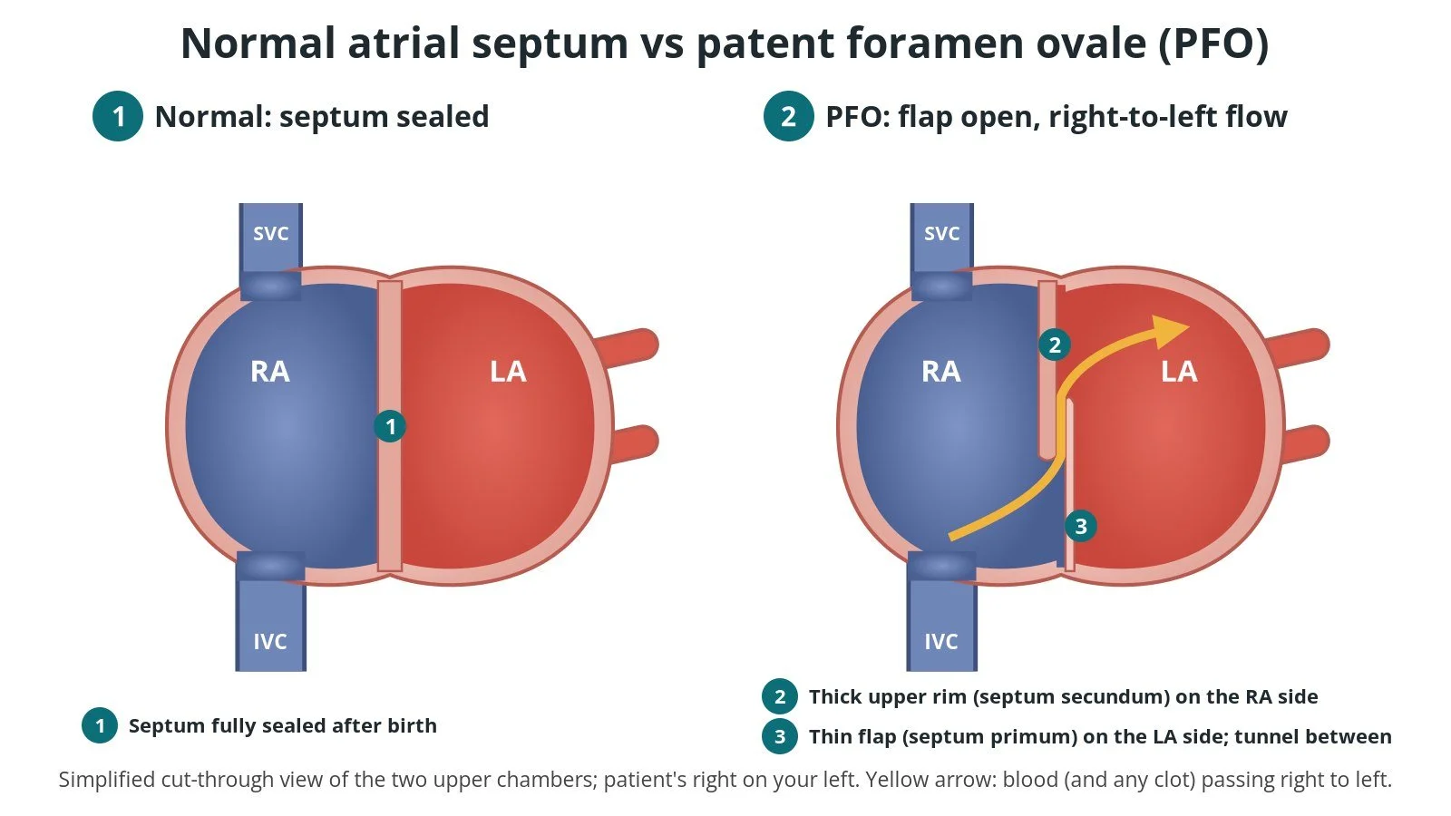
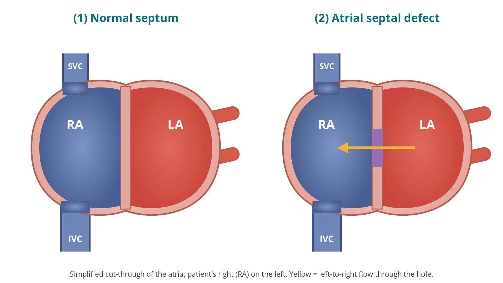
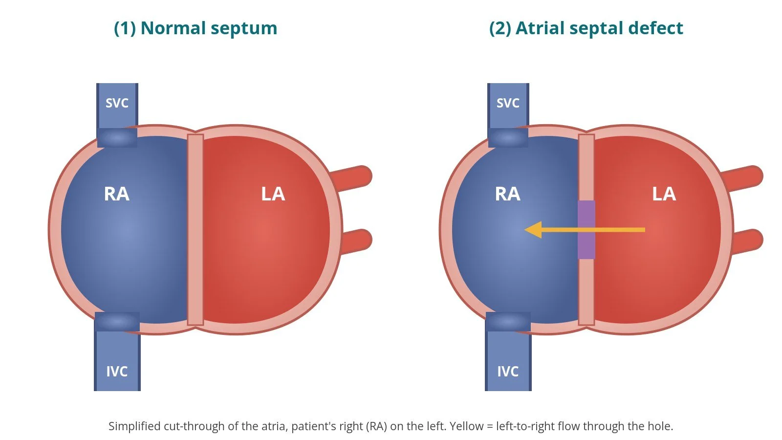
The atrial septum separates the right atrium (RA), which receives blue blood from the body, from the left atrium (LA), which receives red blood from the lungs. An atrial septal defect (ASD) is a true hole in this wall, present from birth. (A PFO is different: a flap-like tunnel rather than a hole. See the PFO guide.)
Because the pressure is a little higher on the left, blood flows through an ASD mainly from left to right. The right side of the heart then pumps extra blood round the lungs. Over years, a significant ASD can stretch the right atrium and right ventricle and may lead to breathlessness, tiredness, palpitations or atrial fibrillation (AF), and sometimes high pressure in the lungs (pulmonary hypertension). Small ASDs often cause no problems, and some close by themselves in early childhood.
There are four main types:
Secundum: in the middle of the septum. The most common type, and the one usually suitable for device closure.
Primum: low in the septum, near the valves, often with valve problems. Closed surgically.
Sinus venosus: high up near the superior vena cava (or low near the inferior vena cava), often with abnormally draining lung veins. Usually surgical, though covered-stent techniques are used in some specialist centres.
Coronary sinus defect: rare; usually surgical.
New to the acronyms? See the Cath Lab Abbreviations A–Z.
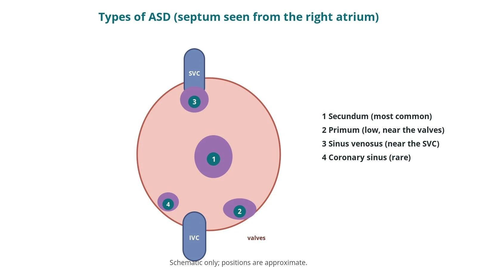
ASD types: (1) secundum, in the middle; (2) primum, low near the valves; (3) sinus venosus, near the superior vena cava; (4) coronary sinus.
Who is it for?
The decision is made by a congenital heart team. In line with the ESC 2020 guidelines for adult congenital heart disease, closure is generally recommended when:
there is evidence of right heart volume overload (an enlarged right ventricle on echo or MRI) from a significant left-to-right shunt
the pressure in the lungs is not too high; this is checked carefully, sometimes with a catheter study, because closing an ASD with severe pulmonary hypertension can be harmful
the defect is a secundum ASD with enough rim of tissue around it to hold a device; otherwise surgery is used
Closure may also be considered after a stroke thought to be caused by a clot crossing the ASD. NICE guidance on endovascular ASD closure (IPG96, now HTG58) supports the procedure with normal arrangements, in units with cardiac surgical support.
When is it done?
It is usually a planned procedure. Before it, the team usually arranges:
echo, including a transoesophageal echo (TOE), to measure the hole and its rims and to check the lung veins drain normally
sometimes MRI or CT, and a check of lung pressures
heart rhythm assessment, blood tests and a review of medicines, as per centre protocol
a check for nickel allergy, as most device frames contain nickel-titanium
a discussion of device closure versus surgery
Step by step
1. Anaesthetic and set-up
Most centres use a general anaesthetic with TOE guidance; some use sedation with intracardiac echo (ICE). X-ray is used throughout, and antibiotics are given as per centre protocol.
2. Femoral venous access
Access is from the right femoral vein, under ultrasound. A sheath is inserted; the delivery sheath size is per device and operator.
Catheters pass up the inferior vena cava (IVC) into the RA. Heparin is given, dose as per centre protocol.
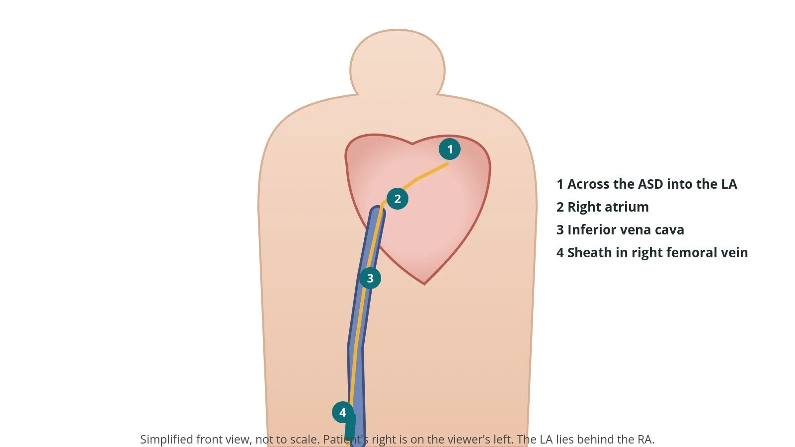
Access: (1) across the ASD into the left atrium; (2) right atrium; (3) inferior vena cava; (4) sheath in the right femoral vein.
3. Crossing, sizing, deploying and releasing
A. Cross the ASD. A multipurpose-shaped catheter and wire are passed across the hole into the LA and the wire is parked in a pulmonary vein, usually the left upper. It is exchanged for a stiff exchange wire.
Sizing. The defect is measured on TOE or ICE. Some operators inflate a soft sizing balloon across it until the flow stops (the "stop-flow" diameter). Balloon use and sizes are per device and operator.
B. Left disc. The delivery sheath is passed into the LA and de-aired carefully. The folded device is pushed out until the left-atrial disc opens; the sheath and disc are pulled back onto the septum.
C. Right disc. With gentle tension, the sheath is withdrawn so the waist fills the hole and the right-atrial disc opens on the right side.
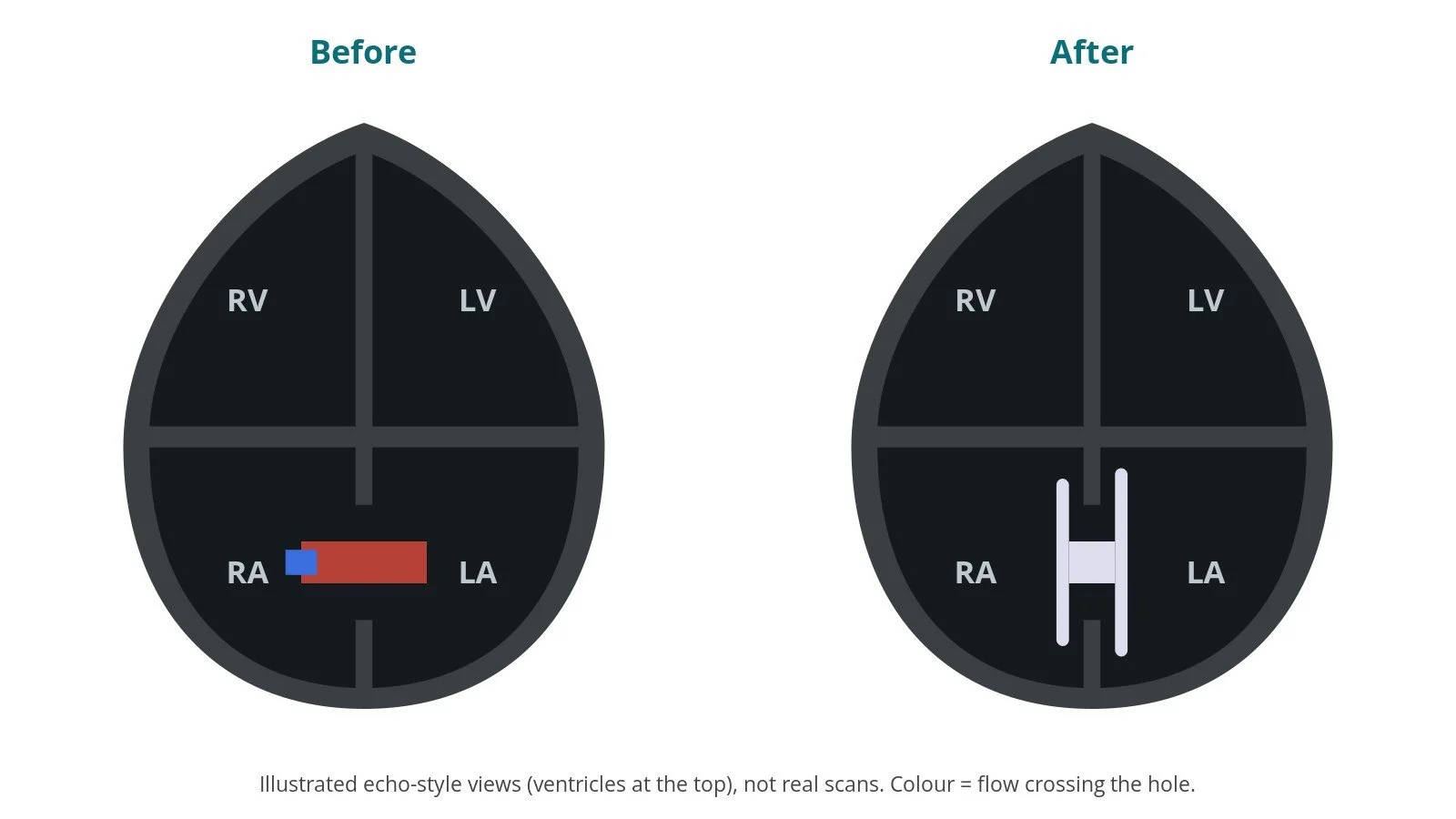
Checks. A gentle push–pull test checks stability. Echo confirms both discs grip the rims, there is little or no leak, and the device is clear of the valves, the aorta and the vein openings.
D. Release. The device is released from its cable. Before release it can be pulled back and repositioned or changed.
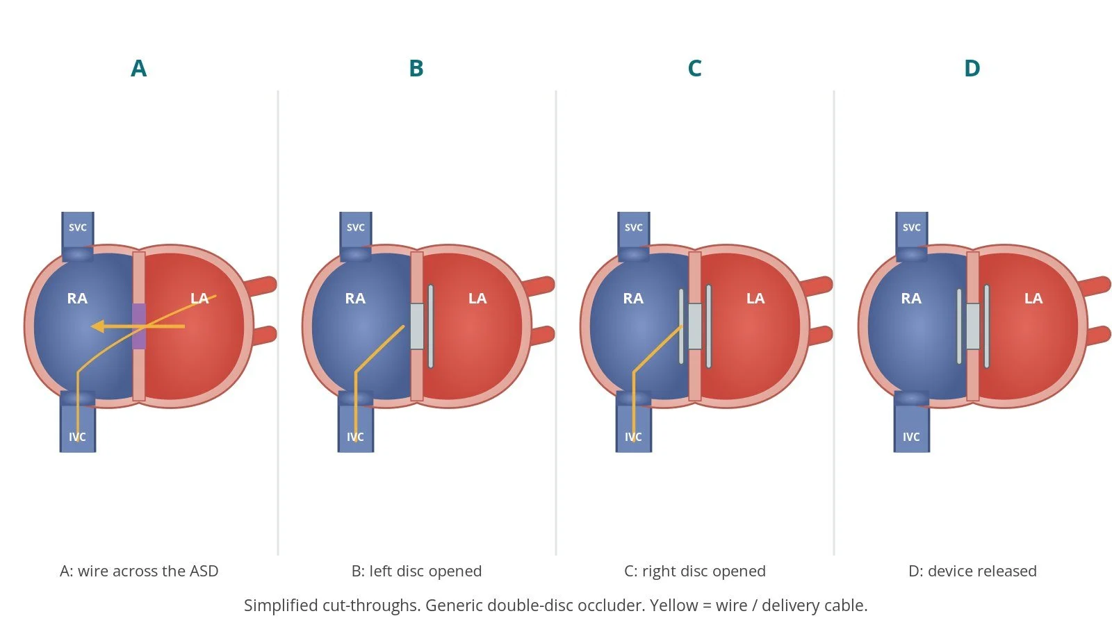
A: cross the ASD. B: open the left disc. C: open the right disc; checks. D: release.
4. Finishing
The sheath is removed and the vein is closed with pressure or a skin stitch, per operator. A final echo checks the device and looks for fluid around the heart.
Recovery and aftercare
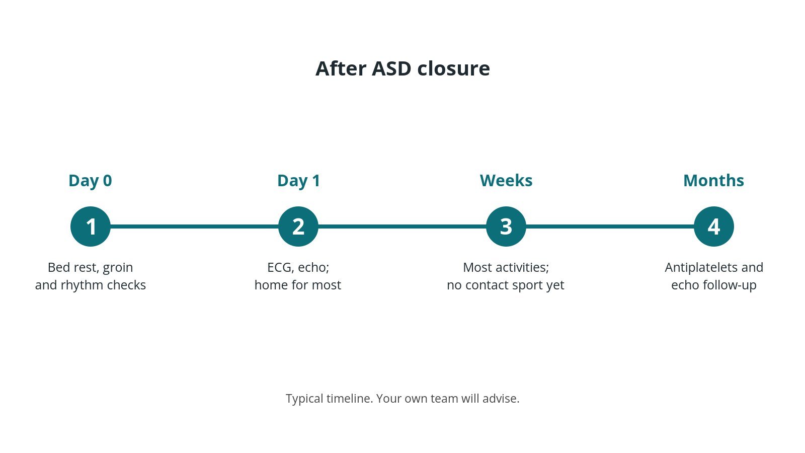
First hours: bed rest with checks of the groin, blood pressure and heart rhythm, as per centre protocol.
Going home: usually the next day after an ECG, an echo and sometimes a chest X-ray.
Medicines: antiplatelet medicine for several months, as advised by your team.
Activity: avoid heavy lifting and contact sports for a few weeks, or as advised.
Follow-up: echo checks over the following months and years.
Dental and medical procedures: tell your dentist and doctors about the device; they follow current guidance on infection prevention.
Driving: ask your team. Do not drive for at least a couple of days, or as advised after a general anaesthetic.
Call 999 if you have chest pain, fainting or sudden breathlessness. Seek urgent advice for new palpitations.
Risks and complications
Your team will discuss your own risks. Possible problems include:
bleeding or bruising at the groin
irregular heart rhythms, including AF, usually short-lived
the device moving out of place (embolisation), which may need retrieval by catheter or surgery
a small leak around the device
rarely, the device wearing through the heart wall (erosion), which can cause fluid around the heart
clots on the device, stroke, infection or headaches in the weeks after
Cath lab kit list (printable)
Barts example kit list
An example from one UK centre, written generically. Every lab has its own protocol, and sizes and doses vary.
Access
femoral venous sheath and delivery sheath, sizes per device and operator
ultrasound probe and cover
local anaesthetics, as per centre protocol
saline flush bag and flush set
heparin, as per centre protocol
non-absorbable suture for a figure-of-eight (Z) skin stitch, per operator
Catheters, wires and device
multipurpose-shaped diagnostic catheter; right coronary-shaped catheter optional
stiff exchange guidewire, J-tip, length per operator
ASD occluder (size per device and operator) and its delivery system
Luer-lock syringes for preparing the device; 3-way tap with extension
Sizing
soft compliant sizing balloon, size per device and operator
contrast and saline mix, as per centre protocol
3-way tap and large Luer-lock syringes
Imaging
TOE probe and echo machine (or ICE), X-ray
Procedural steps (Barts example)
General anaesthetic; TOE probe placed
Right femoral venous access; sheath inserted
Multipurpose catheter and wire across the ASD; wire parked in a pulmonary vein
Wire exchanged for a stiff exchange wire; catheter removed
Sizing on TOE, with a sizing balloon if used
Delivery sheath into the LA and de-aired; device loaded
Left disc, waist and right disc deployed under X-ray and TOE
Push–pull and echo check; device released
Sheath removed; vein closed per operator
First time in the cath lab?
💡 First-time tip
Respect the air. The delivery sheath sits in the left atrium. De-air carefully and keep flush lines bubble-free. Check the device before it opens. Confirm the device type and size with the operator and keep the packaging until the case ends. Have retrieval kit nearby. Know where snares and larger sheaths are kept in case a device moves. Watch the rhythm. Wires and devices in the atria often trigger extra beats or AF. Watch the pressure. A falling blood pressure can mean fluid around the heart. Tell the team straight away.
FAQ
Is an ASD the same as a PFO?
No. An ASD is a true hole; a PFO is a flap-like tunnel left over from before birth. They are treated differently.
Can all ASDs be closed with a device?
No. Device closure is mainly for secundum ASDs with enough tissue around them. Other types are usually closed with surgery.
Will I feel the device?
No. Some people notice palpitations or headaches in the first weeks. Tell your team if you do.
Can I have an MRI scan afterwards?
Most ASD occluders are labelled MR conditional, which means a scan is possible under set conditions. Always tell the scanning team and carry your implant card.
Related Cath Lab 101 guides
Sources
All checked on 10 October 2026. Each page's own date is shown where it gives one.
Baumgartner H, et al. 2020 ESC Guidelines for the management of adult congenital heart disease. Eur Heart J 2021;42(6):563–645. PubMed 32860028
NICE. Endovascular closure of atrial septal defect (IPG96, now HTG58). nice.org.uk/guidance/htg58 (published 27 Oct 2004)
British Heart Foundation. Atrial septal defect. bhf.org.uk
NHS. Congenital heart disease: types. nhs.uk
DVLA / GOV.UK. Cardiovascular disorders: assessing fitness to drive. gov.uk
Disclaimer
Education only. This page is for general education for healthcare students, cath lab staff and curious readers. It isn't medical advice and doesn't replace advice from your own doctor, heart team or local unit protocols. Equipment and medicines are described generically. No product or manufacturer is recommended. If you think someone is having a heart emergency, call 999.
Disclosure: I work for a company that makes a device used in this procedure. This page is independent education, isn't sponsored or reviewed by any company, and doesn't recommend any product.
All diagrams on this page are original illustrations created for rodnieoro.com.