Renal denervation for high blood pressure: a Cath Lab 101 guide
Reviewed by Rodnie Oro, cath lab charge nurse · Last reviewed: October 2026 · Education only, not medical advice
Renal denervation in one minute
What it is: the kidneys and the nerves running in the walls of the kidney (renal) arteries help control blood pressure. Renal denervation is a keyhole procedure in which a catheter from the groin artery delivers energy (radiofrequency or ultrasound) inside the renal arteries to quieten these nerves, aiming to lower blood pressure.
Who it is for: selected adults with high blood pressure that stays high despite several medicines (resistant hypertension), or who cannot take them, as decided by a specialist hypertension team. In the UK it is used with special arrangements.
Main risks: bleeding or a bruise at the groin, damage to the renal artery, and reaction to contrast dye. Your team will explain your own risk.
Usually: under local anaesthetic with sedation and pain relief, about 1 to 1.5 hours, often home the same or next day.
On this page: What is renal denervation? · Who is it for? · When is it done? · Step by step · Recovery · Risks · Kit list · First time in the lab? · FAQ
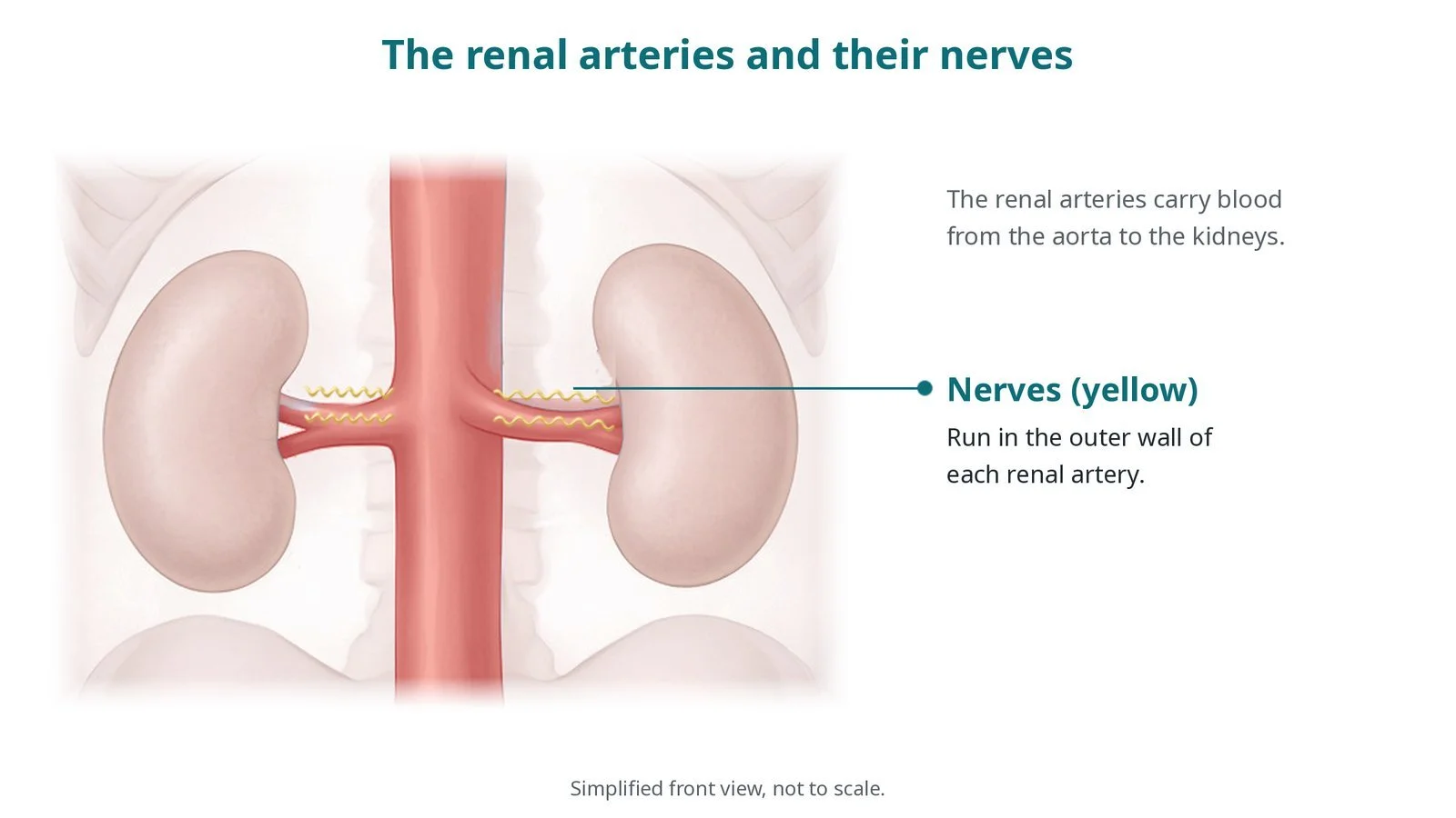
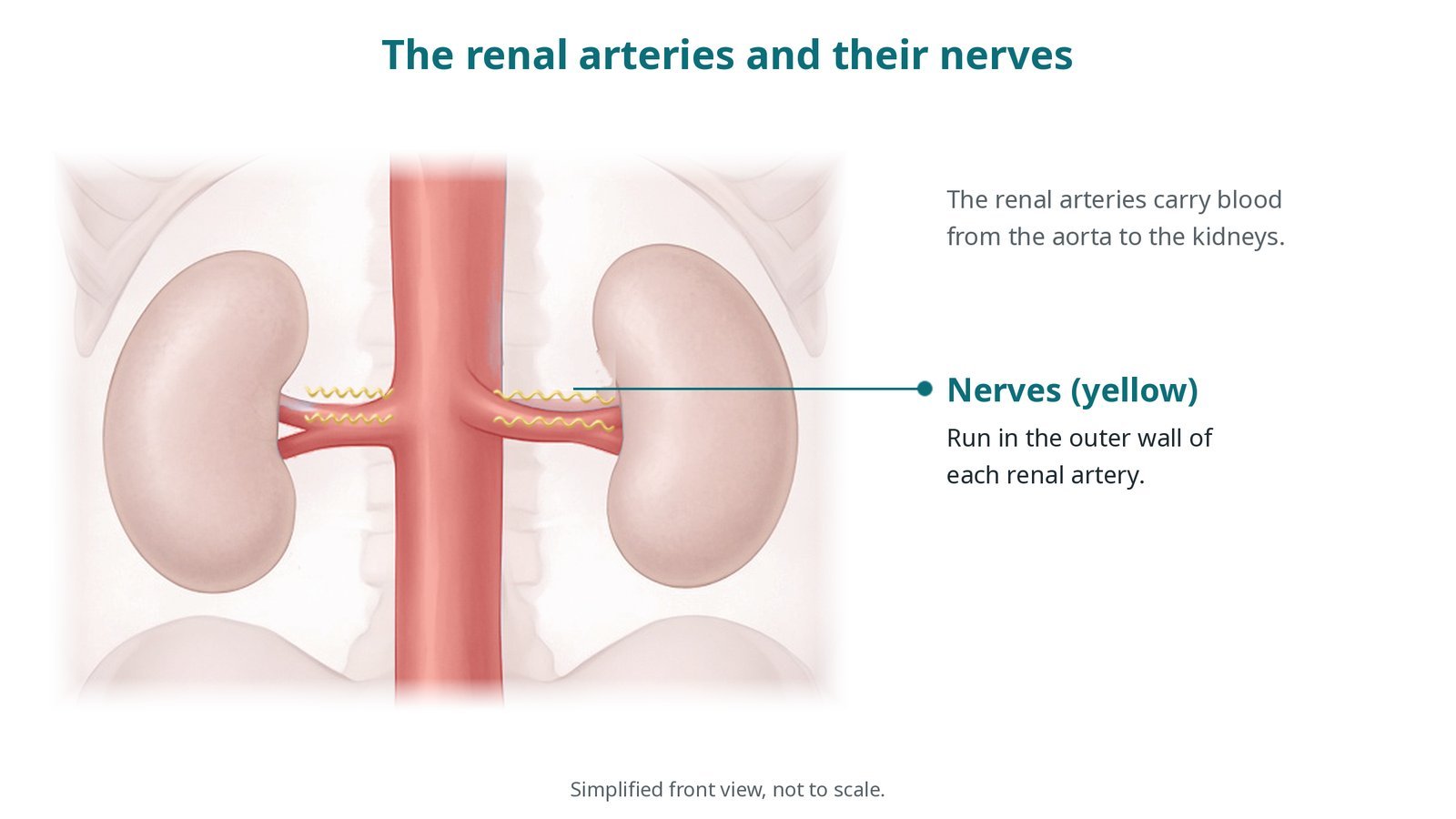
The renal arteries carry blood to the kidneys; nerves (yellow) run in their outer wall.
What is renal denervation?
High blood pressure (hypertension) raises the risk of heart attack, stroke, heart failure and kidney disease. Most people control it with lifestyle changes and medicines, but some have resistant hypertension: blood pressure stays high despite several medicines at good doses, including a diuretic.
The sympathetic nervous system, the "fight or flight" system, helps set blood pressure. Nerves running in the outer wall of each renal artery carry signals between the kidneys and the brain. When they are overactive, the kidneys hold on to salt and water and release hormones that tighten blood vessels.
Renal denervation delivers energy from inside the renal artery to the nerves in its wall, reducing their activity. Two methods are used:
radiofrequency (RF): a catheter with electrodes heats small points of the artery wall, often in a spiral pattern, including branches
ultrasound: a balloon catheter, cooled with circulating fluid, sends ultrasound energy around the artery wall
Renal denervation does not replace lifestyle changes or, usually, medicines. The aim is a lower blood pressure, sometimes with fewer tablets.
New to the acronyms? See the Cath Lab Abbreviations A–Z.
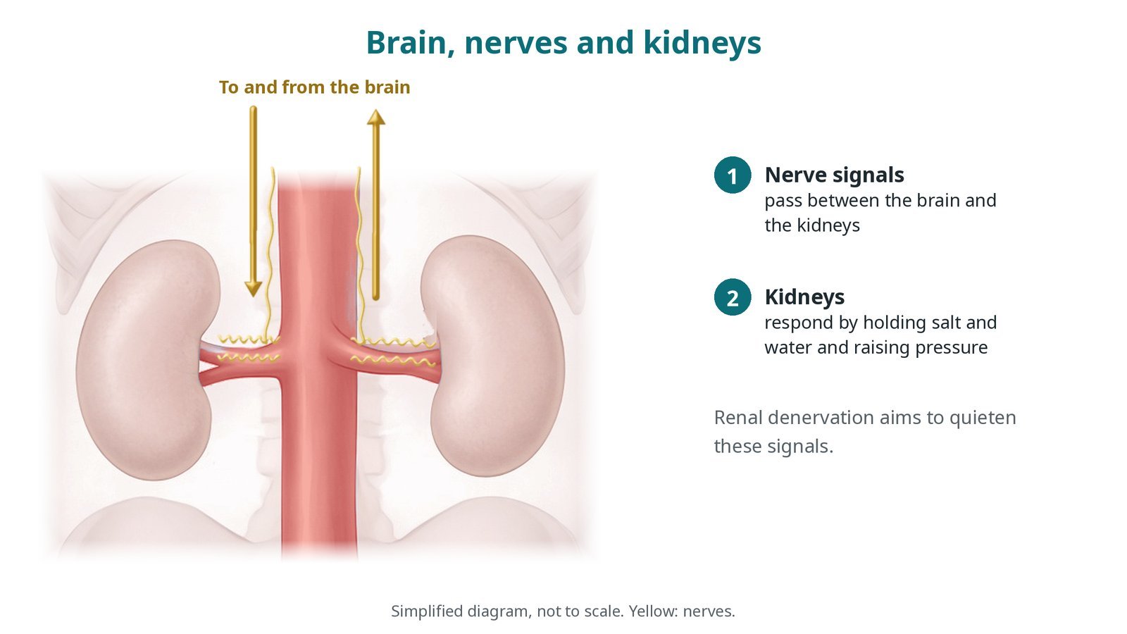
Nerve signals between the brain and kidneys help set blood pressure. Renal denervation aims to quieten them.
Who is it for?
NICE (IPG754, now HealthTech guidance HTG662, March 2023) says renal denervation for resistant hypertension should only be used with special arrangements for clinical governance, consent and audit or research, and that patients should be selected by a specialist team. The ESC 2024 hypertension guidelines say it may be considered in selected people with resistant hypertension, or who cannot tolerate medicines, at medium to high-volume centres, after shared decision-making.
Before it is considered, the team checks:
blood pressure with 24-hour ambulatory monitoring, to confirm it is truly high
that medicines are being taken and are at the right doses
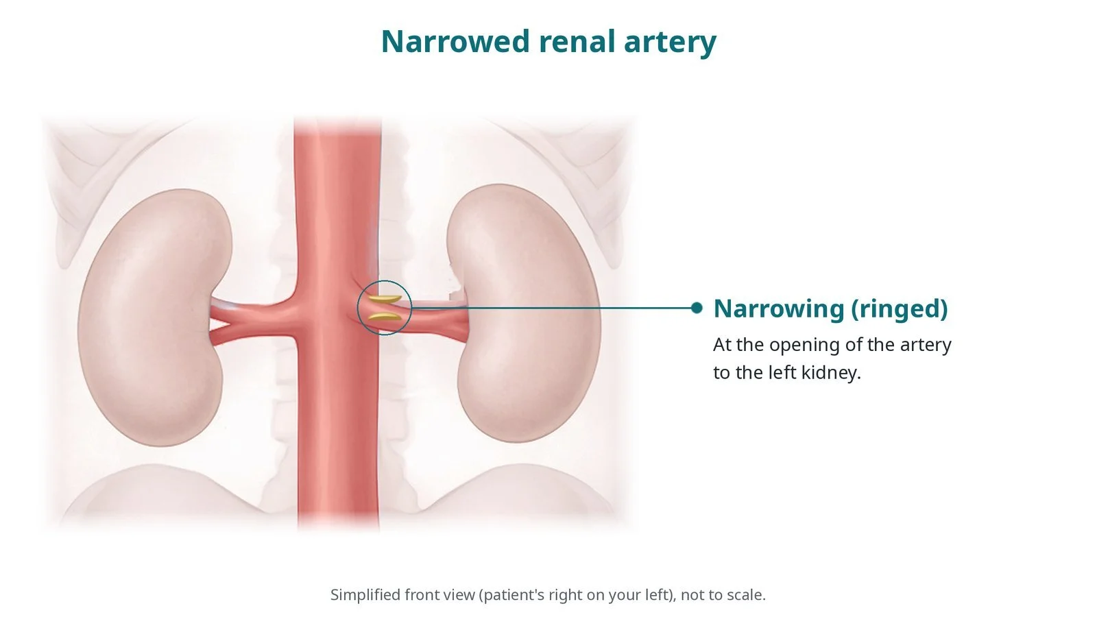
for secondary causes of high blood pressure, such as kidney artery narrowing, hormone problems or sleep apnoea
the anatomy of the renal arteries on CT or MRI, and kidney function
When is it done?
It is a planned procedure. Before it, the team usually:
checks kidney function, allergies (especially to contrast) and blood thinners
confirms the anatomy is suitable
explains what is known and unknown about the benefits, and that blood pressure may take weeks or months to fall
Step by step
1. Getting ready
You lie on the X-ray table. Sedation and strong pain relief are given, as per centre protocol, because the energy delivery can cause brief, deep abdominal or back discomfort. Monitoring includes ECG, blood pressure and oxygen levels.
2. Access
A sheath is placed in the femoral artery in the groin, under ultrasound. Sheath and guide catheter sizes are per device and operator.
Heparin is given, dose as per centre protocol.
A pigtail catheter may be used for an aortogram, then a guide catheter is placed at the opening of each renal artery. The artery size is measured on the angiogram.
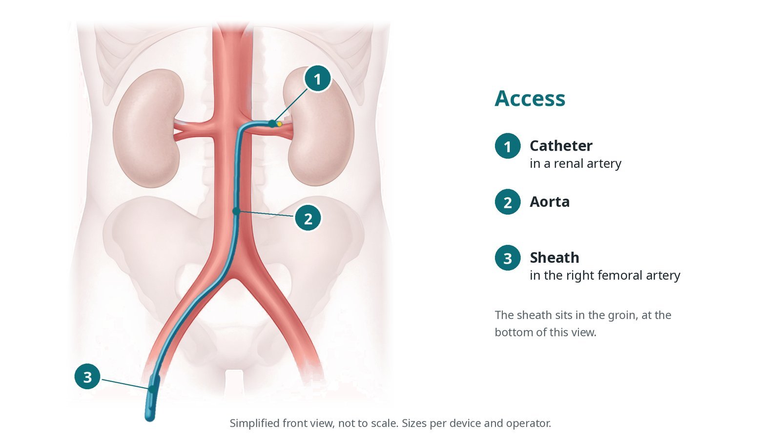
Access: (1) catheter in a renal artery; (2) aorta; (3) sheath in the right femoral artery.
3. Delivering the energy
A. Engage. The guide catheter is placed at the opening of the renal artery.
B. Wire and catheter. A thin wire is passed into the artery and the denervation catheter (RF or ultrasound) is advanced over it to the target position.
C. Energy. Energy is delivered at several points or in short sessions, according to the device and the artery size, per device instructions. Medicines to prevent artery spasm may be given.
D. Other side. The same is done in the other renal artery and, for some methods, its branches. A final angiogram checks the arteries for spasm or damage.
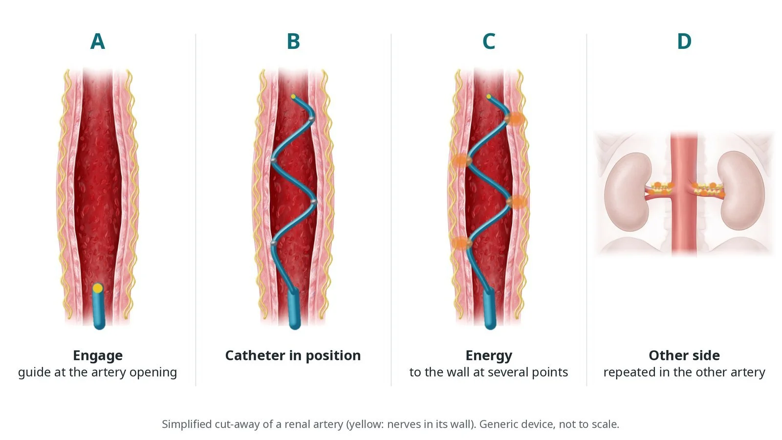
A: engage the artery. B: catheter in position. C: energy to the wall. D: repeat on the other side.
4. Finishing
The sheath is removed and the artery is closed with pressure or a closure device, per operator.
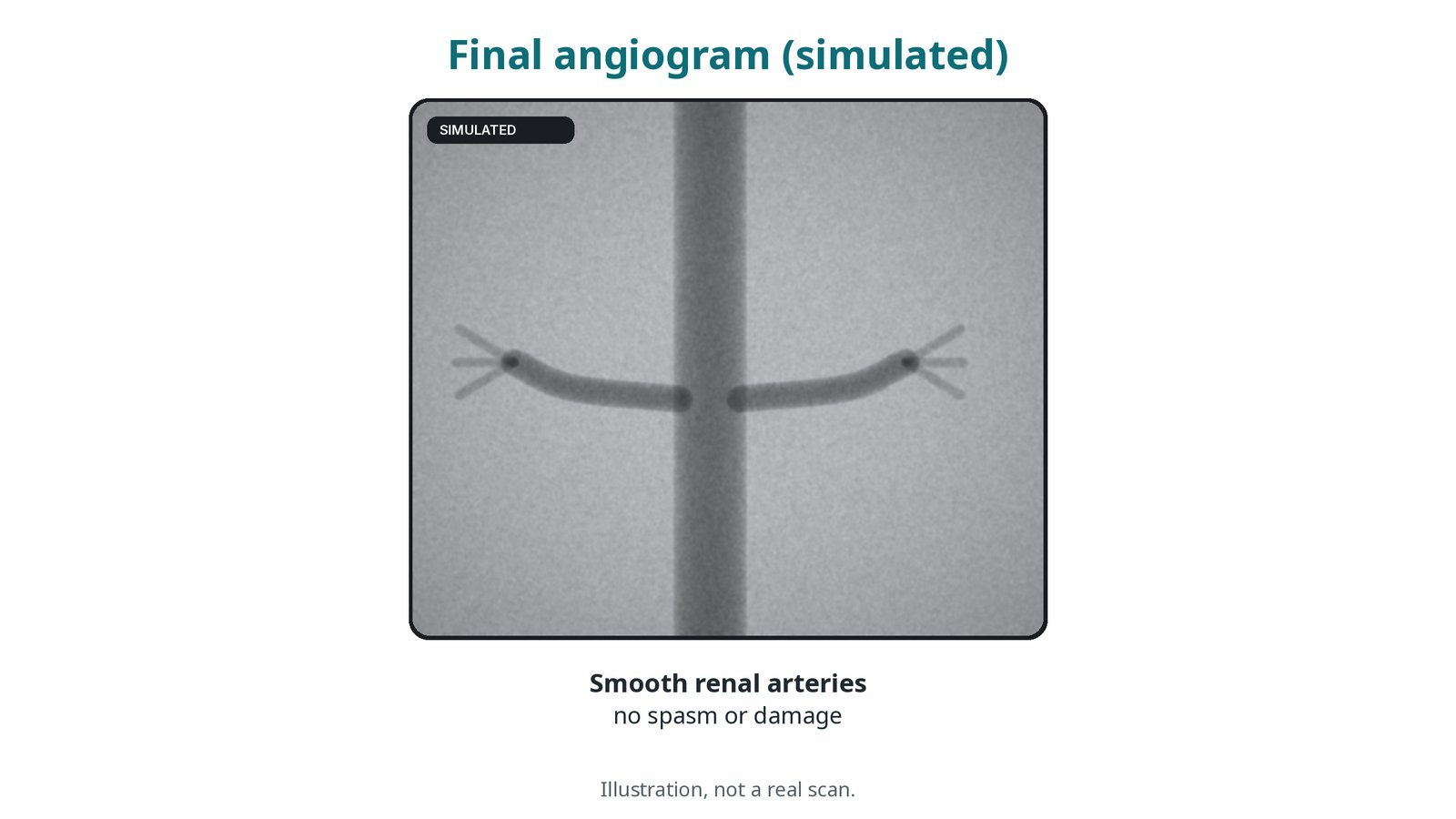
Illustration, not a real scan: a final angiogram checks the renal arteries are smooth, with no spasm or damage.
Recovery and aftercare
First hours: bed rest with checks of the groin, leg pulse and blood pressure, as per centre protocol.
Going home: often the same or next day.
Medicines: keep taking your blood pressure medicines unless your team tells you otherwise; any changes are made gradually.
Monitoring: home and 24-hour blood pressure checks over the following months; kidney function is checked.
Activity: avoid heavy lifting for a few days, or as advised.
Driving: ask your team; do not drive until the groin has healed and the effects of sedation have worn off.
Call 999 if you have heavy bleeding from the groin, a cold, pale or painful leg, or severe abdominal or back pain.
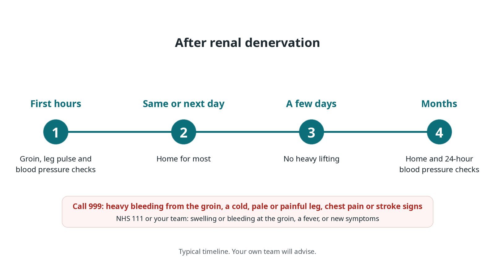
After renal denervation: (1) groin and blood pressure checks; (2) home for most; (3) no heavy lifting for a few days; (4) blood pressure monitoring.
Risks and complications
Your team will discuss your own risks. Possible problems include:
bleeding, bruising or a false aneurysm at the groin
damage to the renal artery, such as a tear or later narrowing (rare)
pain during the energy delivery
slow heart rate or low blood pressure during the procedure
kidney strain from the contrast dye
blood pressure not falling as much as hoped
Cath lab kit list (printable)
Example kit list
Written generically. Every lab has its own protocol, and sizes and doses vary.
angiography pack; femoral arterial sheath, size per device and operator
long sheath and renal guide catheters (different tip shapes), per operator
coronary-type guidewire, per operator
pigtail catheter for an aortogram
renal denervation catheter and generator (RF or ultrasound), per device and operator
contrast; heparin, sedation, analgesia and anti-spasm medicines, as per centre protocol
femoral closure device or manual compression, per operator
radiographer to measure the renal artery diameter on the angiogram
Procedural steps (example)
Sedation and analgesia; femoral arterial access; heparin
Aortogram and renal angiograms; artery diameters measured
Guide into the first renal artery; wire; denervation catheter positioned
Energy delivered per device instructions; repeat in the other artery (and branches if used)
Final angiograms; sheath removed; haemostasis
First time in the cath lab?
💡 First-time tip
Pain relief first. Energy delivery can be very uncomfortable; make sure analgesia has been given before the first treatment. Watch the heart rate. It can slow during treatment. Have atropine available as per centre protocol. Know your generator. Set up and check the console and cables before the patient arrives. Measure carefully. Artery size decides catheter choice; the radiographer's measurement matters. Check the leg. Groin and leg checks continue after the case.
FAQ
Will I be able to stop my tablets?
Most people still need medicines. Some need fewer. Your team will review this over time.
How soon does blood pressure fall?
It can take weeks to months. Regular checks help show the effect.
Is it painful?
You will have sedation and pain relief. Brief abdominal or back discomfort during treatment is common.
Is renal denervation widely available?
In the UK it is offered in selected specialist centres, with special arrangements as advised by NICE.
Related Cath Lab 101 guides
Sources
All checked on 10 October 2026. Each page's own date is shown where it gives one.
NICE. Percutaneous transluminal renal sympathetic denervation for resistant hypertension (IPG754, now HTG662). nice.org.uk/guidance/htg662 (published 1 Mar 2023)
NICE. Hypertension in adults: diagnosis and management (NG136). nice.org.uk/guidance/ng136
McEvoy JW, et al. 2024 ESC Guidelines for the management of elevated blood pressure and hypertension. Eur Heart J 2024;45(38):3912–4018. PubMed 39210715
NHS. High blood pressure (hypertension). nhs.uk
Disclaimer
Education only. This page is for general education for healthcare students, cath lab staff and curious readers. It isn't medical advice and doesn't replace advice from your own doctor, heart team or local unit protocols. Equipment and medicines are described generically. No product or manufacturer is recommended. If you think someone is having a heart emergency, call 999.
Disclosure: I work in the medical device industry. This page is independent education and isn't sponsored or reviewed by any company.
All diagrams on this page are original illustrations created for rodnieoro.com.