Balloon pulmonary valvuloplasty: a Cath Lab 101 guide
Reviewed by Rodnie Oro, cath lab charge nurse · Last reviewed: November 2026 · Education only, not medical advice
Balloon pulmonary valvuloplasty in one minute
What it is: The pulmonary valve lets blood out of the right ventricle into the lung artery. In pulmonary valve stenosis its leaflets are stuck together, so the opening is narrow. In balloon valvuloplasty a balloon catheter is passed from the femoral vein, through the right side of the heart, and inflated across the valve to split the fused edges and widen the opening.
Who it is for: Newborns, children and adults with significant pulmonary valve stenosis, as decided by a congenital heart team. Newborns with critical stenosis need it urgently. It works best on thin, mobile valves; thick (dysplastic) valves respond less well.
Main risks: A leak of the valve afterwards (common, usually mild), a temporary squeeze of the muscle below the valve, abnormal heart rhythms, problems at the groin and, rarely, a tear of the valve ring or outflow tract. Your team will explain your own risk.
Usually: Under general anaesthetic for children and some adults (sedation for others), about 1 to 2 hours. Many children go home the next day; newborns stay longer.
On this page: What is balloon pulmonary valvuloplasty? · Who is it for? · When is it done? · Step by step · Recovery · Risks · Kit list · First time in the lab? · FAQ
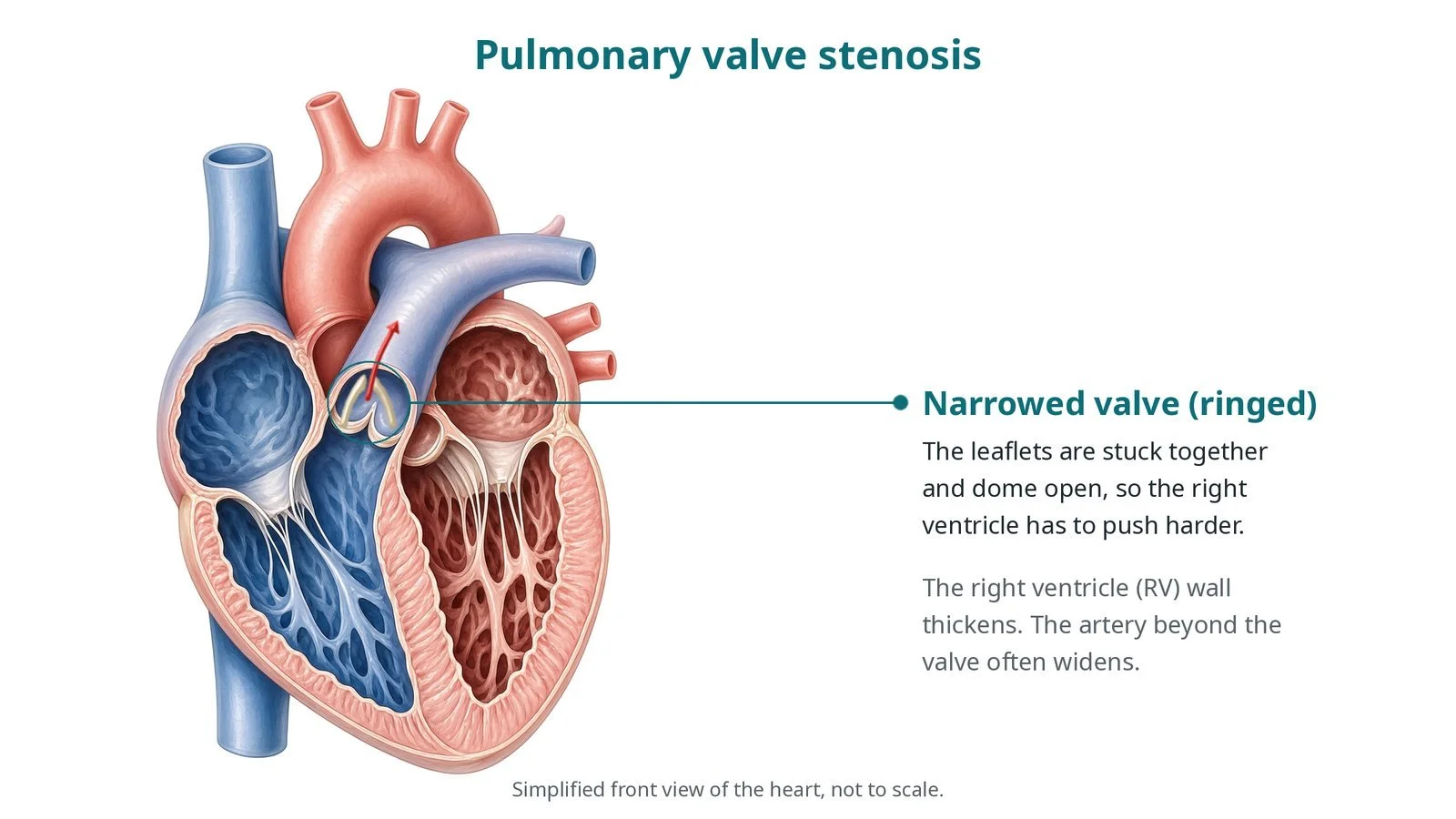
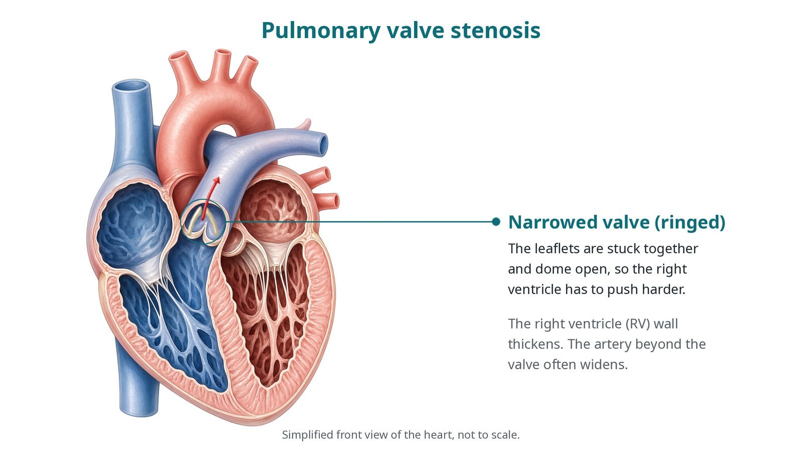
Pulmonary valve stenosis: the narrowed valve (ringed) domes open, so the right ventricle has to push harder; its wall thickens and the artery beyond often widens.
What is balloon pulmonary valvuloplasty?
The pulmonary valve sits at the top of the right ventricular outflow tract, between the right ventricle and the main pulmonary artery. It normally has three thin leaflets that open fully with each beat.
In typical pulmonary valve stenosis, present from birth, the leaflet edges are fused. The valve opens like a dome with a small hole in the middle. The right ventricle has to generate a high pressure to push blood through, so its wall thickens. The artery just beyond the valve often widens (post-stenotic dilatation).
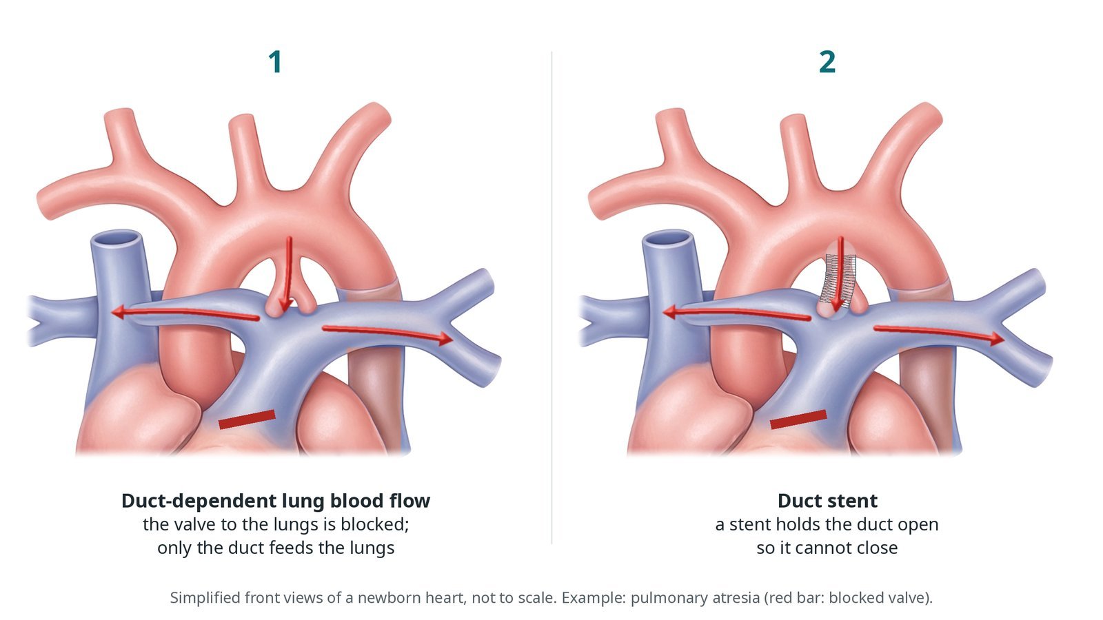
In critical stenosis in a newborn, so little blood gets through that the lungs depend on the arterial duct. A prostaglandin infusion keeps the duct open until the valve is treated.
The balloon tears the fused edges (commissures) apart. Thick, dysplastic valves, seen for example in Noonan syndrome, have little fusion to split and may need surgery instead.
New to the acronyms? See the Cath Lab Abbreviations A–Z.
Who is it for?
The decision is made by a paediatric or adult congenital heart team using current guidelines (for adults, the ESC 2020 and AHA/ACC 2018 adult congenital heart disease guidelines). Usual reasons:
severe narrowing on echo, even without symptoms
moderate narrowing with symptoms, such as breathlessness, or a struggling right ventricle
critical stenosis in a newborn, which is urgent
Who it may not suit: Thick, dysplastic valves, a very small valve ring, or narrowing above or below the valve or in the branch arteries may be better treated with surgery or another procedure.
The decision is made by your heart team, who will talk through the reasons, the alternatives and the risks with you.
When is it done?
Older children and adults have a planned procedure after echo. Newborns with critical stenosis are treated in the first days of life, usually on a prostaglandin infusion, on the neonatal or cardiac unit.
gives you a chance to ask questions and give consent
arranges blood tests, including kidney function and a blood count, and an ECG
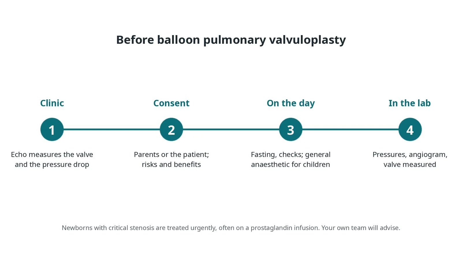
Before balloon pulmonary valvuloplasty: echo and consent; checks and anaesthetic on the day; pressures, angiogram and valve measurement in the lab. Newborns with critical stenosis are treated urgently.
Step by step
1. Getting ready
Children have a general anaesthetic; some adults have sedation. Babies are kept warm on a warming mattress and their temperature is watched. ECG, oxygen saturation and blood pressure are monitored, and antibiotics are given as per centre protocol. In children every drug, flush and contrast volume is worked out by weight, as per centre protocol.
2. Access and pressures
A sheath is placed in the femoral vein under ultrasound. A small arterial line or sheath is often added to watch the blood pressure. Heparin is given, dose as per centre protocol. An end-hole catheter measures pressures in the right ventricle and pulmonary artery. The difference between them (the gradient) shows how tight the valve is.
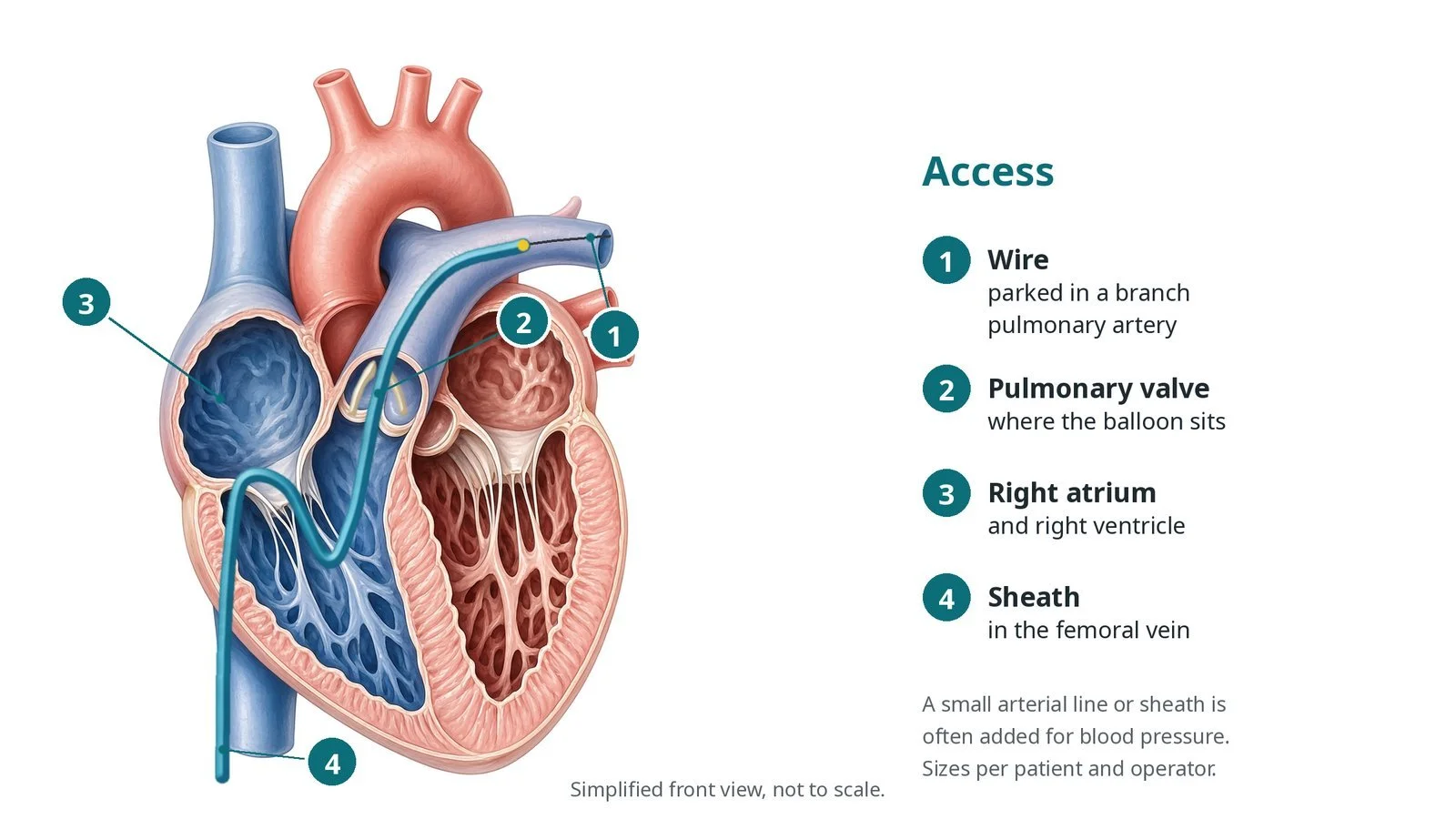
Access: (1) wire parked in a branch pulmonary artery; (2) the pulmonary valve, where the balloon sits; (3) right atrium and ventricle; (4) sheath in the femoral vein.
3. Opening the valve
A. Measure. An angiogram in the right ventricle shows the domed valve, and the valve ring (annulus) is measured. The balloon size is chosen from the annulus, per operator.
B. Wire across. A catheter is passed through the valve and a stiff wire is parked far out in a branch pulmonary artery for support.
C. Inflate. The balloon is centred across the valve and inflated quickly. A waist on the balloon disappears as the valve opens. Blood flow and blood pressure fall briefly while the balloon blocks the valve, so it is deflated straight away. It may be repeated or a larger balloon used.
D. Check. Pressures are measured again on pull-back from the pulmonary artery to the right ventricle, and an angiogram or echo checks the result and any leak.
Watch: Blood pressure and heart rate drop during each inflation. If they do not recover quickly after deflation, tell the operator at once.
4. Watching the right ventricle
Once the valve opens, the thick muscle just below it can squeeze tight and narrow the outflow (reactive infundibular obstruction). In its severe form, sometimes called a "suicide right ventricle", the blood pressure falls even though the valve is now open. It is treated with fluids and, per the team, medicines that slow and relax the heart, such as a beta-blocker. Medicines that make the heart beat harder can make it worse. The muscle usually relaxes over weeks to months.
Watch: Low blood pressure or falling oxygen levels after a good valve result: tell the operator, have fluids ready, and expect the team to avoid drugs that make the heart squeeze harder.
5. Finishing
The sheaths are removed and pressure is applied. In babies and small children the leg's colour, warmth and pulse are checked on both sides, because small vessels can block after a sheath.
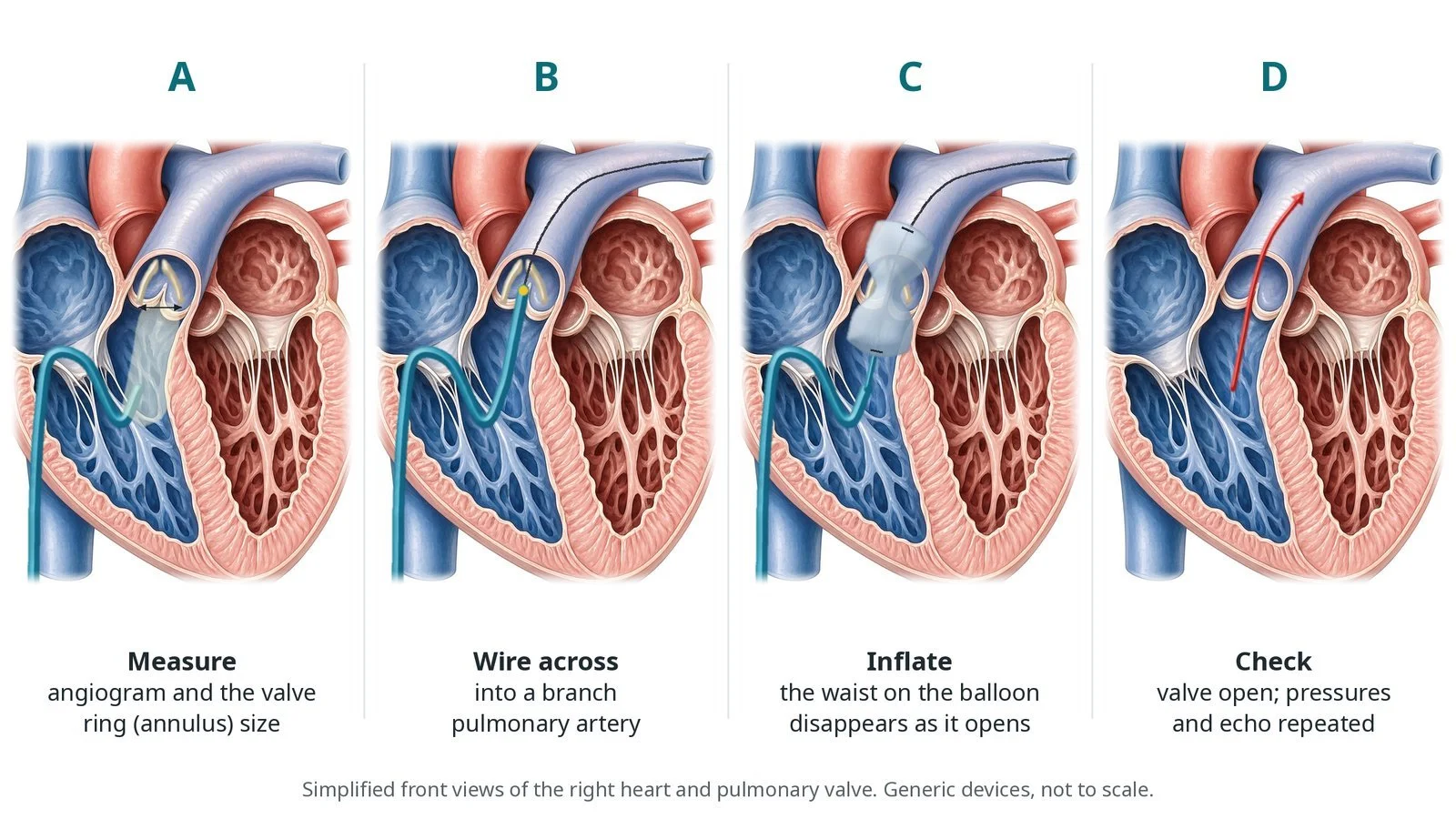
A: angiogram and annulus measurement. B: wire across into a branch pulmonary artery. C: balloon inflated until the waist disappears. D: valve open; pressures and echo repeated.
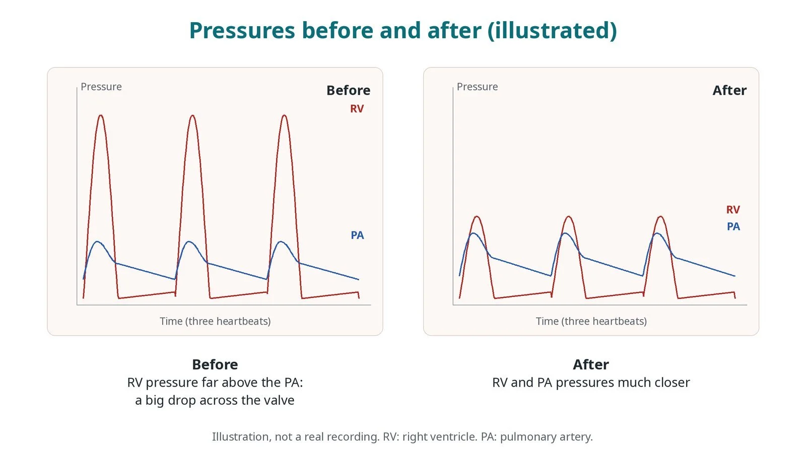
Illustrated pressures, not a real recording. Before: right ventricular pressure far above the pulmonary artery. After: the two are much closer.
Recovery and aftercare
First hours: monitored bed rest with checks of the groin, leg pulses, heart rhythm and oxygen levels, as per centre protocol.
Echo: usually the next day, to check the gradient and any leak.
Going home: many children and adults go home the next day; newborns stay until feeding and oxygen levels are settled.
Activity: avoid heavy lifting and rough play for a few days while the groin heals, or as advised.
Driving (adults): ask your team; do not drive until the groin has healed and any sedation has worn off.
Follow-up: lifelong echo checks with the congenital heart team, because the valve can leak more or narrow again over time.
Call 999 if there is fainting, chest pain, sudden breathlessness, heavy bleeding from the groin, or a baby who is blue, floppy or struggling to breathe.
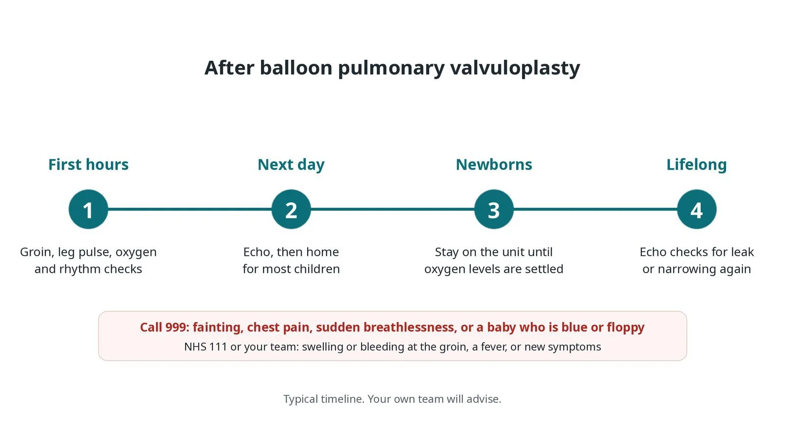
After valvuloplasty: (1) groin, pulse, oxygen and rhythm checks; (2) echo, home the next day for most children; (3) newborns stay until oxygen levels settle; (4) lifelong echo checks.
Risks and complications
Your team will explain the risks that apply to you. Possible problems include:
A leak of the pulmonary valve afterwards (common, usually mild): It is usually well tolerated but is watched on echo over the years.
Temporary squeeze of the muscle below the valve (uncommon)
Abnormal heart rhythms during the procedure
Bleeding or a clot at the groin; in babies, a blocked leg vein or artery (uncommon)
Tear of the valve ring or outflow tract (rare)
Narrowing coming back, needing a repeat procedure or surgery (uncommon)
Cath lab kit list (printable)
Barts example kit list
An example from one UK centre, written generically. Every lab has its own protocol, and sizes and doses vary.
venous sheath, size per patient and operator; small arterial sheath or line
ultrasound for vascular access
end-hole and angiographic catheters (balloon-tipped in small babies), per operator
soft and stiff guidewires
valvuloplasty balloons in a range of sizes, chosen from the annulus, per operator
inflation syringes or device with dilute contrast
pressure transducer set, zeroed and levelled
contrast; heparin and other drugs, weight-based, as per centre protocol
warming mattress and temperature probe for babies
Procedural steps (Barts example)
Getting ready
Access and pressures
Opening the valve
Watching the right ventricle
Finishing
First time in the cath lab?
Know the weight. In children everything is weight-based: drugs, flushes, contrast and balloon size. Check it is written up and use it.
Prepare for the drop. Each inflation briefly stops blood leaving the right ventricle. Watch the monitor and call out a slow recovery.
Gradients are the result. Get clean, zeroed pressure traces before and after; the operator decides the next balloon from them.
Think about the muscle. Low blood pressure after a good result may be the outflow muscle squeezing, not bleeding. Say what you see.
Write the weight on the board. Weight-based doses, contrast limits and balloon choices all come from it.
Zero and level the transducer before the first pressure. The before-and-after gradient is the main result of the case.
Have the inflation syringe or device ready and labelled. Inflations are short, and the team wants to deflate on command.
FAQ
Will the valve need treating again?
Most typical valves stay open well for many years. Some narrow again or leak more, so regular echo checks continue.
Why is the valve leaky afterwards?
Splitting the stuck leaflets often leaves a small leak. It is usually mild and well tolerated.
Why does my baby still need a drip after the procedure?
In critical stenosis the right ventricle can take days or weeks to recover, so some babies need the prostaglandin drip or extra oxygen for a while.
Why is the pressure still a bit high straight afterwards?
The thick muscle below the valve can keep narrowing the outflow until it relaxes over weeks to months.
Related Cath Lab 101 guides
RVOT stenting in tetralogy of Fallot (coming soon)
Ductal (PDA) stenting in newborns (coming soon)
Sources
All checked on 12 November 2026. Each page's own date is shown where it gives one.
Baumgartner H, et al. 2020 ESC Guidelines for the management of adult congenital heart disease. Eur Heart J 2021;42(6):563–645
Stout KK, et al. 2018 AHA/ACC Guideline for the Management of Adults With Congenital Heart Disease. J Am Coll Cardiol 2019;73(12):e81–e192
Feltes TF, et al. Indications for cardiac catheterization and intervention in pediatric cardiac disease: a scientific statement from the American Heart Association. Circulation 2011;123(22):2607–2652
StatPearls (NCBI Bookshelf), search: pulmonic stenosis. ncbi.nlm.nih.gov/books
Disclaimer
Education only. This page is for general education for healthcare students, cath lab staff and curious readers. It isn't medical advice and doesn't replace advice from your own doctor, heart team or local unit protocols. Equipment and medicines are described generically. No product or manufacturer is recommended. If you think someone is having a heart emergency, call 999.
Disclosure: I work in the medical device industry. This page is independent education and isn't sponsored or reviewed by any company.
All diagrams on this page are original illustrations created for rodnieoro.com.