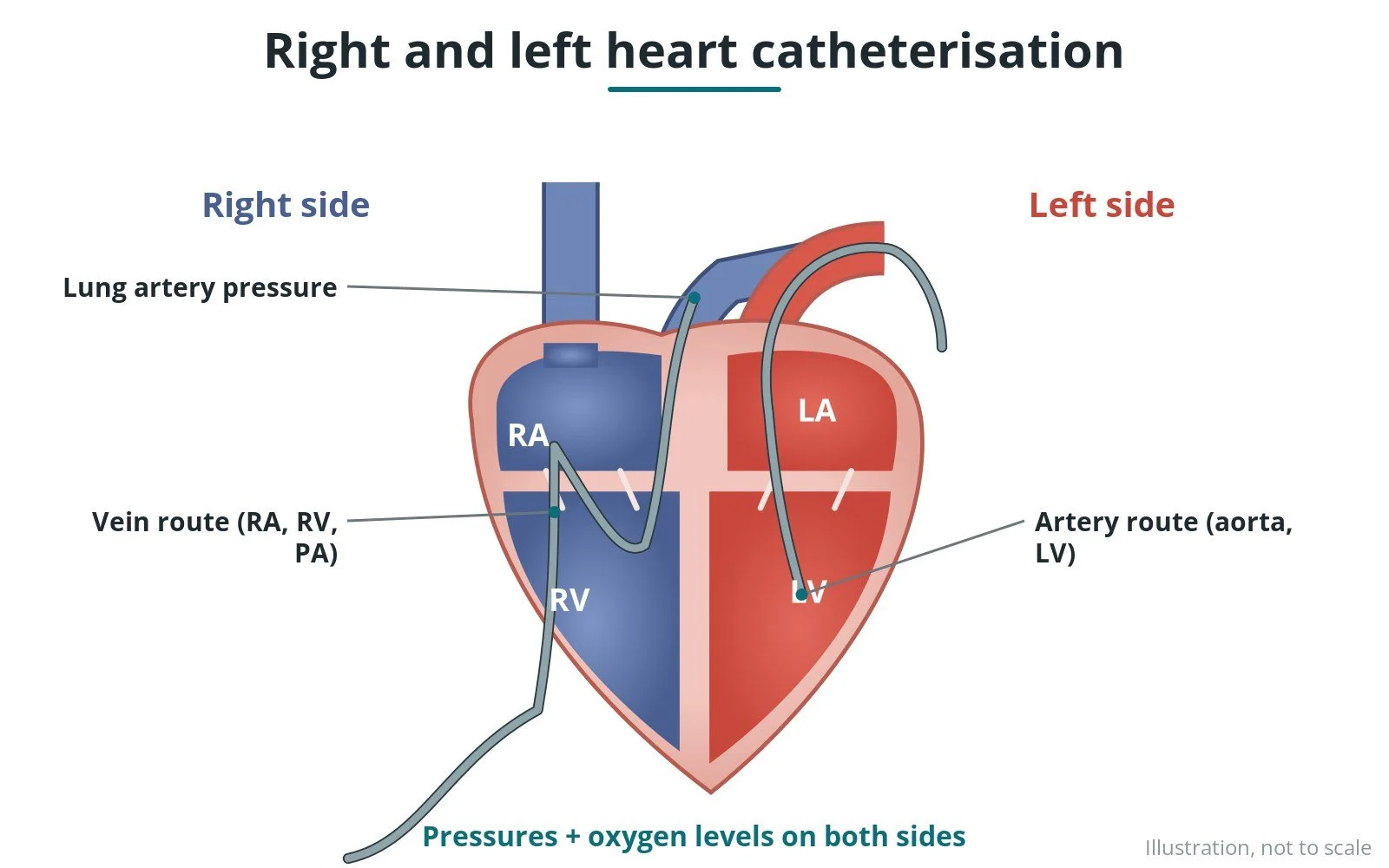
Shunt detection (oximetry run): a Cath Lab 101 guide
Reviewed by Rodnie Oro, cath lab charge nurse · Last reviewed: October 2026 · Education only, not medical advice
Shunt detection in one minute
What it is: checking for a shunt, blood flowing abnormally between the left and right sides of the heart, by taking blood samples from each chamber and measuring their oxygen levels (an oximetry run).
Who it is for: people with a known or suspected hole in the heart, such as an ASD or VSD, or unexplained high lung pressures.
Main risks: those of right heart catheterisation: bruising, brief rhythm disturbances and rarely damage to a vessel.
Usually: part of a heart catheter test, under local anaesthetic, home the same day.
On this page: What is shunt detection? · Who is it for? · When is it done? · Step by step · Recovery · Risks · Kit list · First time in the lab? · FAQ
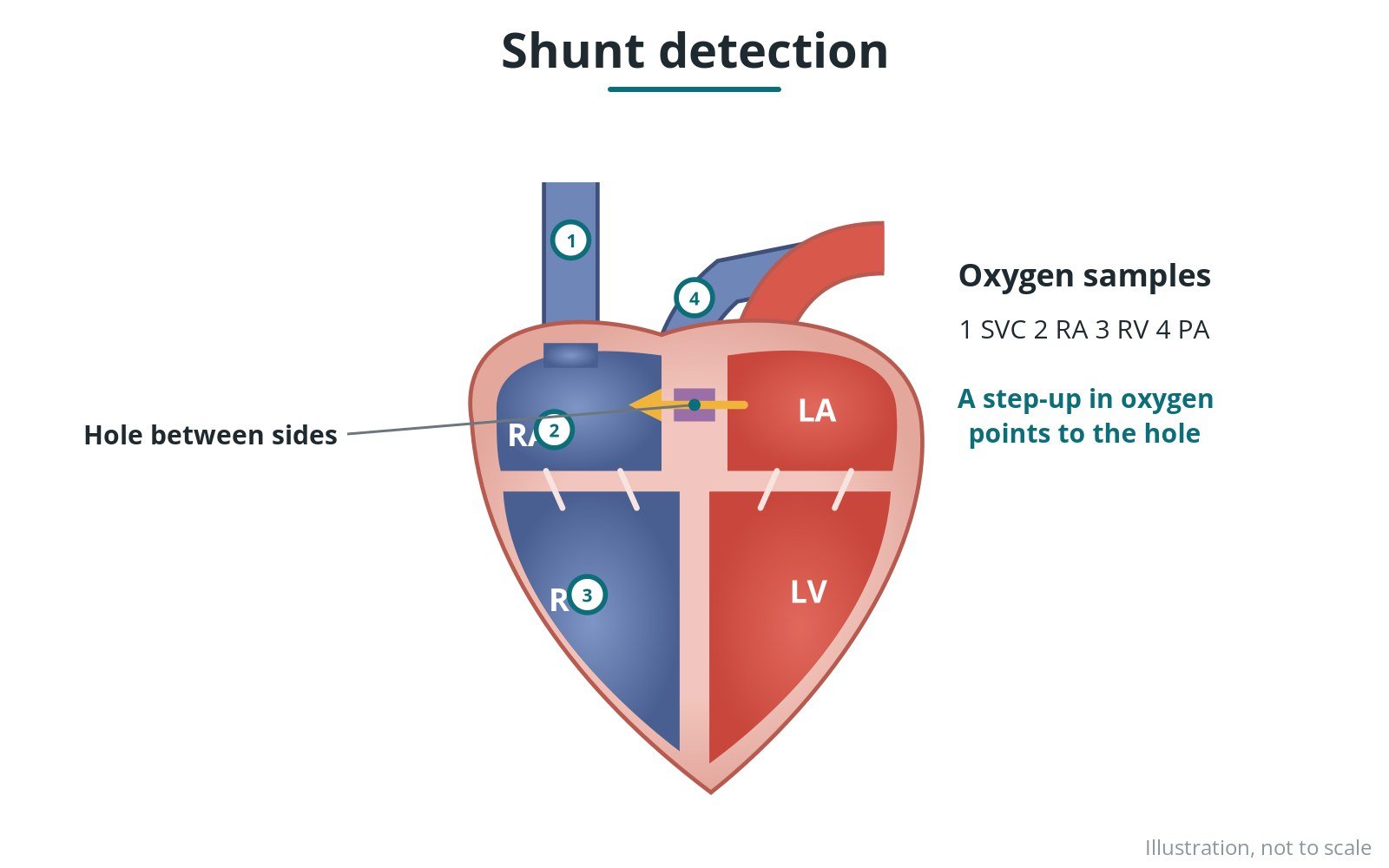
Oxygen samples from each chamber reveal a shunt.
What is shunt detection?
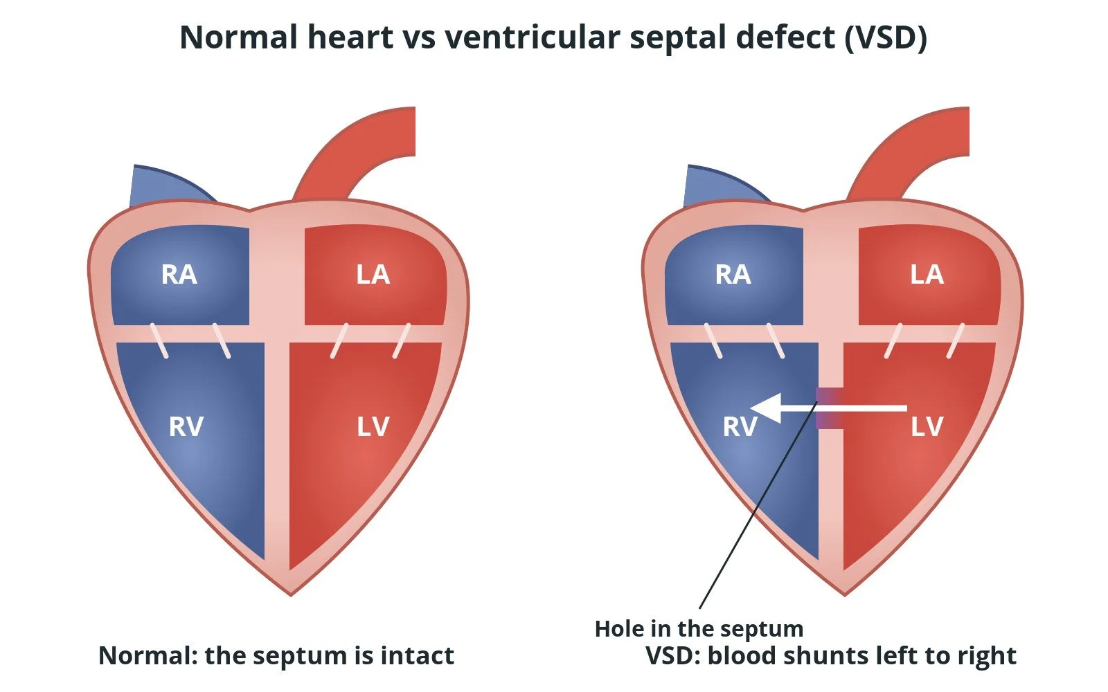
Normally, blood on the right side of the heart is low in oxygen and blood on the left side is high. If there is a hole, blood can leak across.
In an oximetry run:
- samples are taken in a set order from the veins, right atrium, right ventricle and pulmonary artery, and from an artery
- a jump in oxygen level on the right side shows where oxygen-rich blood is entering
- the team calculates the shunt ratio (Qp:Qs), comparing lung flow with body flow
This helps decide whether a hole needs closing.
New to the acronyms? See the Cath Lab Abbreviations A–Z.
Who is it for?
It may be considered for people with:
- atrial or ventricular septal defects (ASD, VSD)
- abnormal vein connections
- suspected shunts with unexplained pulmonary hypertension
- before planned closure procedures
The decision is made by your heart team, who will talk through the reasons, the alternatives and the risks with you.
When is it done?
It can be planned in advance or done urgently. Before it, you usually have:
- blood tests, including kidney function and blood count
- an ECG, and often an echo or other scan
- a review of your medicines, especially blood thinners and diabetes medicines, as per centre protocol
- a chance to ask questions and give consent
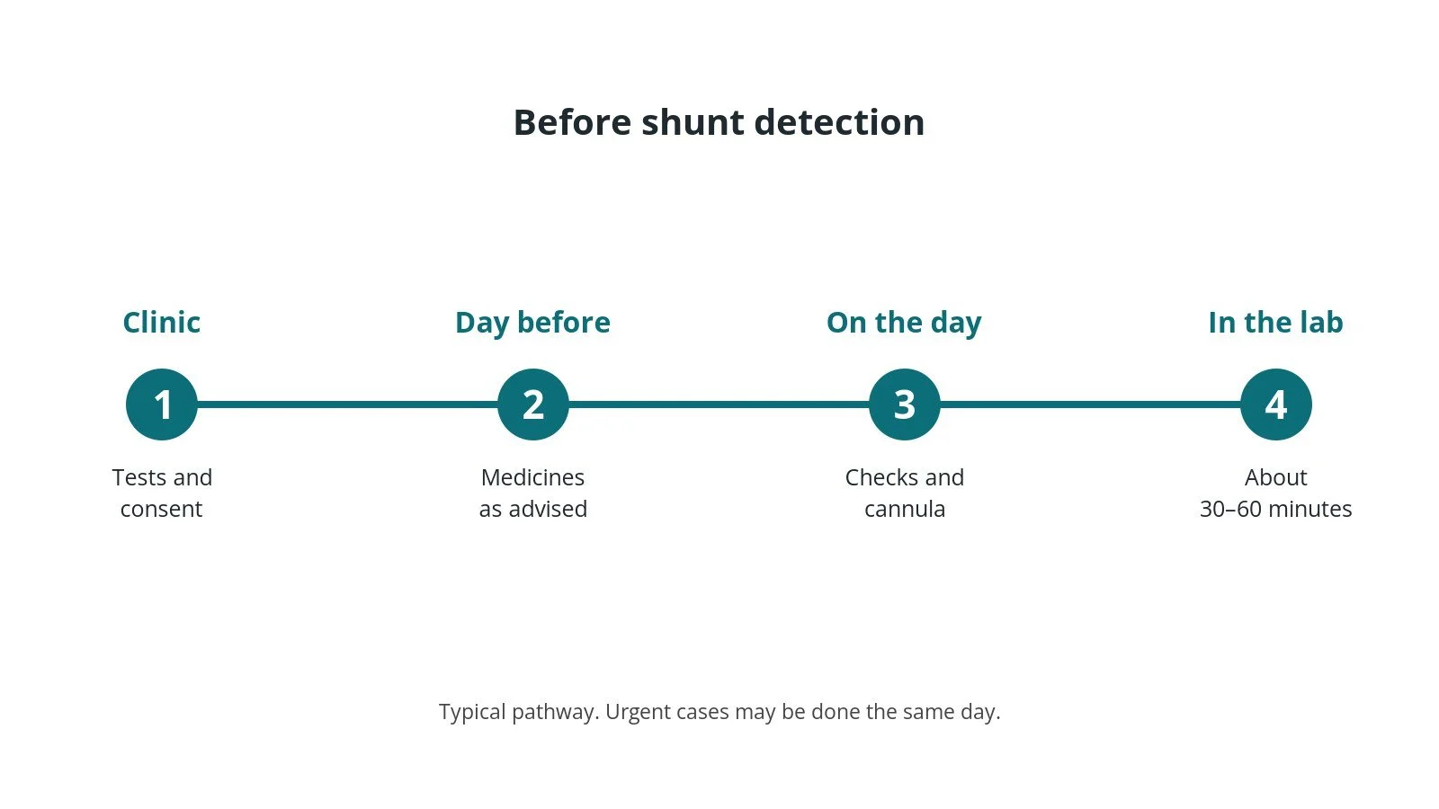
Before the procedure: tests and consent, medicine advice, checks on the day, then the cath lab.
Step by step
1. Getting ready
You lie on the X-ray table. ECG, blood pressure and oxygen levels are monitored. The skin over the access site is cleaned and numbed with local anaesthetic; some people also have light sedation, as per centre protocol.
2. Access
A sheath is placed in the femoral vein, and often an artery for a body sample, sizes per device and operator.
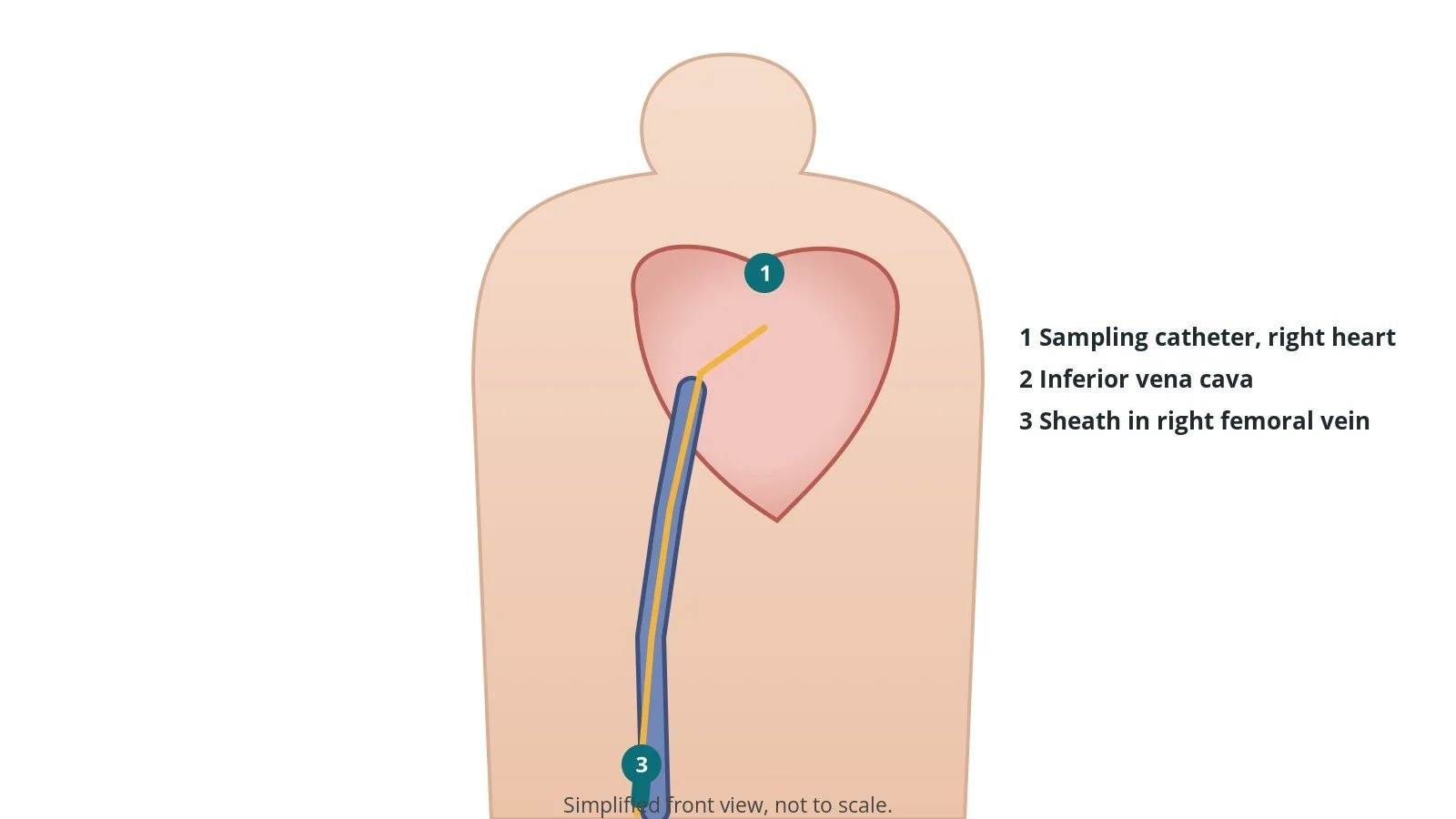
Access: Sampling catheter, right heart; Inferior vena cava; Sheath in right femoral vein.
3. The oximetry run
- A. Veins. Samples from the upper and lower main veins (SVC and IVC).
- B. Right heart. Samples from the right atrium and right ventricle.
- C. Lung artery. Samples from the pulmonary artery.
- D. Artery. An arterial sample for comparison; the shunt ratio is worked out.
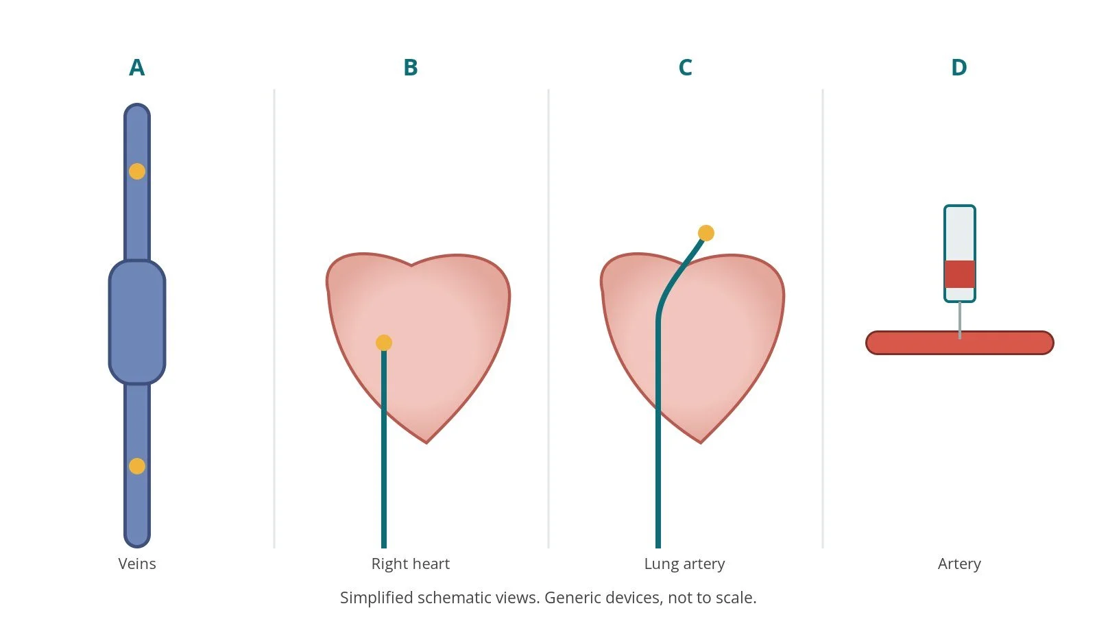
A: veins. B: right heart. C: lung artery. D: artery.
4. Finishing
The catheters are removed. The sheath is taken out and the site closed with pressure, a band or a closure device, per device and operator.
Recovery and aftercare
- First hours: you rest while the nurses check the access site, pulse and blood pressure, as per centre protocol.
- Going home: usually the same day, with someone to take you home.
- Access site: avoid heavy lifting and strenuous activity for a few days; a small bruise is common.
- Results: the team will usually explain the findings before you go home, and send a report to your own doctor.
- Driving: ask your team; for a planned test it is often a couple of days, but it depends on your heart condition.
Call 999 if you have chest pain, breathlessness, fainting, signs of a stroke, or heavy bleeding or swelling at the access site.
Risks and complications
Your team will explain the risks that apply to you. Possible problems include:
- bruising or bleeding at the access site (common, usually minor)
- damage to the artery or vein at the access site (uncommon)
- reaction to the contrast dye, or effects on kidney function
- heart rhythm disturbances, usually brief
- heart attack, stroke or serious complications (rare)
- X-ray exposure, kept as low as possible
Cath lab kit list (printable)
Barts example kit list
An example from one UK centre, written generically. Every lab has its own protocol, and sizes and doses vary.
- venous sheath, size per device and operator
- end-hole or balloon catheter, per operator
- labelled blood gas syringes
- blood gas analyser nearby
- oxygen settings recorded and kept steady
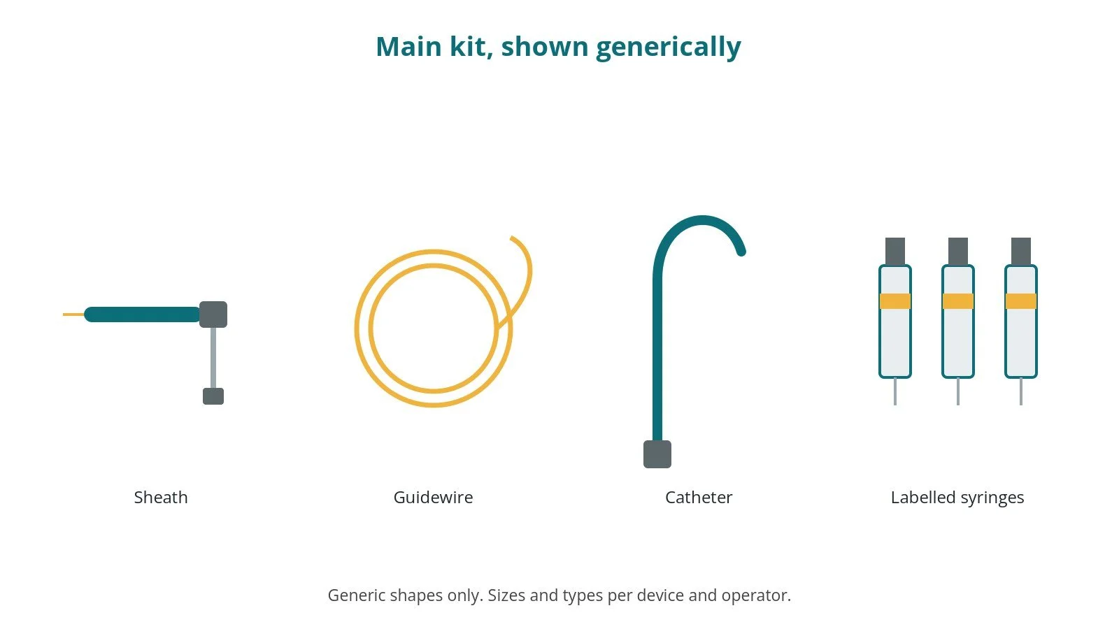
Main kit (generic): Sheath; Guidewire; Catheter; Labelled syringes.
Procedural steps (Barts example)
- Oxygen steady and recorded
- Venous access
- Samples in order: SVC, IVC, RA, RV, PA
- Arterial sample
- Analyse quickly; calculate Qp:Qs
- Catheters out; haemostasis
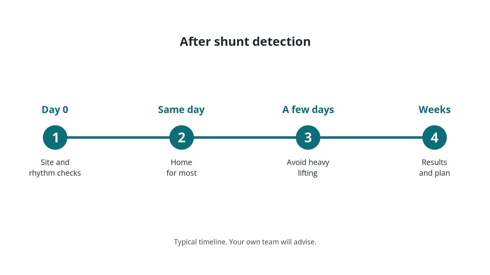
After the procedure: (1) Site and rhythm checks; (2) Home for most; (3) Avoid heavy lifting; (4) Results and plan.
First time in the cath lab?
💡 First-time tip
- Label as you go. Sample order is everything.
- Keep oxygen steady. Changes mid-run spoil the comparison.
- Run samples quickly. Delays change the results.
- Repeat if odd. Repeat any sample that doesn't fit.
FAQ
Do I need to stop eating?
Follow your hospital's fasting advice.
How many samples are taken?
Usually several small samples.
Does this test close the hole?
No. It measures it. Closure is a separate procedure.
Related Cath Lab 101 guides
- ASD closure
- PFO closure (patent foramen ovale)
- Ventricular septal defect (VSD) closure
- Cath Lab Abbreviations A–Z
Sources
All checked on 10 October 2026. Each page's own date is shown where it gives one.
- Baumgartner H, et al. 2020 ESC Guidelines for the management of adult congenital heart disease. Eur Heart J 2021;42(6):563–645. PubMed 32860028
- British Heart Foundation. Tests and procedures. bhf.org.uk/informationsupport/tests
- StatPearls (NCBI Bookshelf), search: cardiac shunt oximetry. ncbi.nlm.nih.gov/books
Disclaimer
Education only. This page is for general education for healthcare students, cath lab staff and curious readers. It isn't medical advice and doesn't replace advice from your own doctor, heart team or local unit protocols. Equipment and medicines are described generically. No product or manufacturer is recommended. If you think someone is having a heart emergency, call 999.
Disclosure: I work in the medical device industry. This page is independent education and isn't sponsored or reviewed by any company.
All diagrams on this page are original illustrations created for rodnieoro.com.