Extravascular ICD: a Cath Lab 101 guide
Reviewed by Rodnie Oro, cath lab charge nurse · Last reviewed: November 2026 · Education only, not medical advice
Extravascular ICD in one minute
What it is: An implantable cardioverter defibrillator (ICD) protects against dangerous fast heart rhythms. In the extravascular ICD (EV-ICD), the lead is tunnelled under the breastbone (substernal), outside the heart and veins, and the generator sits on the left side under the arm.
Who it is for: Selected people who need an ICD, particularly younger people or those at higher risk from leads inside the veins, who do not need regular pacing for a slow heart rate.
Main risks: Wound infection or bleeding, the lead moving, inappropriate shocks, and rarely injury to structures behind the breastbone, such as the heart or a blood vessel.
Usually: Under general anaesthetic or deep sedation, about 1 to 2 hours, often home the next day.
On this page: What is extravascular ICD? · Who is it for? · When is it done? · Step by step · Recovery · Risks · Kit list · First time in the lab? · FAQ
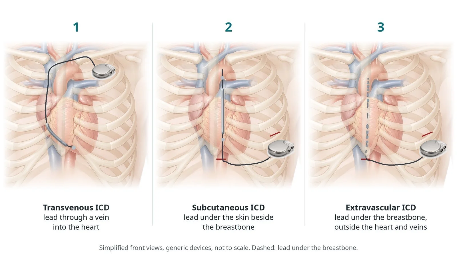
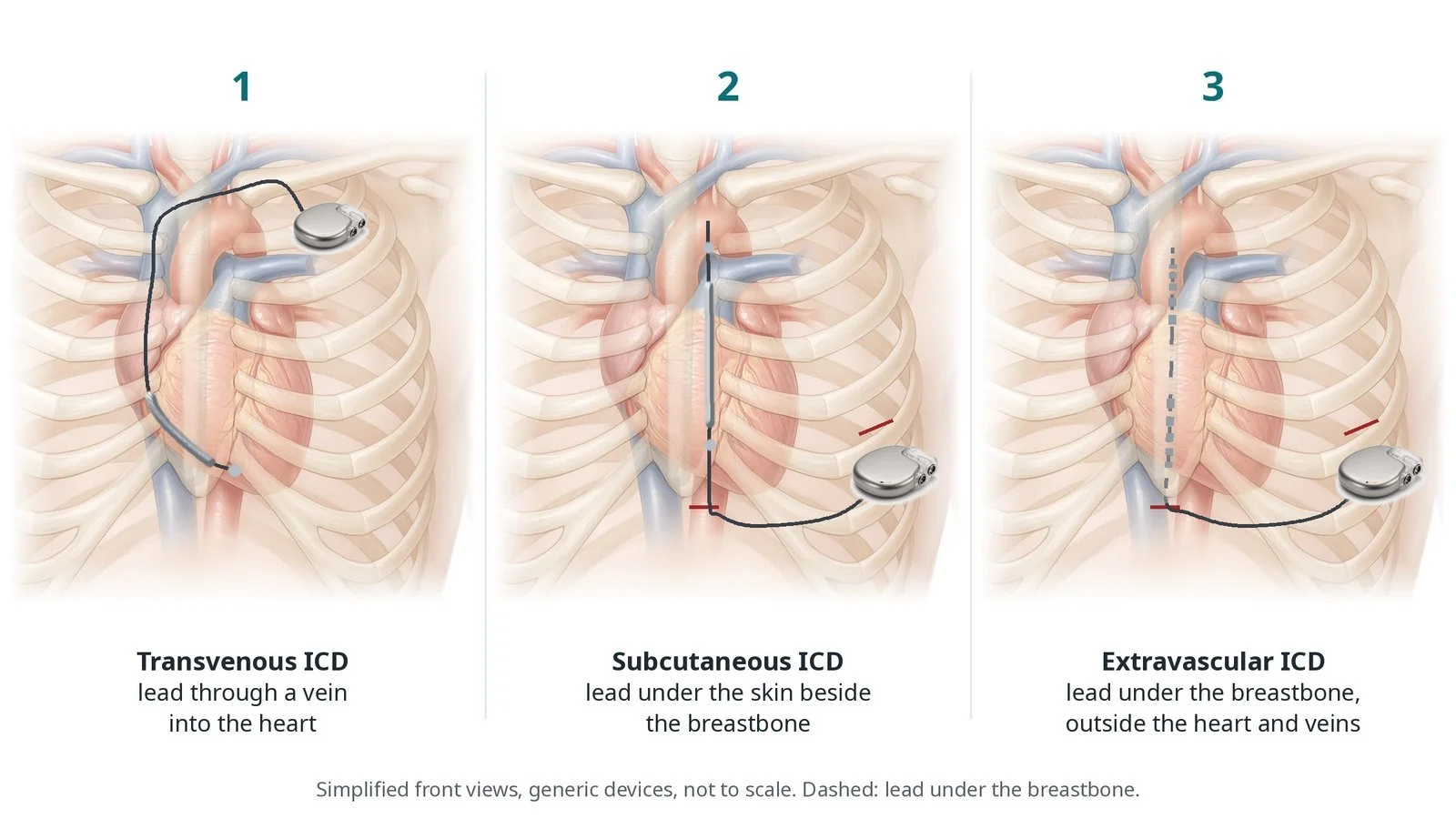
(1) Transvenous ICD: lead through a vein into the heart. (2) Subcutaneous ICD: lead under the skin beside the breastbone. (3) Extravascular ICD: lead under the breastbone, outside the heart and veins.
What is extravascular ICD?
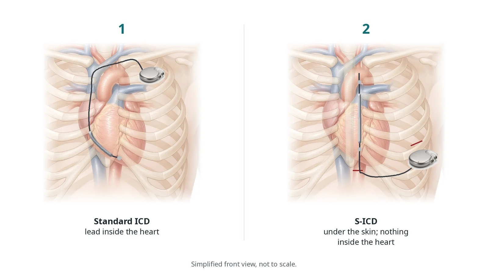
A standard (transvenous) ICD has a lead that runs through a vein into the heart. A subcutaneous ICD (S-ICD) avoids the veins: its lead lies under the skin beside the breastbone. The S-ICD can shock, but it cannot give anti-tachycardia pacing, and it gives only brief pacing after a shock.
The extravascular ICD puts the lead in a third place: under the breastbone, in the front of the chest (the anterior mediastinum). It lies just in front of the heart but outside the pericardium, the heart and the veins. Being closer to the heart lets it shock with less energy than an S-ICD, and lets it pace.
It can deliver anti-tachycardia pacing (ATP), a short burst of fast pacing that can stop some VT without a shock, and pause prevention pacing for short pauses. It is not designed for people who need regular, long-term pacing.
The lead enters through a small cut just below the lower end of the breastbone and is tunnelled up behind it. It then runs under the skin across to the generator, on the left side of the chest wall at about the mid-axillary line.
New to the acronyms? See the Cath Lab Abbreviations A–Z.
Who is it for?
It may be considered for people who need an ICD to protect against sudden cardiac death, and who:
do not need pacing for a slow heart rate, CRT or regular pacing
are at higher risk of problems from leads in the veins, for example younger people, or those with difficult vein access or a past device infection
may benefit from anti-tachycardia pacing, which an S-ICD cannot give
It is a newer option, available in selected centres. The choice between a transvenous, subcutaneous or extravascular ICD is made with your team.
Who it may not suit: People who need regular pacing or CRT, people who have had surgery through the breastbone (sternotomy) or have scarring behind it, and some people with an unusual chest shape.
The decision is made by your heart team, who will talk through the reasons, the alternatives and the risks with you.
When is it done?
It is planned.
Before it, you usually have:
an ECG and blood tests
a chest X-ray or CT to check the space behind the breastbone, as per centre protocol
advice on blood thinners
a check for previous surgery through the breastbone, which usually rules it out
gives you a chance to ask questions and give consent
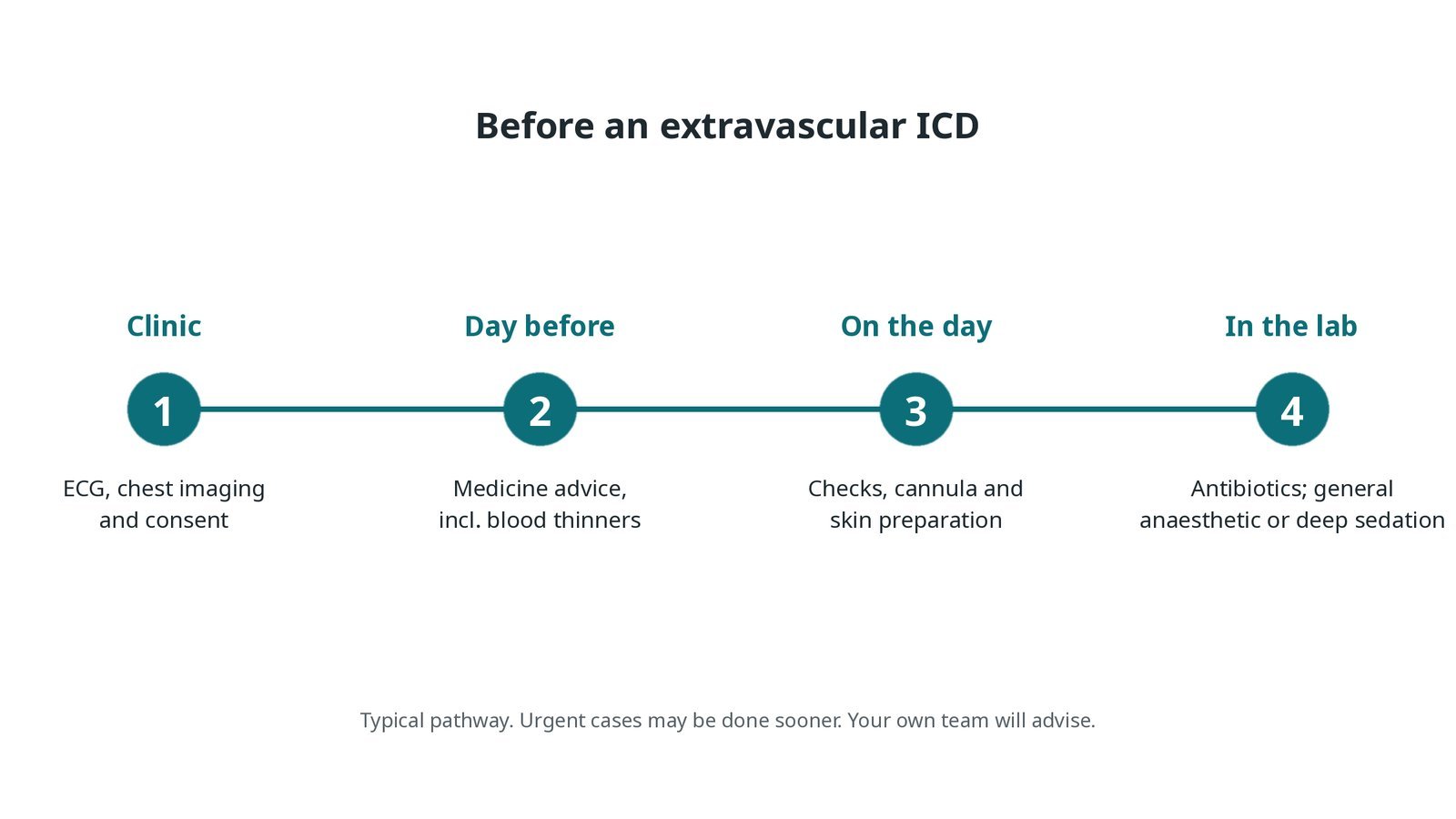
Before an extravascular ICD: ECG, chest imaging and consent; medicine advice; checks and skin preparation on the day; antibiotics and anaesthetic or deep sedation in the lab.
Step by step
1. Getting ready
You lie on the table with ECG, blood pressure and oxygen monitoring, and external defibrillator pads. Antibiotics are given before the first cut, as per centre protocol. A general anaesthetic or deep sedation is used, because tunnelling under the breastbone is uncomfortable.
2. Incisions
A small cut is made just below the lower end of the breastbone (the xiphoid). A second cut is made on the left side of the chest, at about the mid-axillary line, for the generator pocket.
3. Placing the lead
A. Incision. A small cut is made below the breastbone and the tissue is gently opened to reach the space behind it.
B. Tunnel up. A blunt tunnelling tool is passed up behind the breastbone, kept close to the bone, under X-ray. An introducer sheath is left in the tunnel.
C. Lead and generator. The lead goes in through the sheath and is positioned on X-ray, then tunnelled under the skin across to the pocket and connected to the generator.
D. Test. Sensing and pacing are checked. Defibrillation testing (starting a dangerous rhythm on purpose so the device can treat it) may be done, as per centre protocol.
Watch: Falling blood pressure after tunnelling: possible injury behind the breastbone. Tell the operator and get the echo ready.
4. Finishing
The wounds are closed with dissolvable stitches or glue. The device physiologist programmes the device, including whether ATP is used, as per centre protocol.
Watch: During defibrillation testing: pads on, the defibrillator charged, and a named person ready to give an external shock if the device does not stop the rhythm.
5. Before going home
A chest X-ray checks the lead position and a device check confirms sensing, pacing and settings. Most people go home the next day.
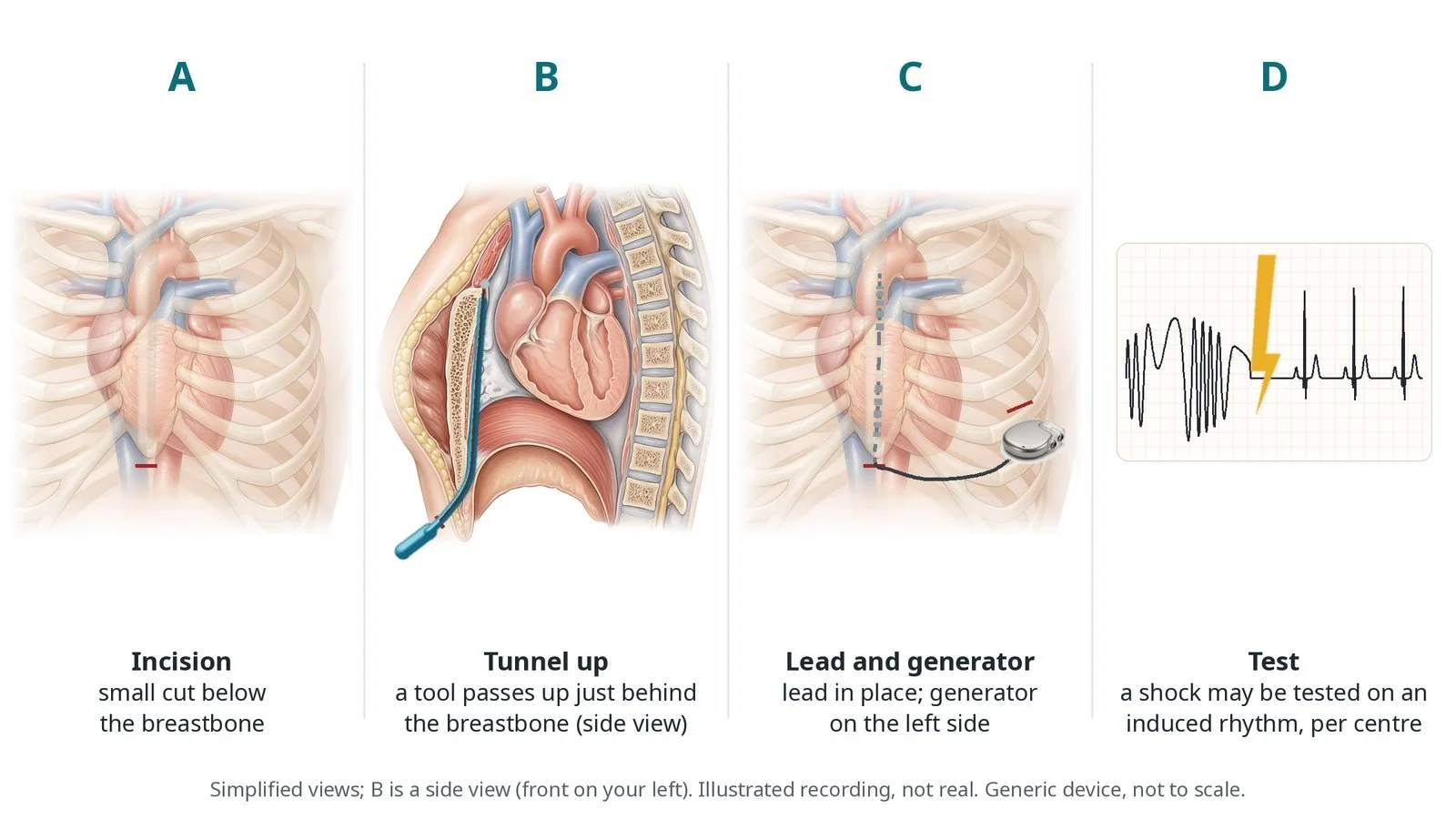
A: small incision below the breastbone. B: a tool passes up just behind the breastbone (side view). C: lead in place, generator on the left side. D: a shock may be tested on an induced rhythm, per centre.
Recovery and aftercare
First hours: checks of the wounds, rhythm and blood pressure, as per centre protocol; pain relief as needed.
Going home: usually the next day, after a chest X-ray and device check.
Wounds: keep them clean and dry; avoid heavy lifting and strenuous arm movement for a few weeks, as advised.
Medicines: pain relief as needed; restart blood thinners only as advised.
Driving: DVLA rules for ICDs apply, and you cannot drive for a period after the implant. Your team will tell you how long and whether to tell the DVLA.
Follow-up: device clinic and remote monitoring.
Call 999 if you have more than one shock, chest pain, fainting, or feel very unwell.
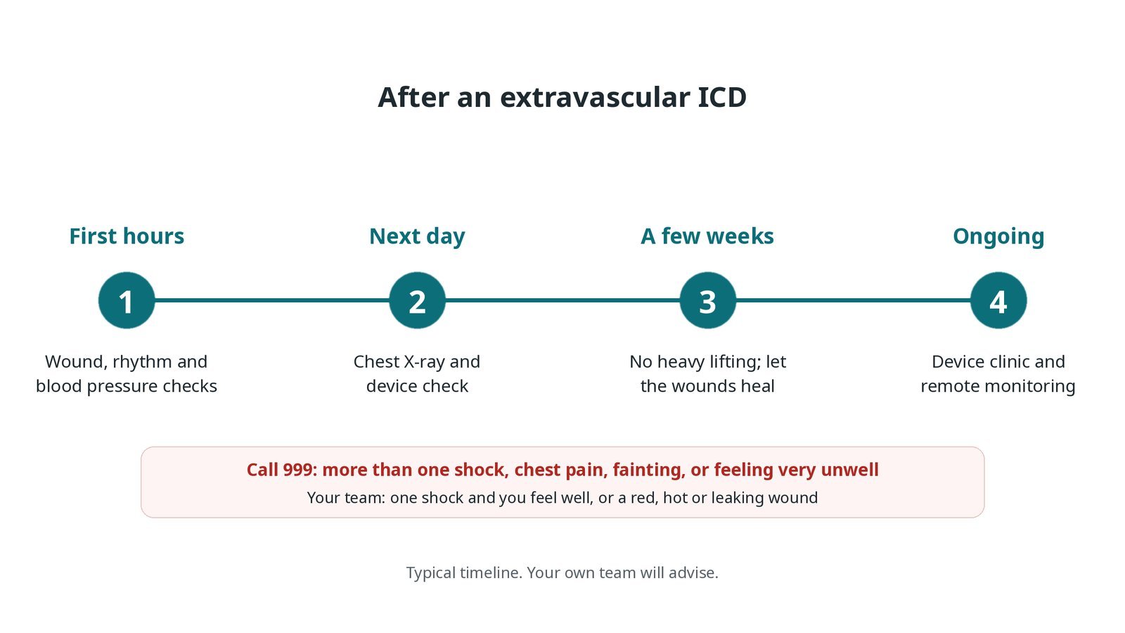
After an extravascular ICD: (1) wound, rhythm and blood pressure checks; (2) chest X-ray and device check; (3) no heavy lifting for a few weeks; (4) device clinic and remote monitoring.
Risks and complications
Your team will explain the risks that apply to you. Possible problems include:
Wound infection or bleeding (uncommon): Infection can mean removing the system.
The lead moving out of place (uncommon): May need another procedure.
Inappropriate shocks, for example from sensing noise (uncommon)
Soreness behind the breastbone for a few weeks
Injury to the heart, a blood vessel or a lung during tunnelling (rare)
Fluid around the heart or lungs (rare)
Cath lab kit list (printable)
Barts example kit list
An example from one UK centre, written generically. Every lab has its own protocol, and sizes and doses vary.
extravascular ICD generator, per device
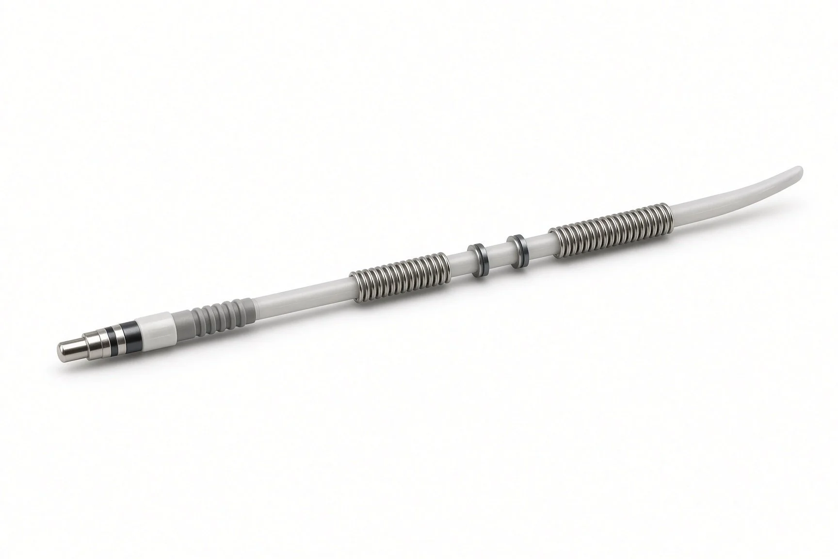
substernal defibrillation lead
substernal tunnelling tool and introducer sheath, per device
device programmer and device physiologist
external defibrillator pads and defibrillator
antibiotics and skin preparation, as per centre protocol
general anaesthetic or deep sedation set-up, as per centre protocol
device theatre set: drapes, instruments, diathermy and sutures
echo and pericardiocentesis kit, available
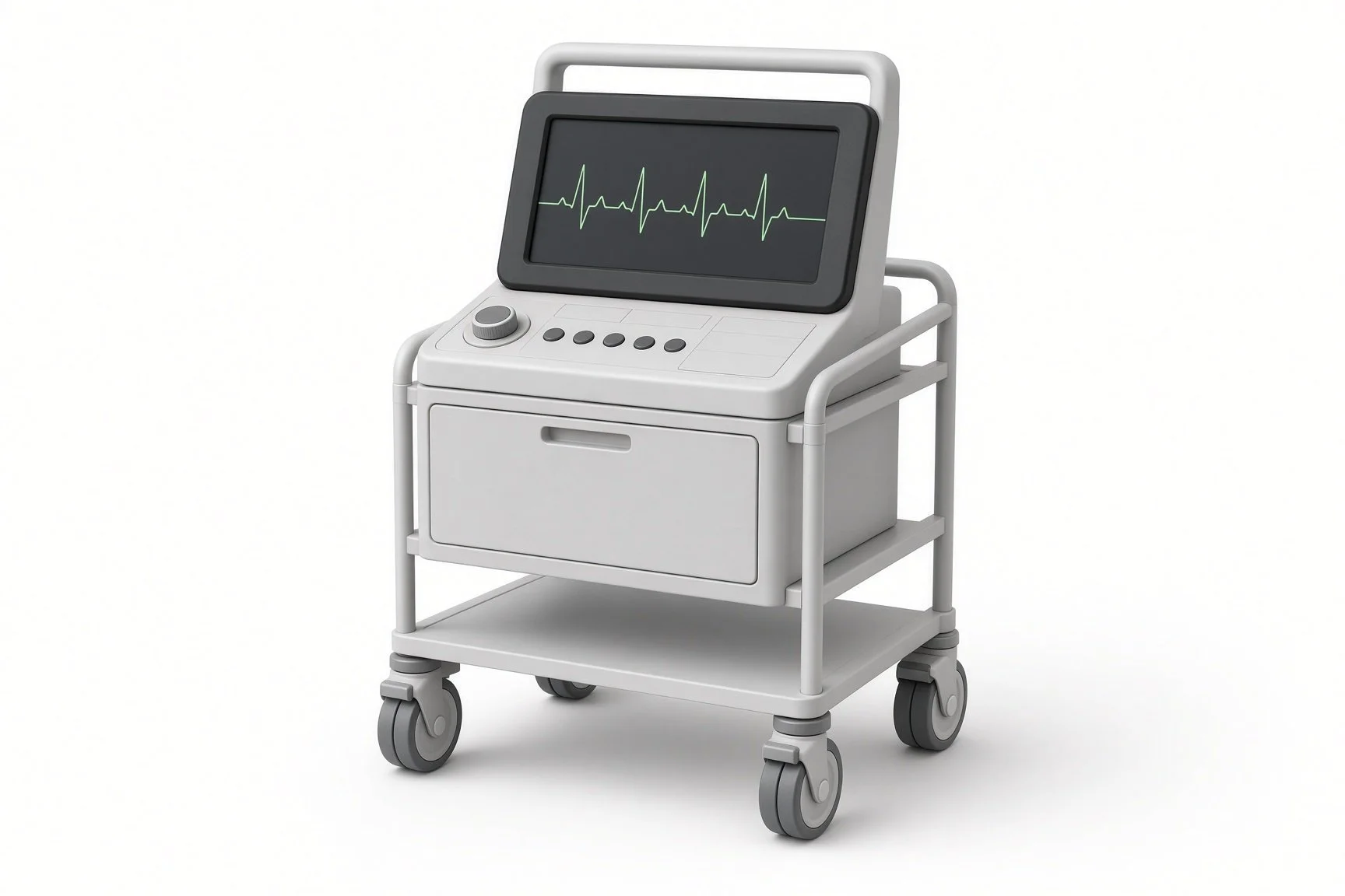
Device programmer.
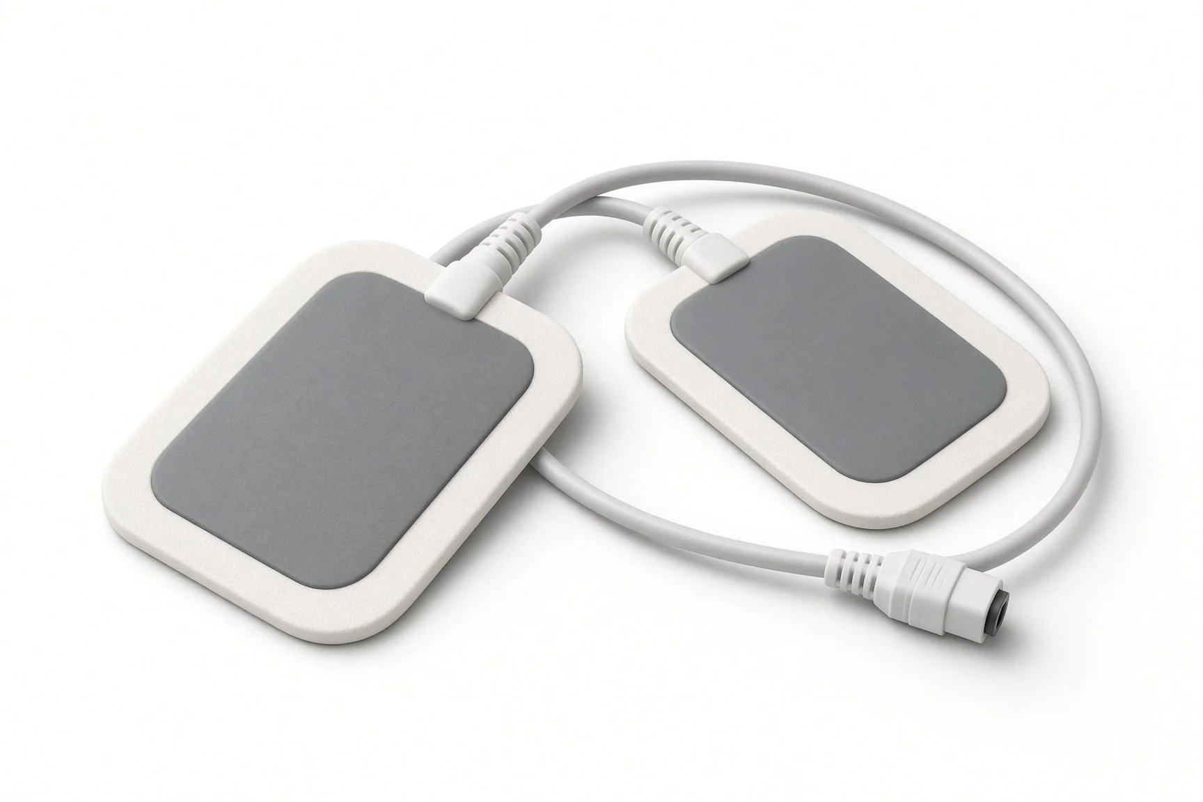
External defibrillator pads.
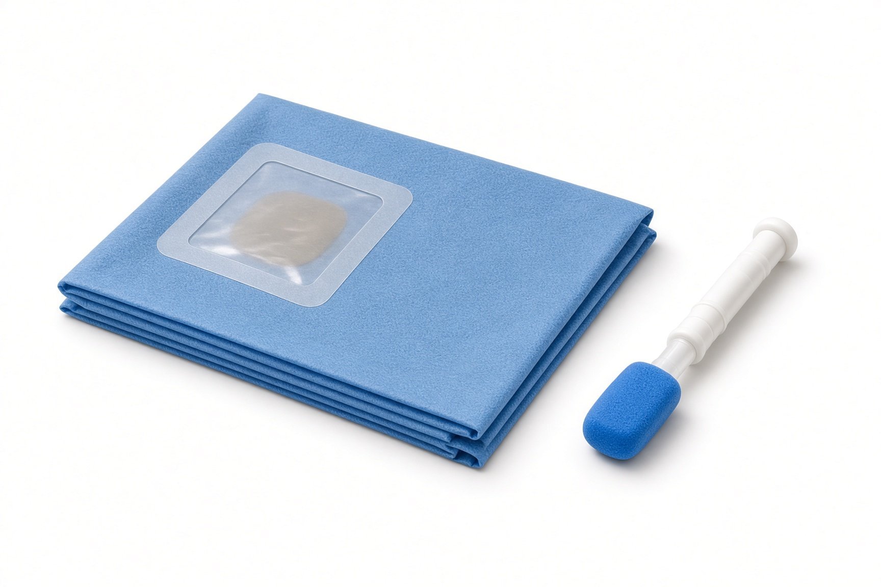
Sterile drapes.
Extravascular ICD generator.
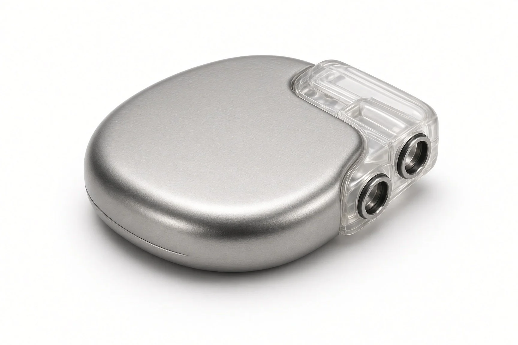
Extravascular ICD generator.
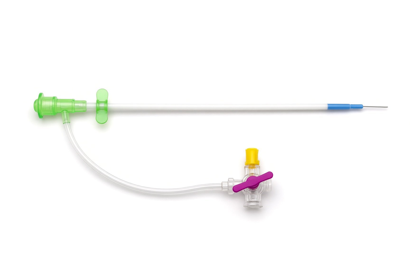
Introducer sheath.
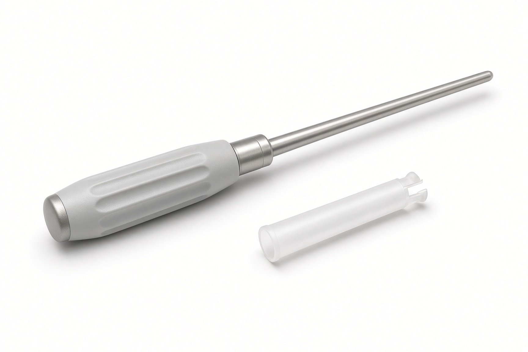
Tunnelling tool.
Antibiotics and skin preparation.
Procedural steps (Barts example)
Getting ready
Incisions
Placing the lead
Finishing
Before going home
First time in the cath lab?
Know the three ICD types. Transvenous: lead in the heart. S-ICD: lead under the skin. EV-ICD: lead under the breastbone. Patients often ask how theirs is different.
Watch the tunnelling. The step behind the breastbone carries the most risk. Keep the room quiet, keep the image steady and watch the blood pressure.
Be ready for testing. If defibrillation testing is planned, external pads, a charged defibrillator and a named person to shock are needed before the rhythm is started.
Keep the X-ray position steady during tunnelling. The operator relies on it to stay close to the breastbone.
FAQ
How is this different from an S-ICD?
Both keep wires out of the heart and veins. The EV-ICD lead sits under the breastbone, closer to the heart, so it can also pace to stop some fast rhythms and to cover short pauses.
Can it work as a pacemaker?
Only for short-term needs, such as stopping VT with pacing or covering pauses. It is not meant for people who need regular pacing.
Will it hurt?
You will have a general anaesthetic or deep sedation. The chest can be sore for a few weeks.
What if I get a shock?
One shock and you feel well: contact your device clinic. More than one shock, or you feel unwell: call 999.
Related Cath Lab 101 guides
VT ablation (coming soon)
Transvenous lead extraction (coming soon)
Sources
All checked on 12 November 2026. Each page's own date is shown where it gives one.
Friedman P, et al. Efficacy and safety of an extravascular implantable cardioverter–defibrillator. N Engl J Med 2022.
Zeppenfeld K, et al. 2022 ESC Guidelines for the management of patients with ventricular arrhythmias and the prevention of sudden cardiac death. Eur Heart J 2022.
DVLA. Assessing fitness to drive: a guide for medical professionals. gov.uk
StatPearls (NCBI Bookshelf), search: implantable cardioverter defibrillator. ncbi.nlm.nih.gov/books
Disclaimer
Education only. This page is for general education for healthcare students, cath lab staff and curious readers. It isn't medical advice and doesn't replace advice from your own doctor, heart team or local unit protocols. Equipment and medicines are described generically. No product or manufacturer is recommended. If you think someone is having a heart emergency, call 999.
Disclosure: I work in the medical device industry. This page is independent education and isn't sponsored or reviewed by any company.
All diagrams on this page are original illustrations created for rodnieoro.com.