Cardioneuroablation: a Cath Lab 101 guide
Reviewed by Rodnie Oro, cath lab charge nurse · Last reviewed: November 2026 · Education only, not medical advice
Cardioneuroablation in one minute
What it is: Catheter ablation of the ganglionated plexi: clusters of nerve cells in fat pads around the atria that carry the vagus nerve's "slow down" signals to the heart. Reducing their effect can stop the heart from suddenly slowing or pausing.
Who it is for: Selected, usually younger, people with frequent, severe reflex (vasovagal) fainting in which the heart slows or stops (cardioinhibitory), or with heart block caused by the vagus nerve (functional AV block), when other measures have not helped.
Main risks: Bruising at the groin, fluid around the heart, stroke (rare), a faster resting heart rate, and the fainting not stopping. It is a newer treatment and long-term results are still being studied.
Usually: Under sedation or general anaesthetic, about 2 to 3 hours, home the same or next day; done in specialist centres.
On this page: What is cardioneuroablation? · Who is it for? · When is it done? · Step by step · Recovery · Risks · Kit list · First time in the lab? · FAQ
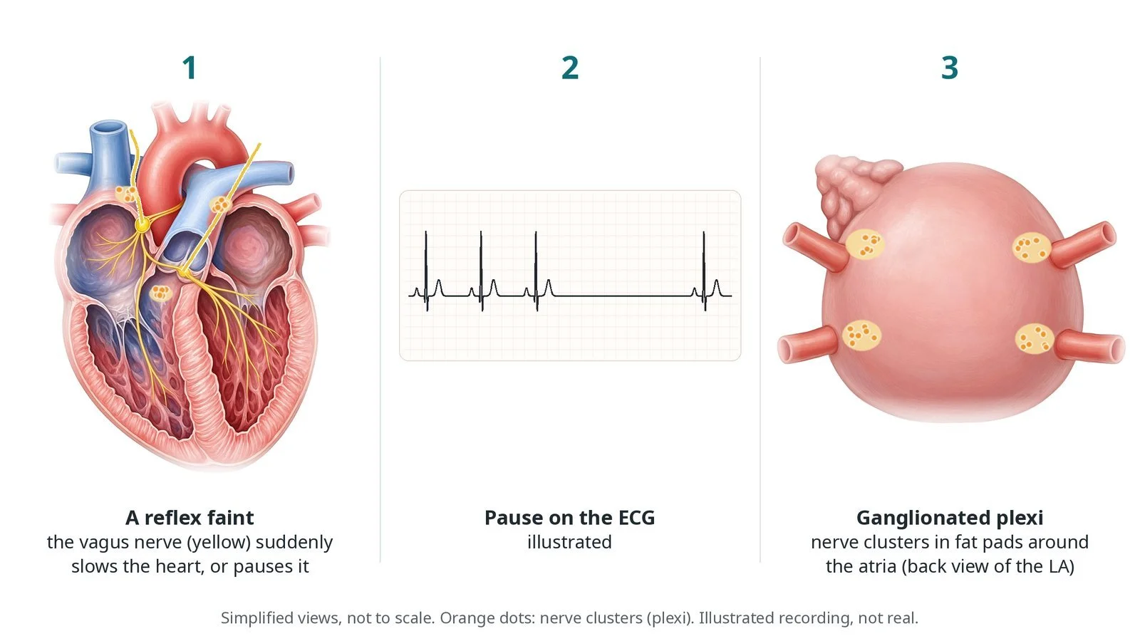
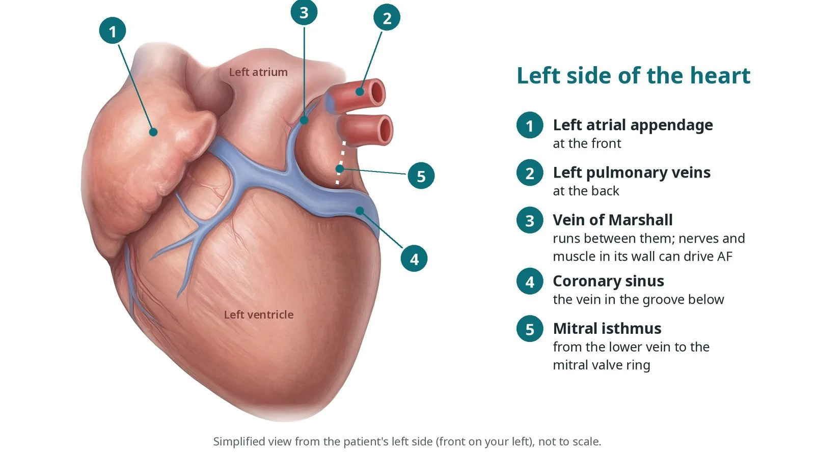
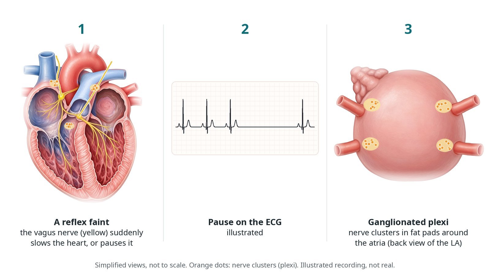
(1) A reflex faint: the vagus nerve suddenly slows or pauses the heart. (2) A pause on the ECG (illustrated). (3) Ganglionated plexi: nerve clusters in fat pads around the atria.
What is cardioneuroablation?
The vagus nerve is the body's brake on the heart. Its fibres reach the heart through clusters of nerve cells called ganglionated plexi (GP). These sit in fat pads on the outside of the atria, mainly around the openings of the pulmonary veins and between the atria, and they supply the sinus node and the AV node.
In reflex (vasovagal) syncope, a trigger such as standing for a long time, pain or heat sets off a sudden surge of vagal activity. In the cardioinhibitory type, the heart slows sharply or pauses for several seconds and the person faints. In functional AV block, vagal surges block the AV node even though the conduction system itself is healthy.
Cardioneuroablation treats the plexi from inside the atria, usually the left atrium (reached across the septum) and sometimes the right atrium too. The plexi are found from their usual sites and, depending on the centre, by their signals or by high-frequency stimulation that briefly slows the heart. Afterwards the resting heart rate usually rises and the heart responds less to vagal surges.
It is an emerging treatment in specialist centres. It does not treat the fall in blood pressure that is also part of many faints, so patients are chosen carefully.
New to the acronyms? See the Cath Lab Abbreviations A–Z.
Who is it for?
It may be considered for people with:
severe, recurrent reflex syncope of the cardioinhibitory type, shown on tilt testing or by a monitor recording pauses
functional (vagal) AV block, where tests show the AV node itself is healthy
symptoms despite advice on fluids, salt and avoiding triggers, and other measures
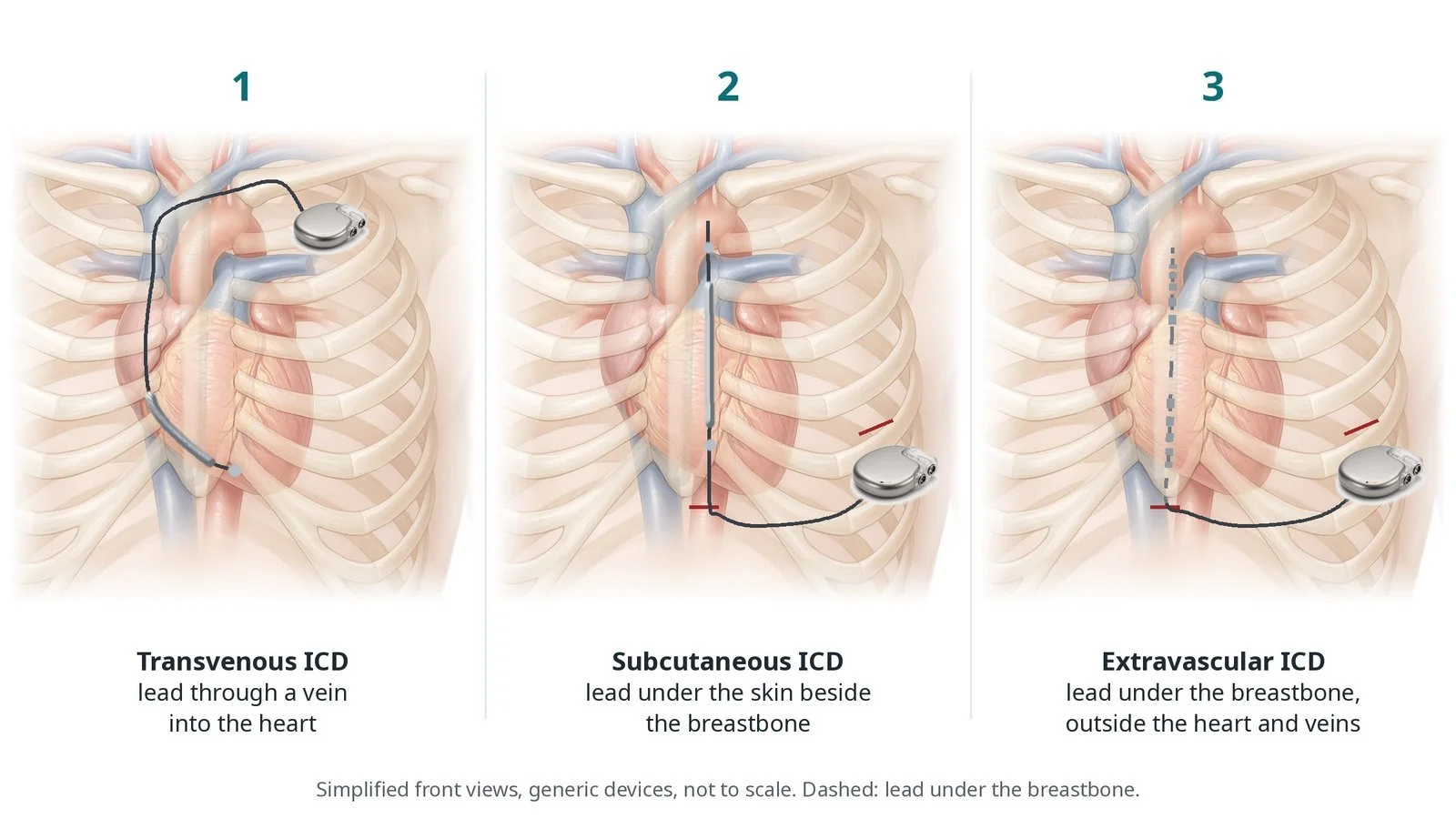
Younger people, for whom a pacemaker would mean decades of device and lead care, are the main group. The decision is made with a specialist syncope or electrophysiology team.
Who it may not suit: People whose faints are mainly due to low blood pressure rather than a slow heart, people with disease of the conduction system itself, and older people, for whom a pacemaker is the usual choice.
The decision is made by your heart team, who will talk through the reasons, the alternatives and the risks with you.
When is it done?
It is planned.
Before it, you usually have:
tilt testing, Holter monitoring or an implantable loop recorder, to show the heart slowing or pausing during symptoms
in some centres, an atropine test: atropine blocks the vagus nerve, and a clear rise in heart rate (or the AV block clearing) suggests the vagus is the cause
an ECG, echo and blood tests
a talk about the alternatives, including a pacemaker
gives you a chance to ask questions and give consent
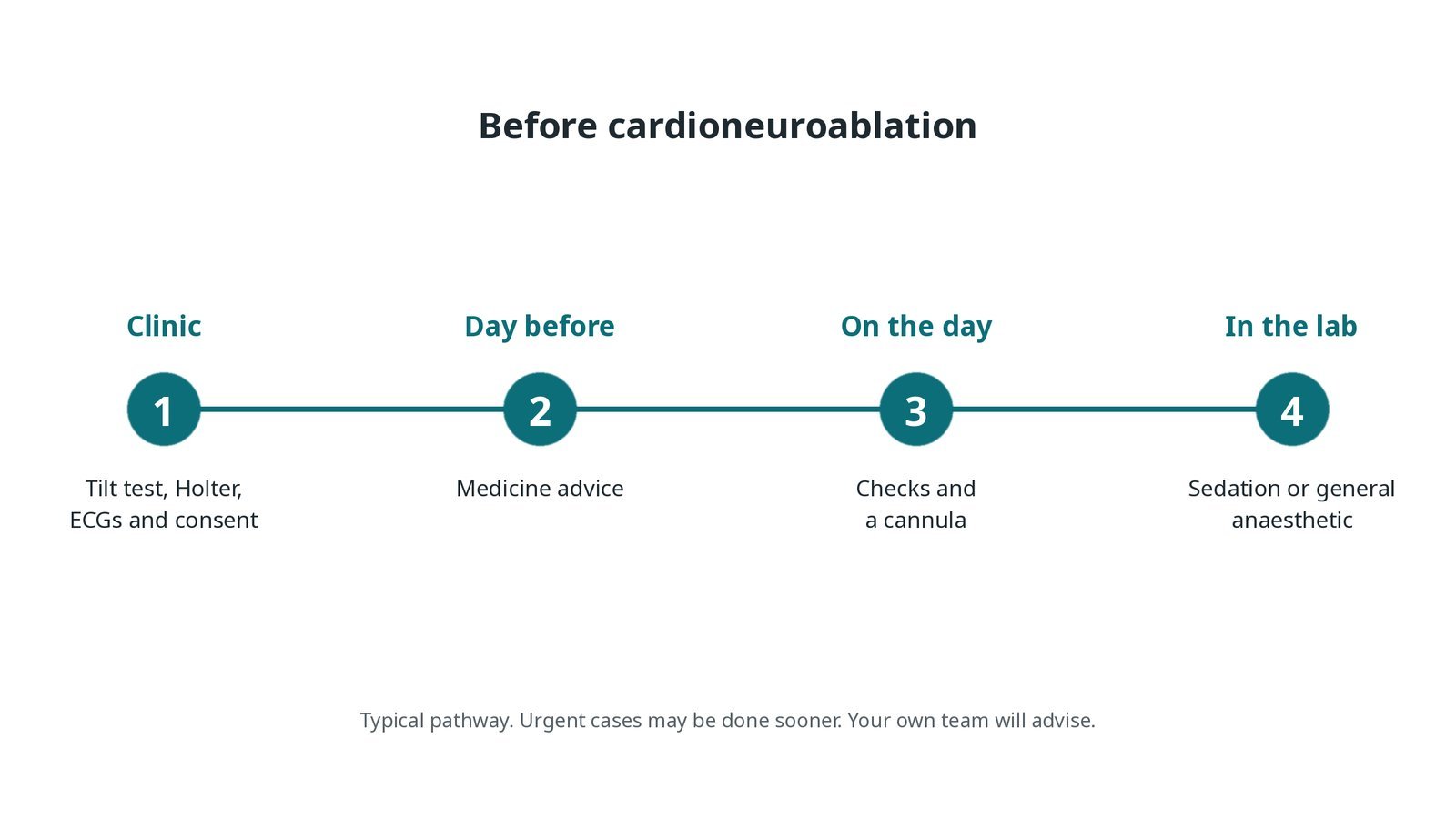
Before cardioneuroablation: tilt test, Holter, ECGs and consent; medicine advice; checks on the day; sedation or general anaesthetic in the lab.
Step by step
1. Getting ready
You lie on the X-ray table with ECG, blood pressure and oxygen monitoring, and external pads. Sedation or a general anaesthetic is given, as per centre protocol. Baseline heart rate and intervals are recorded first.
2. Access
Sheaths are placed in the femoral vein under ultrasound, sizes per device and operator. A transseptal puncture gives access to the left atrium, and heparin is given, as per centre protocol. A 3D mapping system is usually used.
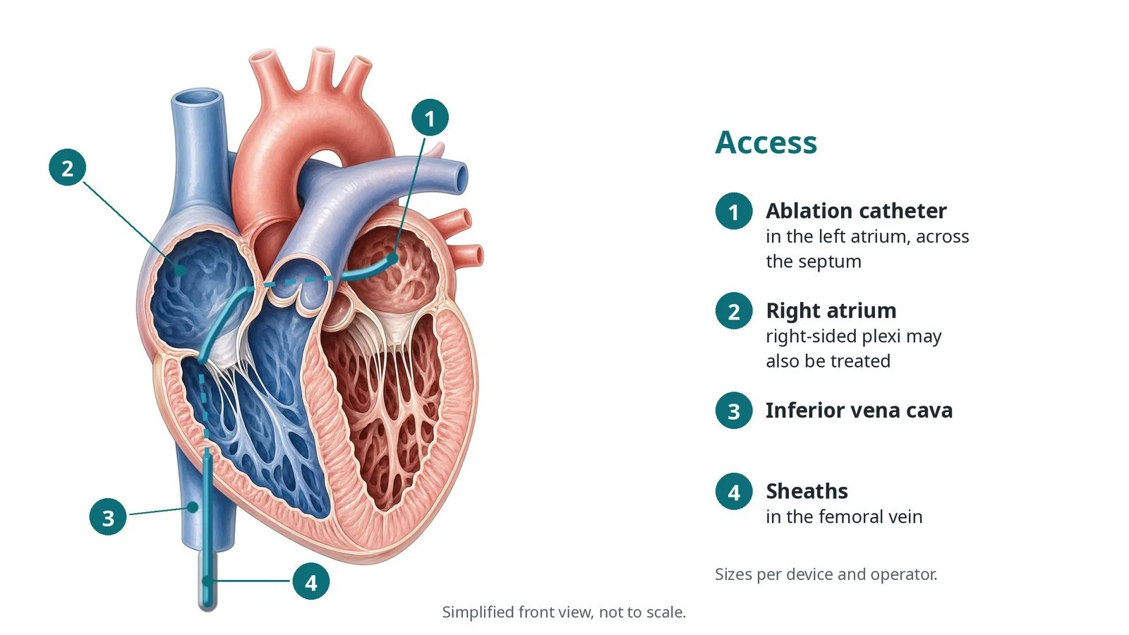
Access: (1) ablation catheter in the left atrium, across the septum; (2) right atrium, where right-sided plexi may also be treated; (3) inferior vena cava; (4) sheaths in the femoral vein.
3. Ablating the plexi
A. Locate. The plexi are found from their usual sites around the pulmonary veins and atria, and by signals or stimulation, per operator.
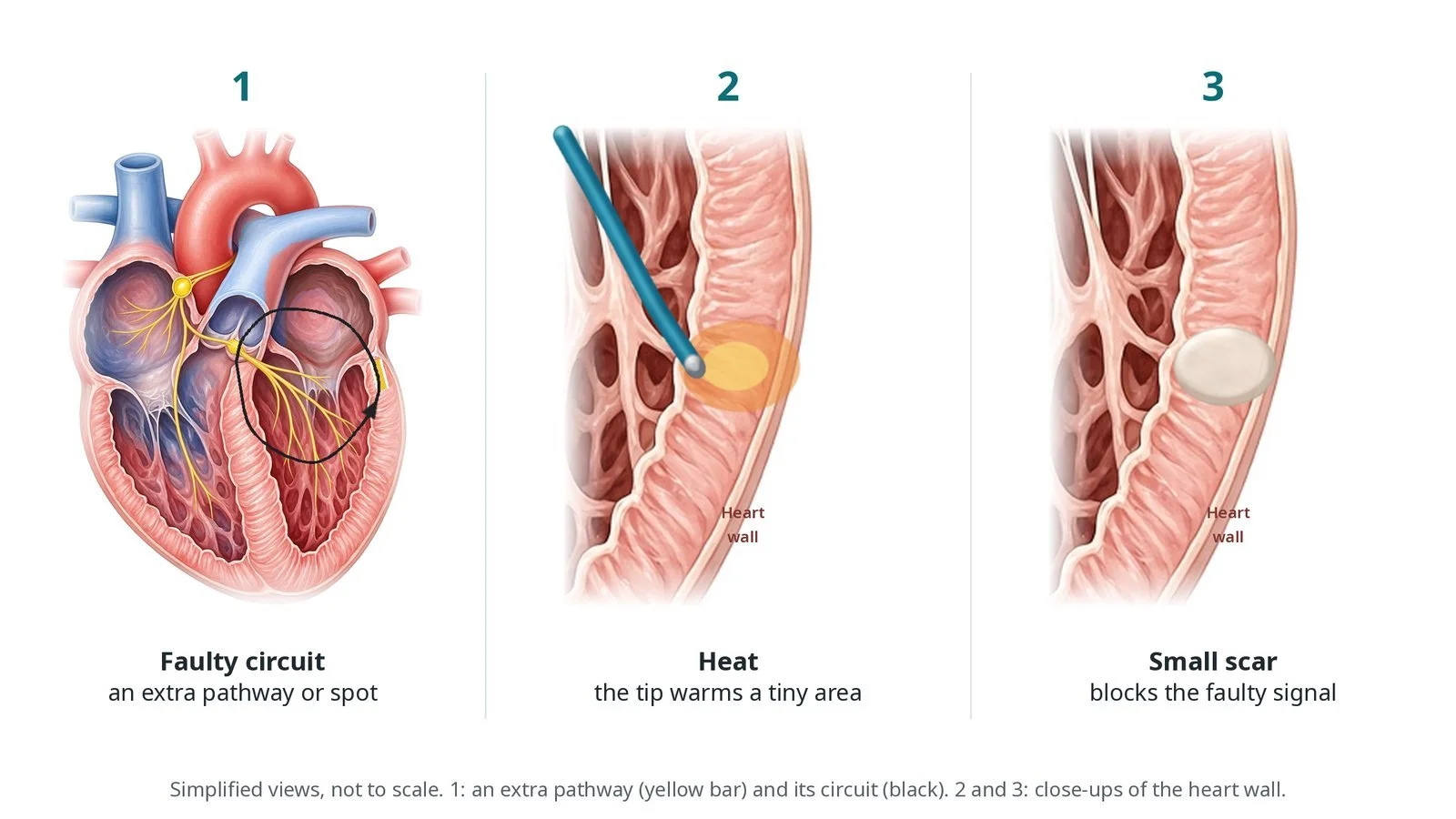
B. Ablate. Radiofrequency energy is delivered around each plexus, usually starting in the left atrium; right-sided plexi may also be treated.
C. Heart rate rises. The heart may slow briefly during energy (a vagal response), then the resting rate usually rises as the plexi are treated.
D. Atropine test. Atropine is given at the end, as per centre protocol. Little or no further rise in heart rate suggests the vagal effect on the heart has been removed.
Watch: A sudden pause or very slow rhythm during energy: this is a vagal response. Be ready to pace and tell the operator; it usually settles when energy stops.
4. Finishing
The catheters and sheaths are removed. The groin is closed with pressure or a stitch, per operator.
5. Follow-up monitoring
You are monitored for a few hours. Follow-up usually includes a Holter or an existing loop recorder to look for pauses, and sometimes a repeat tilt test.
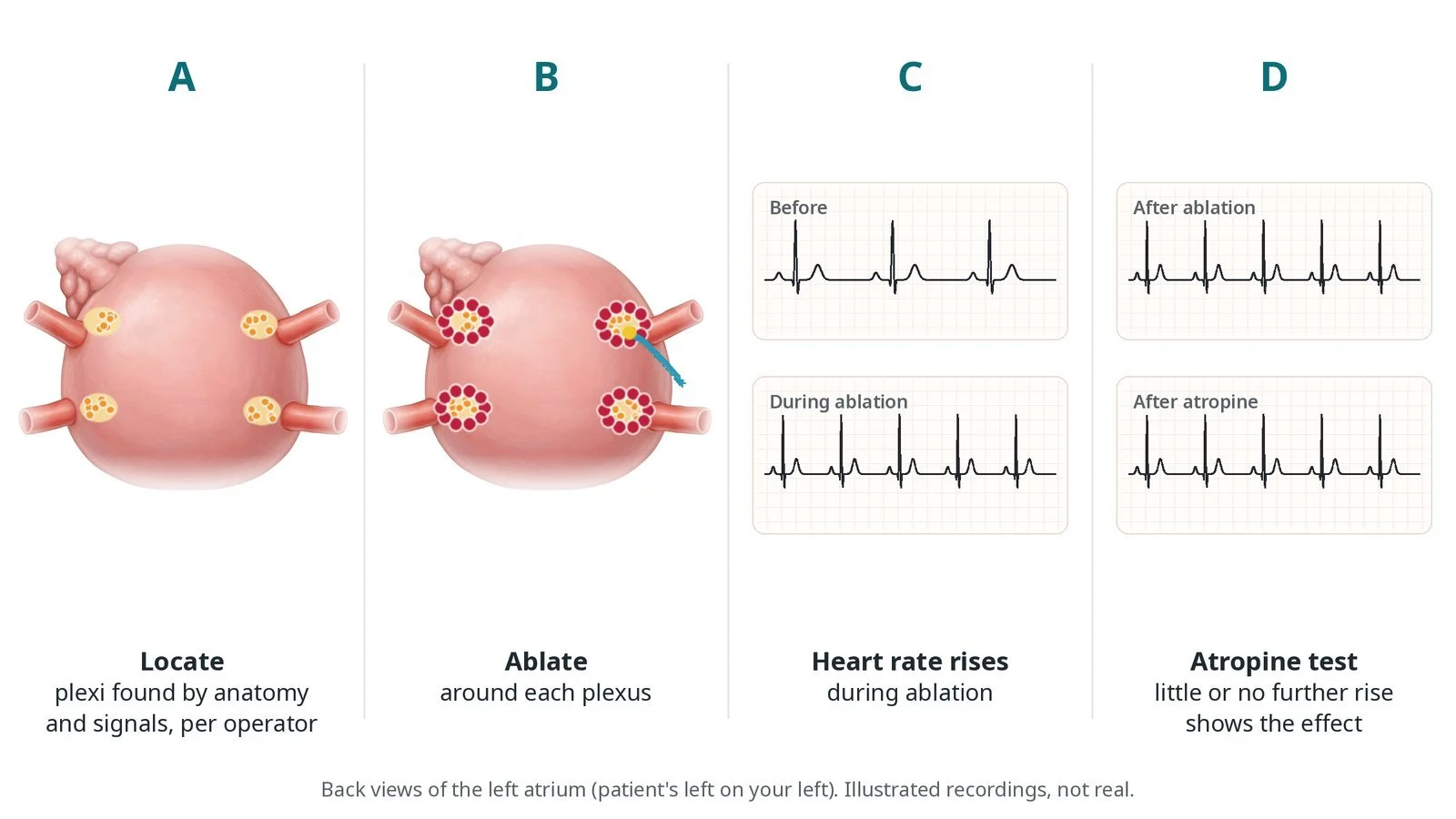
A: locate the plexi by anatomy and signals. B: ablate around each plexus. C: the heart rate rises during ablation. D: an atropine test: little or no further rise shows the effect.
Recovery and aftercare
First hours: bed rest while the groin settles; checks of the groin, rhythm and blood pressure, as per centre protocol.
Going home: often the same or next day.
Activity: avoid heavy lifting and strenuous exercise for about a week.
Medicines: a blood thinner may be given for a short time afterwards, as per centre protocol.
Driving: DVLA rules for fainting apply and depend on your history; ask your team before you drive.
Follow-up: clinic review with a Holter or loop recorder; sometimes a repeat tilt test.
Call 999 if you have chest pain, sudden breathlessness, signs of a stroke, or heavy bleeding or swelling at the groin.
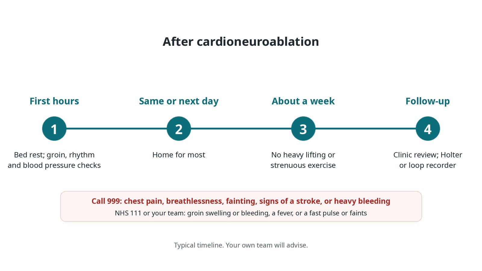
After cardioneuroablation: (1) bed rest and checks; (2) home for most the same or next day; (3) no heavy lifting for about a week; (4) clinic review with a Holter or loop recorder.
Risks and complications
Your team will explain the risks that apply to you. Possible problems include:
Bruising or bleeding at the groin
Fluid or bleeding around the heart (tamponade) (uncommon)
Stroke or a clot (rare): Because the left atrium is entered.
A faster resting heart rate or palpitations afterwards: Often settles with time.
Damage to nearby structures, such as the oesophagus or the phrenic nerve (rare)
The fainting not stopping, or coming back: Some people still need a pacemaker later.
Cath lab kit list (printable)
Barts example kit list
An example from one UK centre, written generically. Every lab has its own protocol, and sizes and doses vary.
femoral venous sheaths, sizes per device and operator
ultrasound for vein access
transseptal needle and steerable sheath, per operator
irrigated radiofrequency ablation catheter, per operator
3D mapping system and skin patches
diagnostic EP catheters and recording system
stimulator for high-frequency stimulation, if used, per operator
atropine, as per centre protocol
back-up pacing and external pads
heparin, as per centre protocol
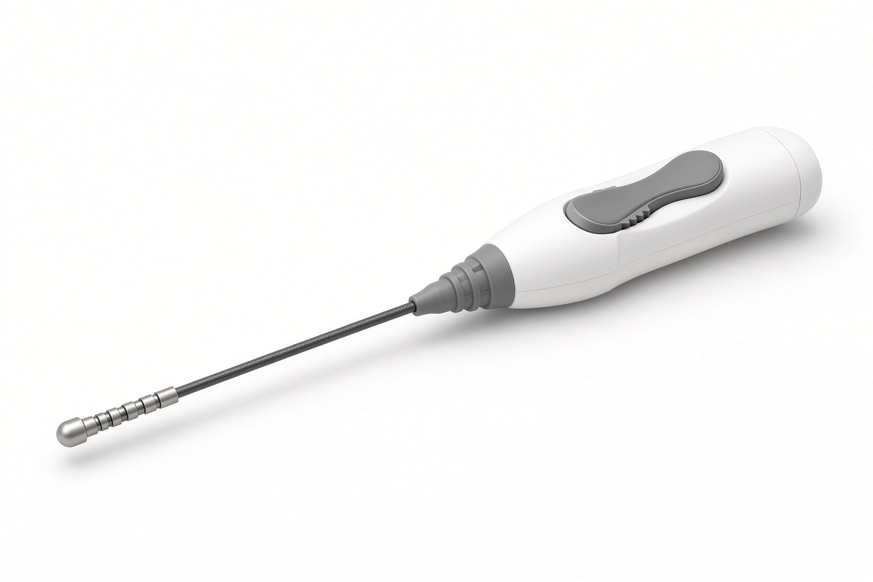
Ablation catheter.
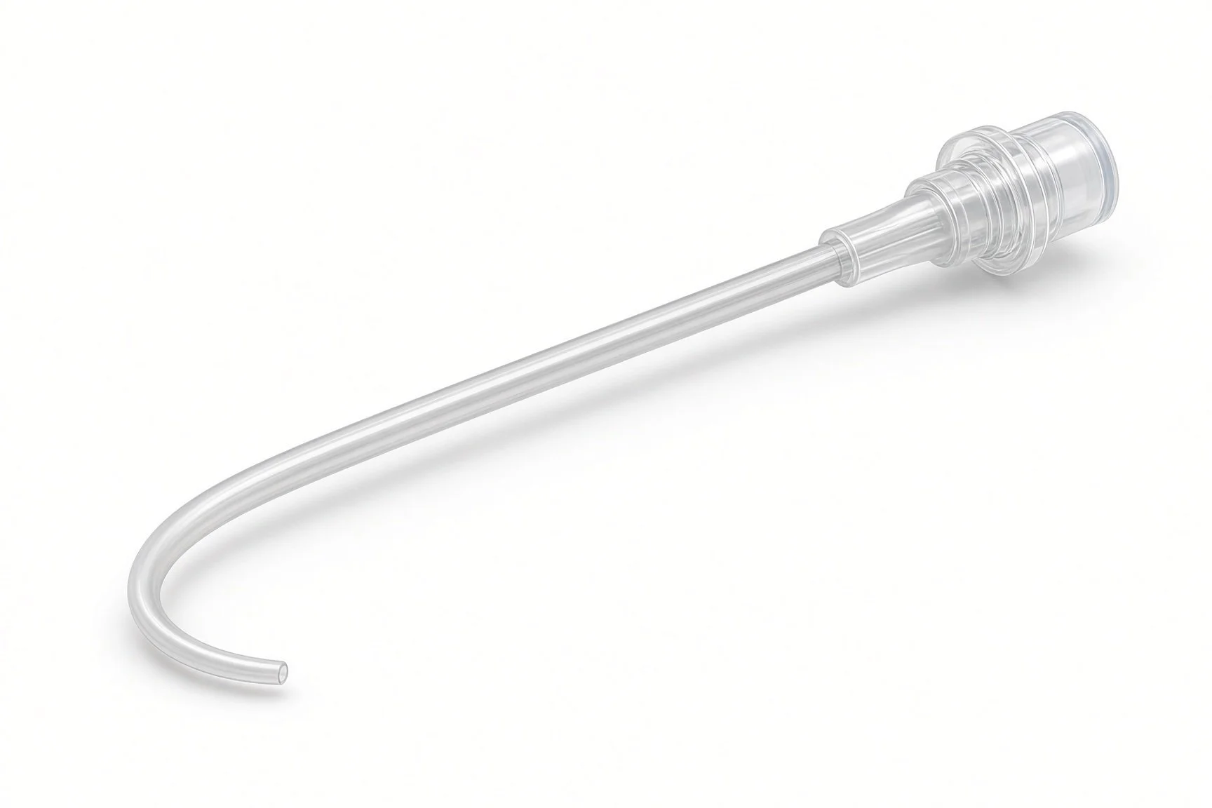
Diagnostic EP catheters.
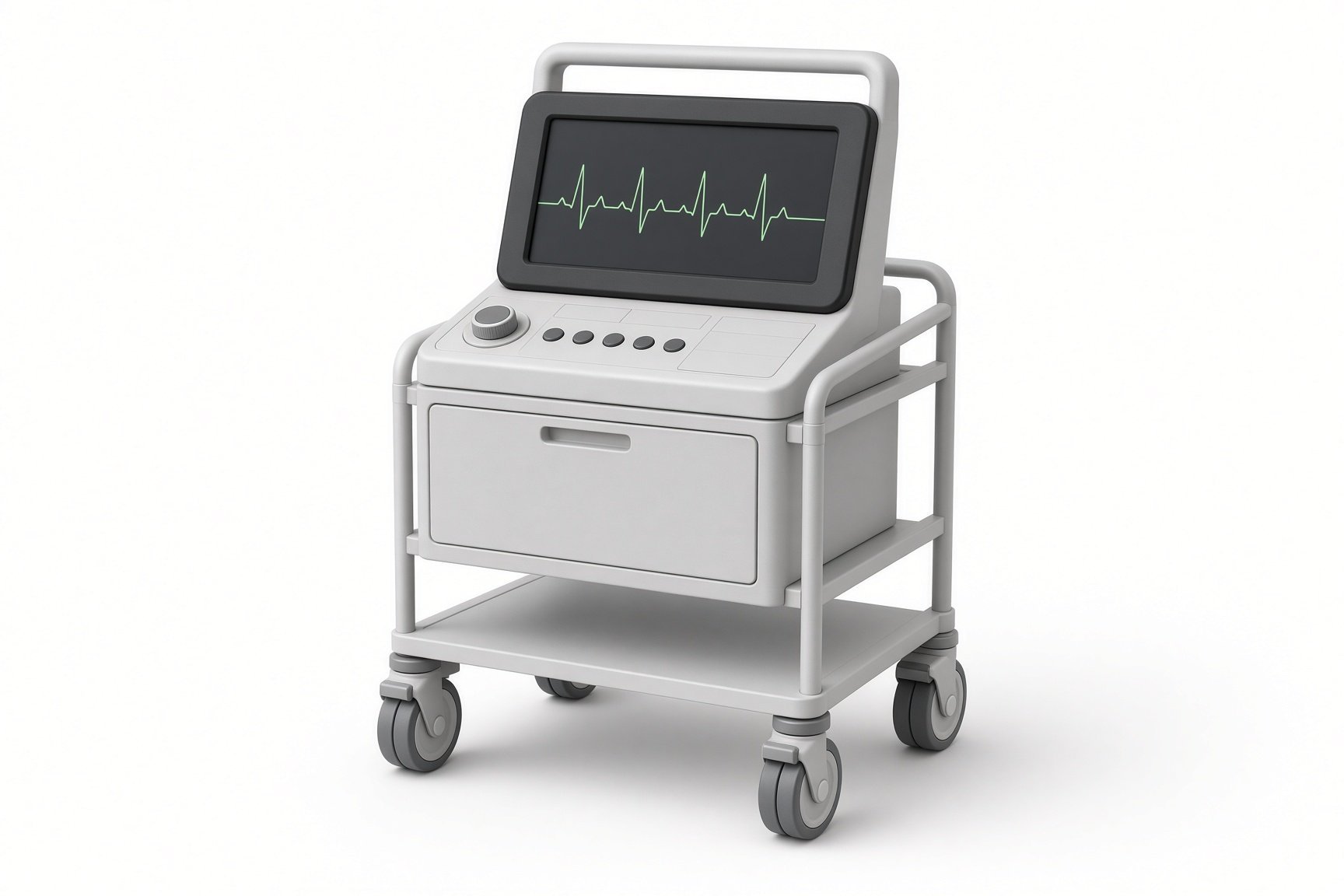
3D mapping system.
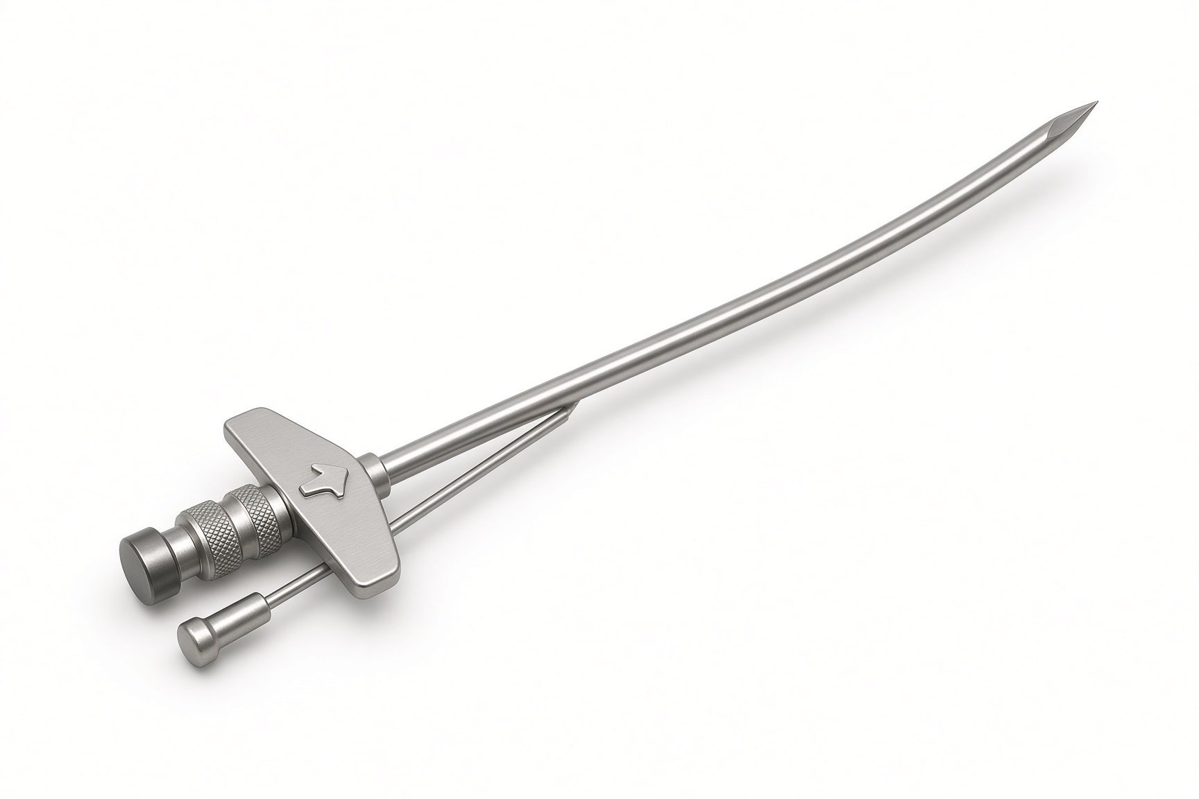
Transseptal needle.
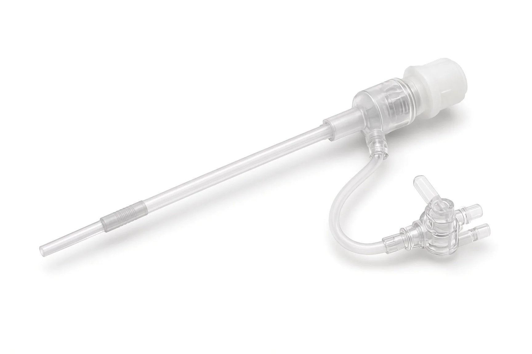
Steerable sheath.
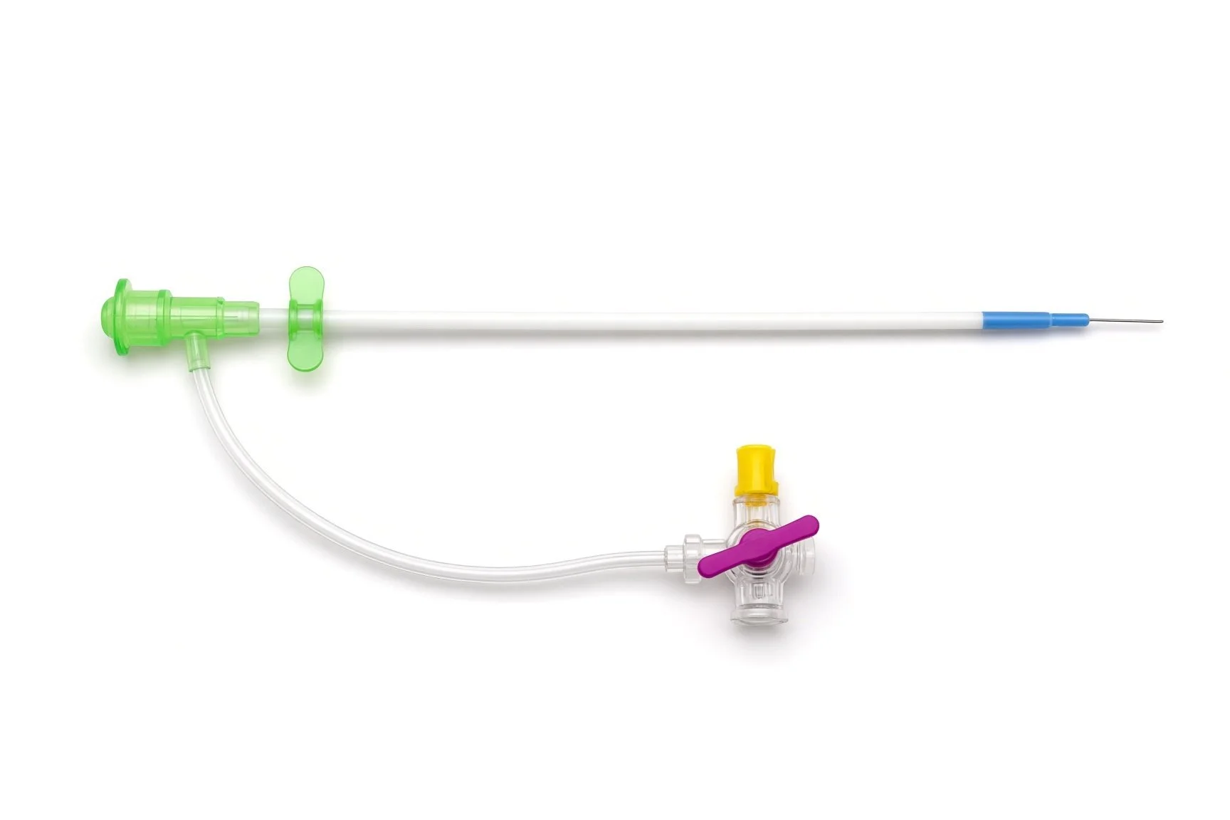
Femoral venous sheaths.
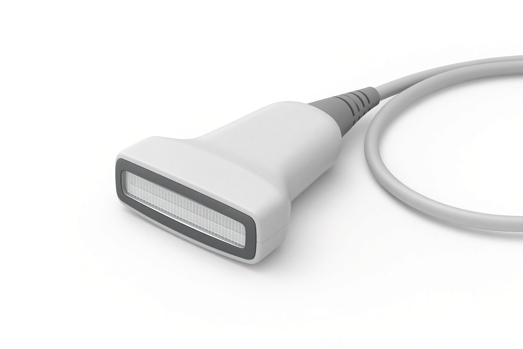
Ultrasound for vein access.
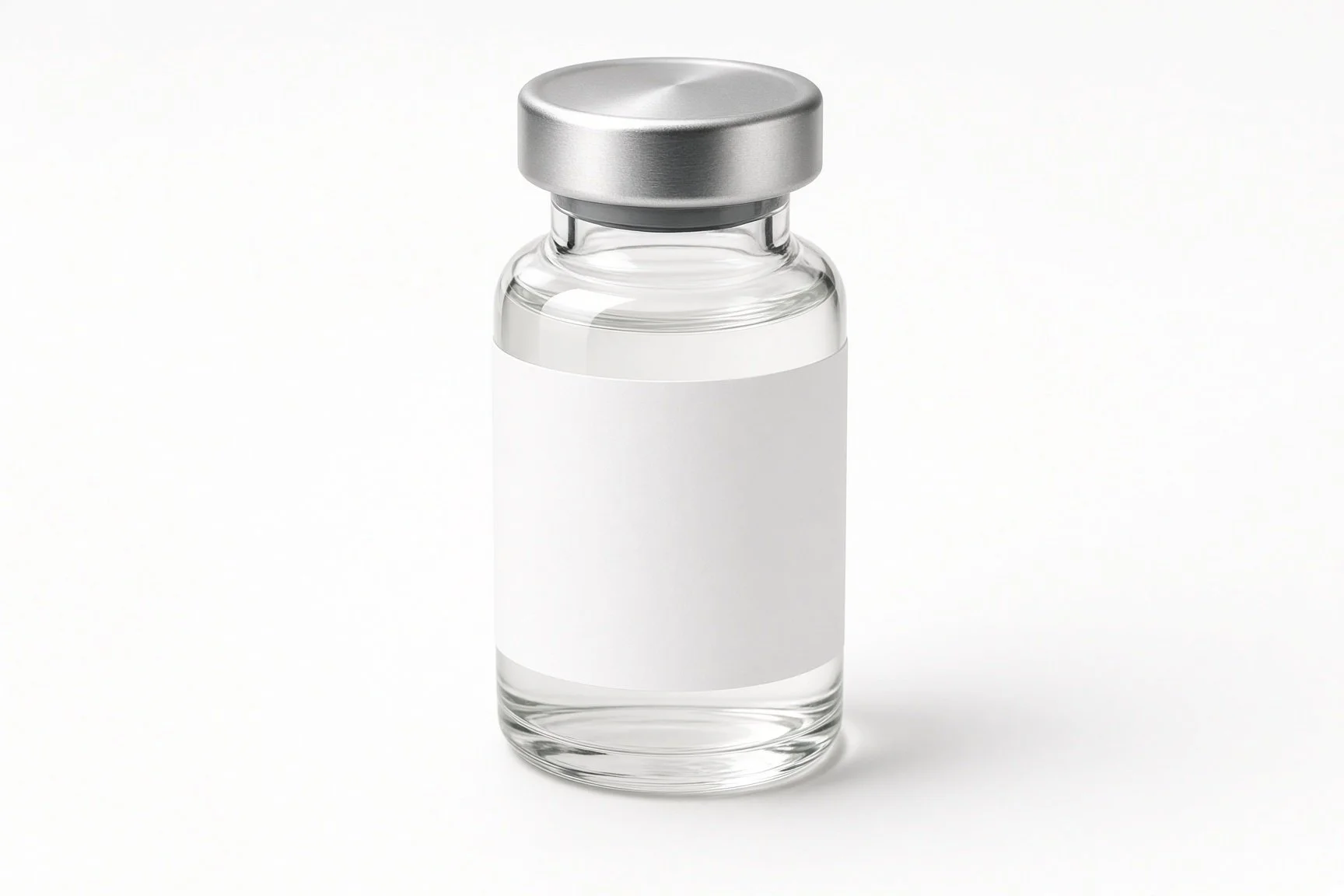
Atropine.
Procedural steps (Barts example)
Getting ready
Access
Ablating the plexi
Finishing
Follow-up monitoring
First time in the cath lab?
Expect slow rhythms. Vagal responses during energy can cause pauses. Back-up pacing should be ready before ablation starts.
Time the atropine. Atropine changes the vagal response, so it is usually given at the end. Check when the operator wants it.
Record the heart rates. Baseline and end-of-case heart rates, before and after atropine, are part of how success is judged.
Patients are often young. Many have been fainting for years. Explain each step simply; this is often their first heart procedure.
Agree before the case when atropine will be given, so it does not hide the vagal responses the operator is looking for.
Note the time and heart rate at each stage on the record. The change is part of the result.
FAQ
Is this instead of a pacemaker?
For selected people it may avoid or delay one. It is not right for everyone, and a pacemaker remains an option.
Will I stop fainting?
Many people faint less, but not everyone. The blood pressure part of a faint is not treated by this procedure.
Is it new?
Yes. It is an emerging treatment done in specialist centres, and long-term results are still being studied.
Why is my heart rate faster afterwards?
The vagal brake on the heart is reduced. This is expected and often settles.
Related Cath Lab 101 guides
Implantable loop recorder (coming soon)
Sources
All checked on 12 November 2026. Each page's own date is shown where it gives one.
Brignole M, et al. 2018 ESC Guidelines for the diagnosis and management of syncope. Eur Heart J 2018.
Glikson M, et al. 2021 ESC Guidelines on cardiac pacing and cardiac resynchronization therapy. Eur Heart J 2021.
StatPearls (NCBI Bookshelf), search: vasovagal syncope. ncbi.nlm.nih.gov/books
Disclaimer
Education only. This page is for general education for healthcare students, cath lab staff and curious readers. It isn't medical advice and doesn't replace advice from your own doctor, heart team or local unit protocols. Equipment and medicines are described generically. No product or manufacturer is recommended. If you think someone is having a heart emergency, call 999.
Disclosure: I work for a company that makes a device used in this procedure. This page is independent education, isn't sponsored or reviewed by any company, and doesn't recommend any product.
All diagrams on this page are original illustrations created for rodnieoro.com.