AV node ablation (ablate and pace): a Cath Lab 101 guide
Reviewed by Rodnie Oro, cath lab charge nurse · Last reviewed: November 2026 · Education only, not medical advice
AV node ablation in one minute
What it is: A two-part treatment for atrial fibrillation (AF) with a fast heart rate. A pacemaker is fitted first. Then a catheter from the groin vein ablates the AV node, the electrical gateway between the top and bottom chambers, so the pacemaker sets a steady rate.
Who it is for: People with AF whose heart rate stays too fast, or causes symptoms or heart failure, despite medicines, and for whom other rhythm treatments are not suitable or have not worked.
Main risks: Bruising at the groin, problems with the pacemaker lead, and lifelong reliance on the pacemaker. Rarely, serious rhythm problems in the first weeks, which is why the pacing rate is set higher at first.
Usually: Under local anaesthetic, sometimes with light sedation, often under an hour; many go home the same or next day.
On this page: What is AV node ablation? · Who is it for? · When is it done? · Step by step · Recovery · Risks · Kit list · First time in the lab? · FAQ
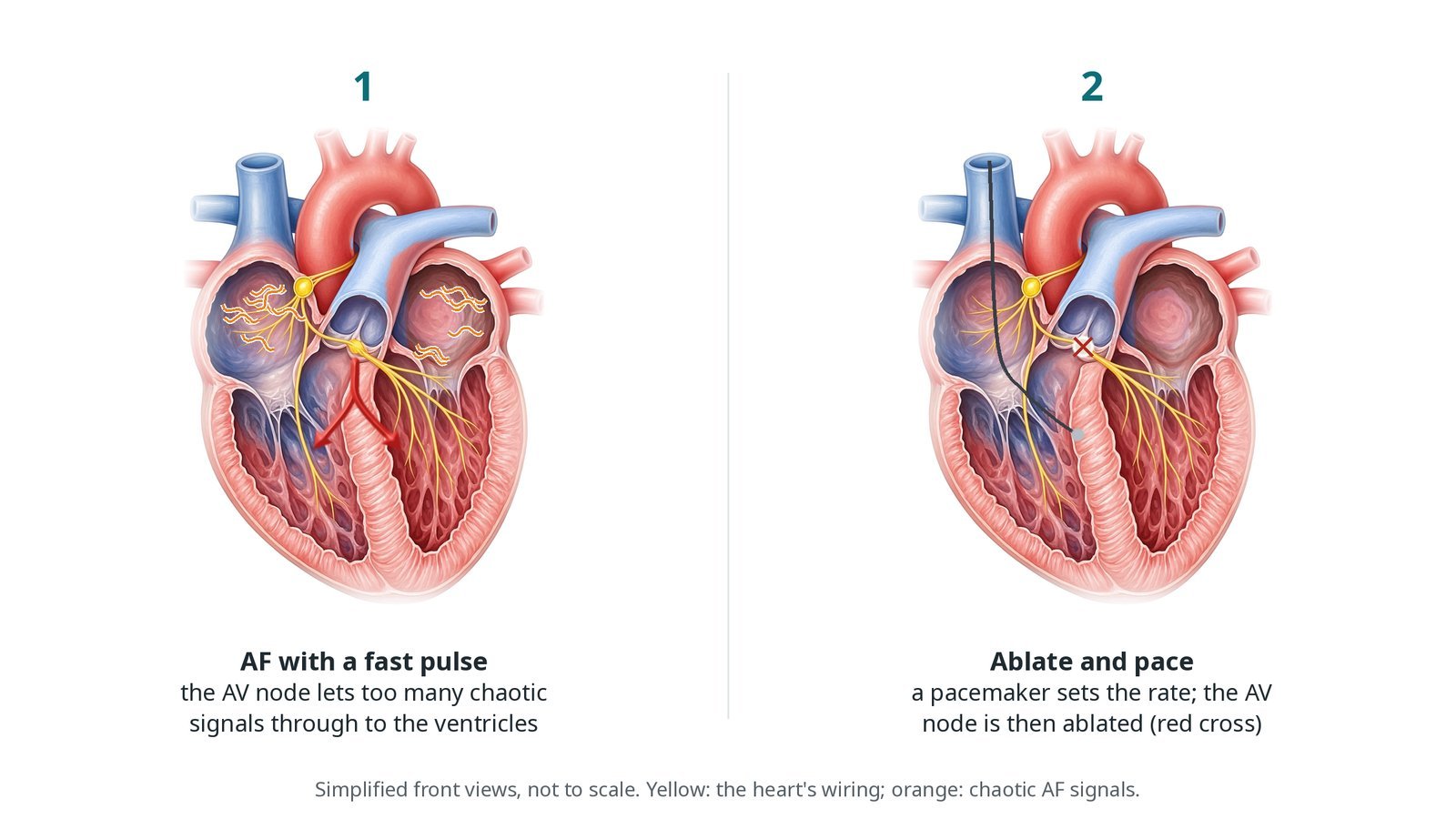
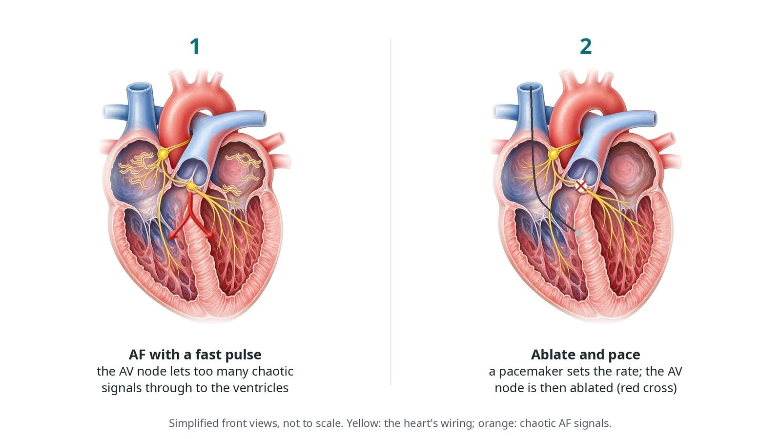
(1) AF with a fast pulse: the AV node lets too many chaotic signals through to the ventricles. (2) Ablate and pace: a pacemaker sets the rate and the AV node is ablated.
What is AV node ablation?
In atrial fibrillation (AF) the top chambers (atria) fire chaotically, hundreds of times a minute. The AV node, a small cluster of cells low in the septum between the atria, filters these signals before they reach the ventricles. In some people too many still get through, so the pulse is fast and irregular. This can cause breathlessness and tiredness, and can weaken the heart over time.
AV node ablation, often called "ablate and pace", cuts that link on purpose. Radiofrequency energy makes a small scar at the AV node, usually from the right side of the heart, close to the His bundle. The atria may still fibrillate, but the ventricles no longer follow them.
Because the heart's own connection is gone, a pacemaker must set the rate from then on. It is fitted first, often weeks before. It is usually a conduction system pacing device (pacing the heart's own wiring, such as the left bundle branch area) or a biventricular (CRT) device, so both ventricles squeeze together.
The ablation does not cure AF, and it does not change the need for blood thinners. It controls the rate, permanently.
New to the acronyms? See the Cath Lab Abbreviations A–Z.
Who is it for?
It may be considered for people with:
permanent or long-standing AF with a fast rate despite rate-control medicines
symptoms or heart failure linked to the fast rate
AF where rhythm control (ablation of the AF itself, cardioversion or medicines) is not suitable or has not worked
NICE and ESC guidance both include it as a rate-control option when other treatments fail. Because the result is permanent, the heart team talks it through carefully.
Who it may not suit: People who could still be offered ablation of the AF itself, younger people for whom lifelong pacemaker dependence is a big step, and anyone who has not yet tried the usual rate-control medicines.
The decision is made by your heart team, who will talk through the reasons, the alternatives and the risks with you.
When is it done?
It is planned. Usually:
the pacemaker is fitted first and checked to be working well; some centres do both on the same day
blood thinners and other medicines are reviewed, as per centre protocol
the pacemaker is checked again on the day, before the ablation
gives you a chance to ask questions and give consent
arranges blood tests, including kidney function and a blood count, and an ECG
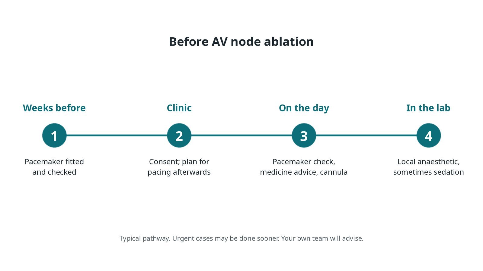
Before AV node ablation: pacemaker fitted and checked weeks before; consent and a plan for pacing; pacemaker check and medicine advice on the day; local anaesthetic in the lab.
Step by step
1. Getting ready
You lie on the X-ray table. ECG, blood pressure and oxygen are monitored, and external defibrillator pads may be placed, as per centre protocol. The pacemaker is checked first with its programmer. The groin is cleaned and numbed; sedation is light or not needed.
2. Access
A sheath is placed in the femoral vein in the groin, using ultrasound; size per device and operator. An ablation catheter is passed up through the inferior vena cava into the right atrium under X-ray. Rarely, if the AV node cannot be blocked from the right, it is reached from the left side through the femoral artery and aorta.
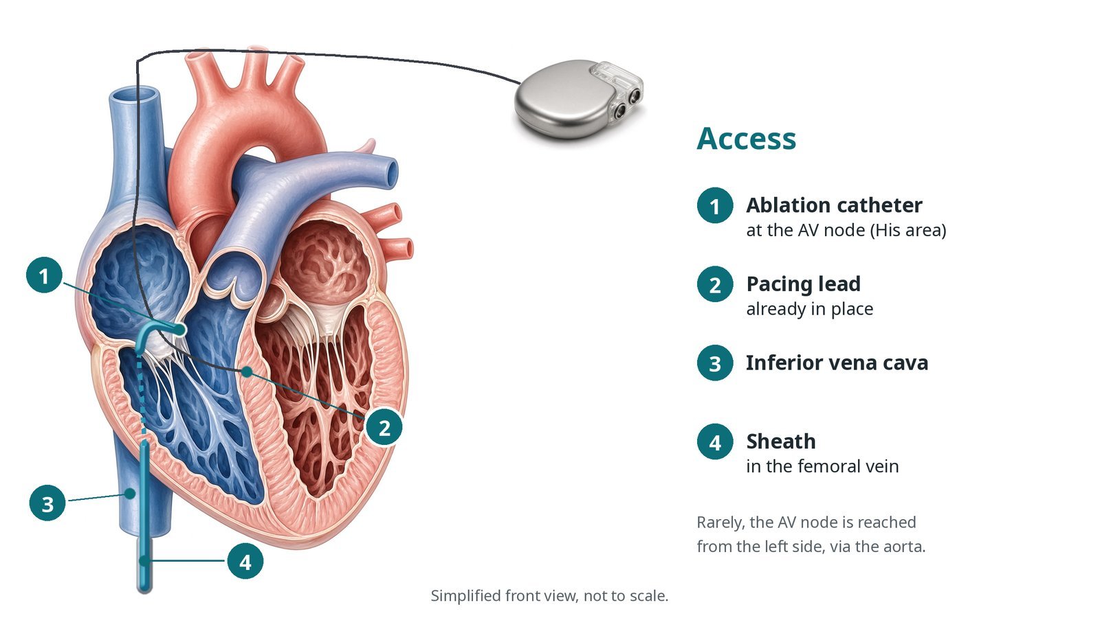
Access: (1) ablation catheter at the AV node (His area); (2) pacing lead already in place; (3) inferior vena cava; (4) sheath in the femoral vein.
3. Ablating the AV node
A. Pacemaker first. The pacemaker is tested and programmed so it will pace the heart reliably once the AV node is gone.
B. Find the His signal. The catheter tip is placed on the septum, just behind the tricuspid valve, where the His bundle signal is recorded. The target is usually slightly towards the atrium, at the compact AV node.
C. Ablate. Radiofrequency energy is delivered, per operator. A run of junctional beats is often seen, then complete heart block.
D. Paced rhythm. Every beat now comes from the pacemaker. The team waits and watches to make sure the block lasts.
Watch: No paced beats after block (the pacemaker is not capturing): start back-up pacing at once, as planned, and tell the operator.
4. Programming and finishing
The sheath is removed and pressure is held on the groin. The pacemaker is reprogrammed, as per centre protocol. The lower rate is usually set higher for the first weeks, because the heart is more prone to dangerous rhythms just after the ablation. It is turned down at a later check.
Watch: Groin swelling or a falling blood pressure: press over the site and tell the team.
5. Before going home
A device check confirms the leads are working and every beat is paced. You are told that you now rely on the pacemaker (pacemaker-dependent) and given an appointment to lower the rate.
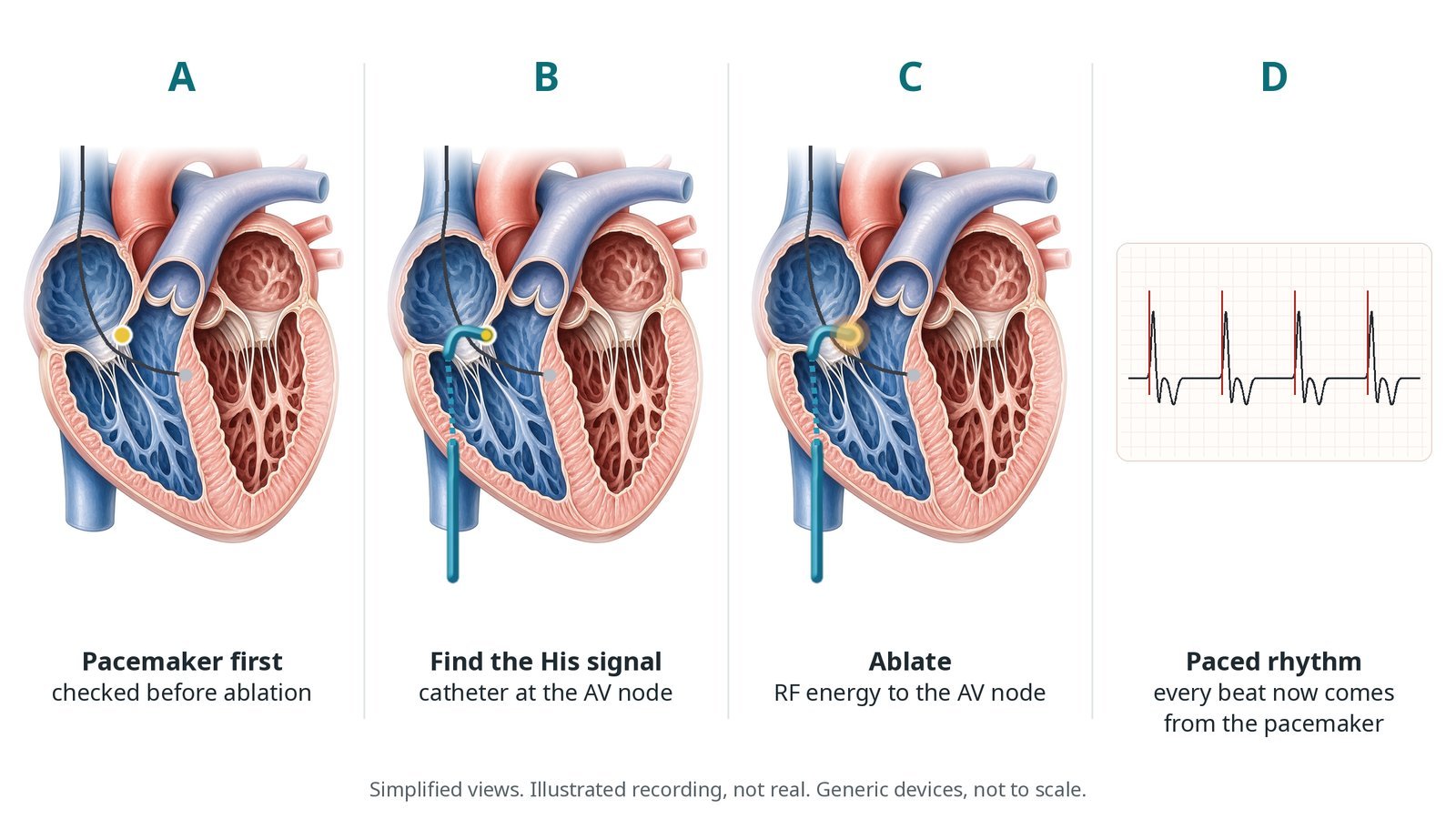
A: pacemaker first, checked before ablation. B: the catheter finds the His signal at the AV node. C: RF energy ablates the AV node. D: every beat now comes from the pacemaker.
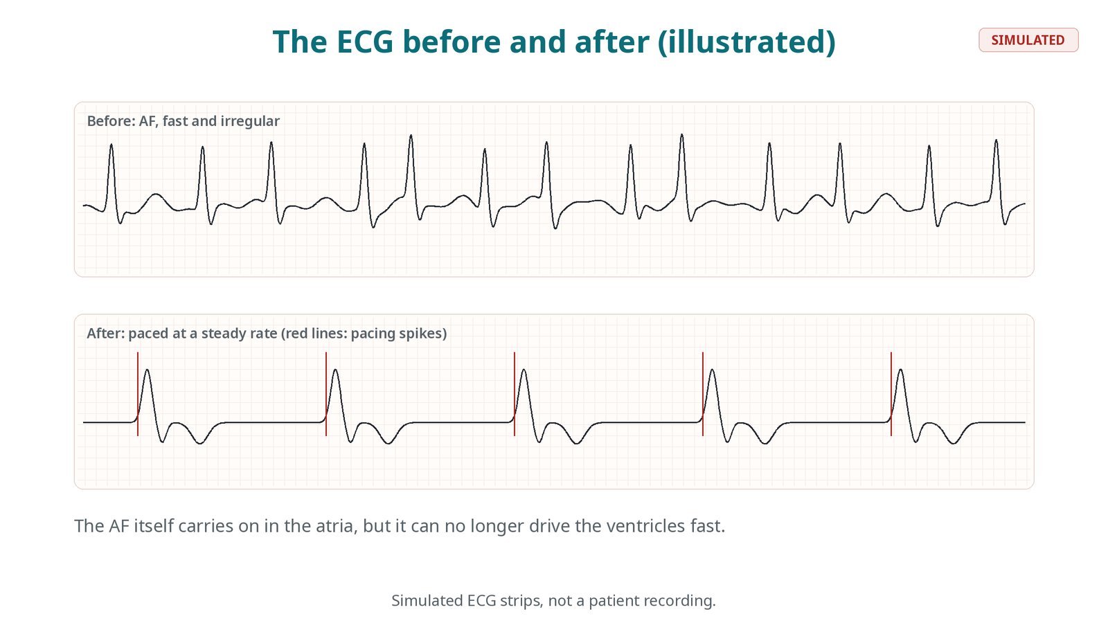
Simulated ECG strips, not a patient recording. Before: AF, fast and irregular. After: paced at a steady rate.
Recovery and aftercare
First hours: bed rest while the groin settles, with checks of the groin, heart rhythm and blood pressure, as per centre protocol.
Going home: often the same or next day, after a pacemaker check.
Pacemaker: the rate is set higher at first and lowered at a check a few weeks later, as per centre protocol.
Medicines: some rate-control medicines may be stopped; keep taking blood thinners if you were on them, unless told otherwise.
Driving: DVLA rules depend on your pacemaker and symptoms; ask your team before you drive.
Follow-up: device clinic, often with remote monitoring, for life.
Call 999 if you have chest pain, fainting, sudden breathlessness, or a very slow or very fast pulse.
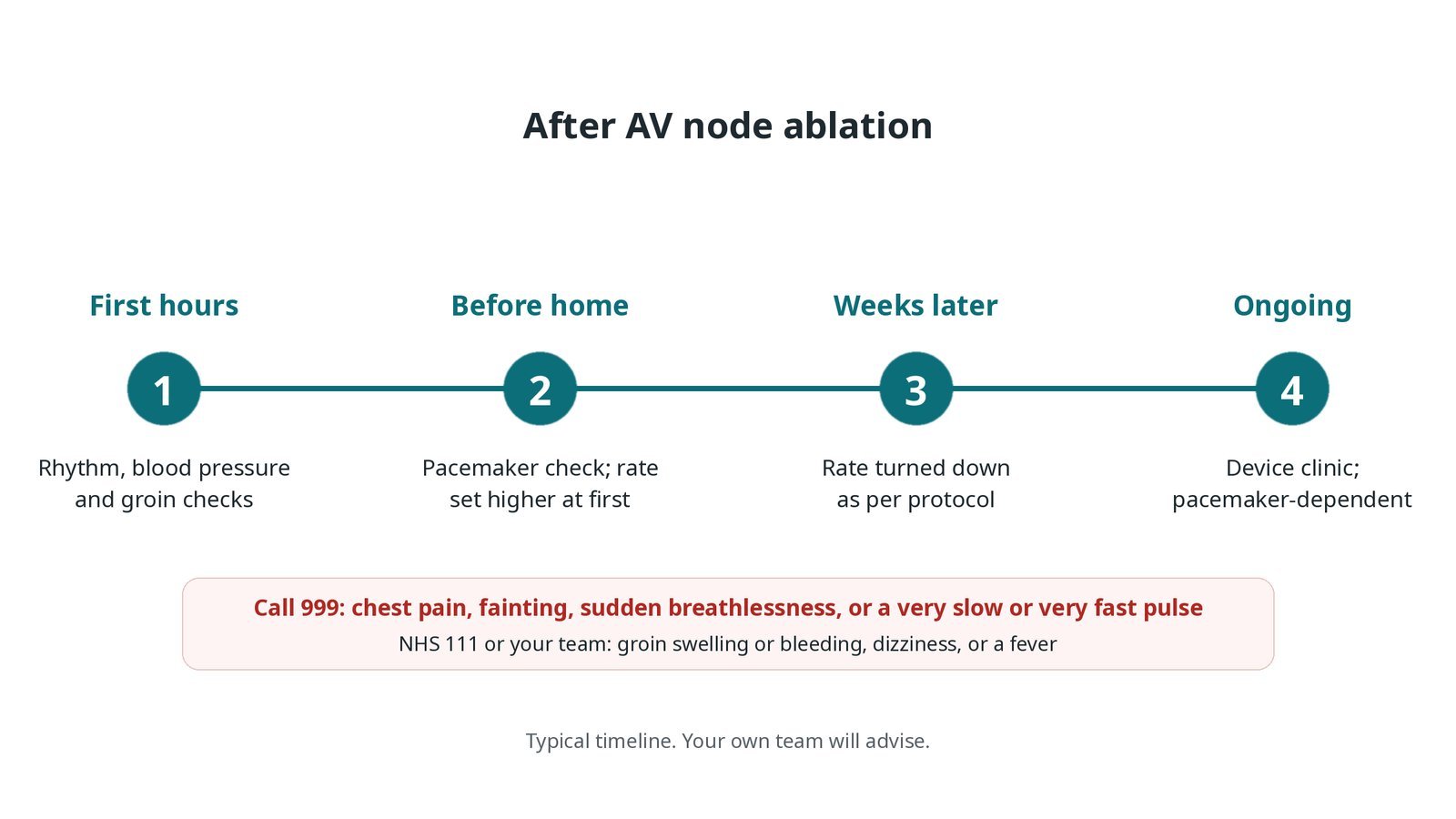
After AV node ablation: (1) rhythm, blood pressure and groin checks; (2) pacemaker check, rate set higher at first; (3) rate turned down weeks later as per protocol; (4) device clinic, pacemaker-dependent.
Risks and complications
Your team will explain the risks that apply to you. Possible problems include:
Bruising or bleeding at the groin
Pacemaker lead problems needing another procedure (uncommon): Usually found at a device check.
Lifelong reliance on the pacemaker: This is expected, not a complication.
Serious rhythm problems in the first weeks (rare): The pacing rate is set higher at first to lower this risk.
The AV node recovering, needing a repeat ablation (uncommon)
Heart failure getting worse with right ventricular pacing alone (uncommon): Conduction system or CRT pacing is often chosen to avoid this.
Cath lab kit list (printable)
Barts example kit list
An example from one UK centre, written generically. Every lab has its own protocol, and sizes and doses vary.
femoral venous sheath, size per device and operator
ultrasound for vein access
steerable radiofrequency ablation catheter, per operator
radiofrequency generator and grounding patch
recording system for the His signal
pacemaker programmer and device physiologist
external defibrillator pads and a back-up pacing plan, as per centre protocol
local anaesthetic and light sedation, as per centre protocol
manual compression or groin closure, per operator
Procedural steps (Barts example)
Getting ready
Access
Ablating the AV node
Programming and finishing
Before going home
First time in the cath lab?
Check the pacemaker first. Nobody ablates the AV node until the pacemaker has been tested and the physiologist and operator are happy with it.
Know your back-up pacing. Ask before the case how the team will pace if the pacemaker fails to capture: through the ablation catheter, a temporary wire or the pads.
Expect junctional beats. A run of regular junctional beats during energy is a good sign the target is right; complete block usually follows.
Explain dependence kindly. Patients often ask if they will rely on the pacemaker. The honest answer is yes, so device follow-up really matters.
Have the pacemaker programmer and the device physiologist in the room before you start.
Watch the ECG during energy delivery: junctional beats are expected. Call out heart block when it happens.
FAQ
Does this cure my AF?
No. The atria may still fibrillate, but the ventricles beat at the steady rate set by the pacemaker, so most people feel better.
Will I still need blood thinners?
Often yes. The stroke risk from AF stays the same, so anticoagulation is decided as before.
Will I depend on the pacemaker?
Yes. That is why it is fitted and checked first, and why regular device checks matter.
Why is my heart rate set higher at first?
Just after the ablation the heart is more prone to dangerous rhythms. A higher pacing rate for the first weeks lowers that risk; it is reduced at a later check.
Related Cath Lab 101 guides
Conduction system pacing (His bundle and left bundle branch area pacing) (coming soon)
Cardiac resynchronisation therapy (CRT-P and CRT-D) (coming soon)
Sources
All checked on 12 November 2026. Each page's own date is shown where it gives one.
Van Gelder IC, et al. 2024 ESC Guidelines for the management of atrial fibrillation. Eur Heart J 2024;45(36):3314–3414. PubMed 39210723
Glikson M, et al. 2021 ESC Guidelines on cardiac pacing and cardiac resynchronization therapy. Eur Heart J 2021.
NICE. Atrial fibrillation: diagnosis and management (NG196). nice.org.uk/guidance/ng196
StatPearls (NCBI Bookshelf), search: atrioventricular node ablation. ncbi.nlm.nih.gov/books
Disclaimer
Education only. This page is for general education for healthcare students, cath lab staff and curious readers. It isn't medical advice and doesn't replace advice from your own doctor, heart team or local unit protocols. Equipment and medicines are described generically. No product or manufacturer is recommended. If you think someone is having a heart emergency, call 999.
Disclosure: I work for a company that makes a device used in this procedure. This page is independent education, isn't sponsored or reviewed by any company, and doesn't recommend any product.
All diagrams on this page are original illustrations created for rodnieoro.com.