VT ablation: a Cath Lab 101 guide
Reviewed by Rodnie Oro, cath lab charge nurse · Last reviewed: November 2026 · Education only, not medical advice
VT ablation in one minute
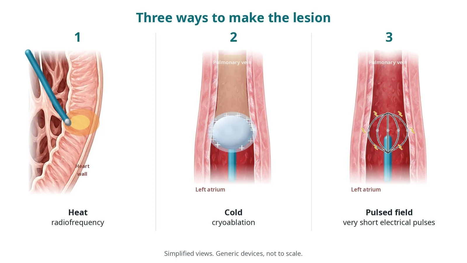
What it is: Ventricular tachycardia (VT) is a fast rhythm from the lower chambers. In VT ablation, catheters build a 3D map of the ventricle, find the circuit (often in scar) or the spot that drives the VT, and ablate it with heat (radiofrequency).
Who it is for: People with recurrent VT, especially with an ICD that keeps giving shocks or other therapies, with electrical storm, or with VT that medicines do not control.
Main risks: Bleeding at the groin, fluid around the heart (tamponade), stroke, damage to a heart valve or the conduction system, and the VT coming back. Risks are higher in very unwell patients.
Usually: Under general anaesthetic or sedation, often several hours, with a few days in hospital.
On this page: What is VT ablation? · Who is it for? · When is it done? · Step by step · Recovery · Risks · Kit list · First time in the lab? · FAQ
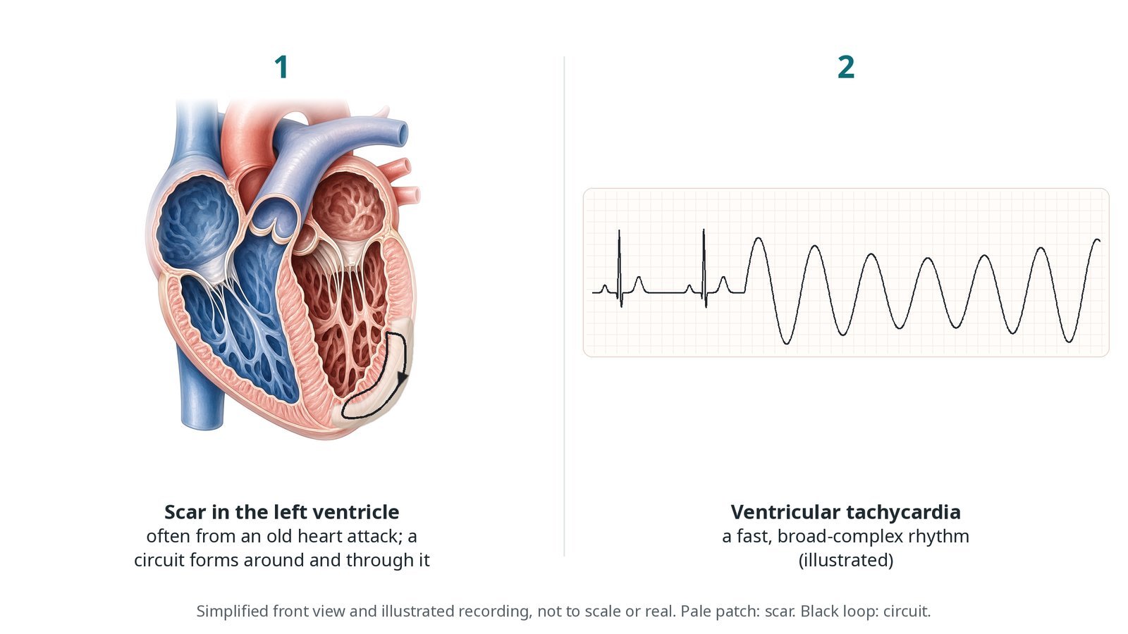
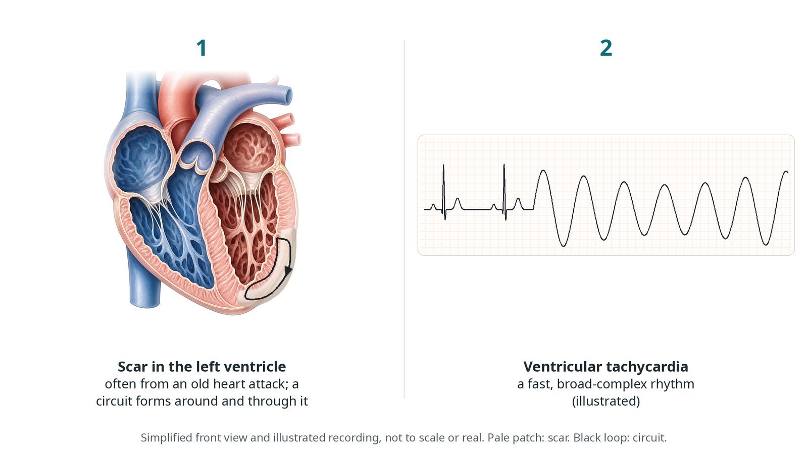
(1) Scar in the left ventricle, often from an old heart attack: a circuit forms around and through it. (2) Ventricular tachycardia: a fast, broad-complex rhythm (illustrated).
What is VT ablation?
Most VT that is ablated in adults comes from scar in the left ventricle. After a heart attack, or in some heart muscle diseases (cardiomyopathies), patches of scar form. Thin channels of surviving muscle run through the scar. A signal can travel slowly along a channel, come out the other side and loop back round. This re-entry circuit drives the ventricle very fast.
Some VT has no scar. It comes from a single irritable spot, often in the outflow tract of the right or left ventricle, and is usually simpler to treat.
During ablation, a 3D mapping system tracks the catheters and builds a model of the ventricle. A voltage map shows healthy muscle and scar. If the VT can be started and the blood pressure holds, the circuit can be followed while it runs (activation mapping). If not, the team targets abnormal signals in and around the scar in normal rhythm (substrate mapping), and uses pace mapping to match the shape of the VT on the ECG.
The left ventricle is reached backwards across the aortic valve from the femoral artery (retrograde), or across the atrial septum and mitral valve (transseptal). Some circuits lie on the outer surface of the heart and need epicardial access: a needle under the breastbone into the sac around the heart.
New to the acronyms? See the Cath Lab Abbreviations A–Z.
Who is it for?
It may be considered for people with:
scar-related VT that keeps coming back despite medicines
repeated ICD shocks or anti-tachycardia pacing
electrical storm (three or more VT episodes within 24 hours)
VT from a single spot, such as the outflow tract, causing symptoms
The 2022 ESC ventricular arrhythmia guidelines support ablation in these groups. The decision is made by an electrophysiologist and the heart team.
Who it may not suit: People with a clot in the left ventricle until it is treated, very frail people in whom the risks outweigh the benefits, and VT with a reversible cause, such as a heart attack happening now or a very low potassium.
The decision is made by your heart team, who will talk through the reasons, the alternatives and the risks with you.
When is it done?
It can be planned or urgent.
Before it, the team usually:
reviews ECGs of the VT and the ICD recordings
looks at scans: echo, and often cardiac MRI or CT to show the scar
checks for clot in the left ventricle
plans the anaesthetic, and support for the circulation (a heart pump or ECMO) in very unwell patients
gives you a chance to ask questions and give consent
arranges blood tests, including kidney function and a blood count, and an ECG
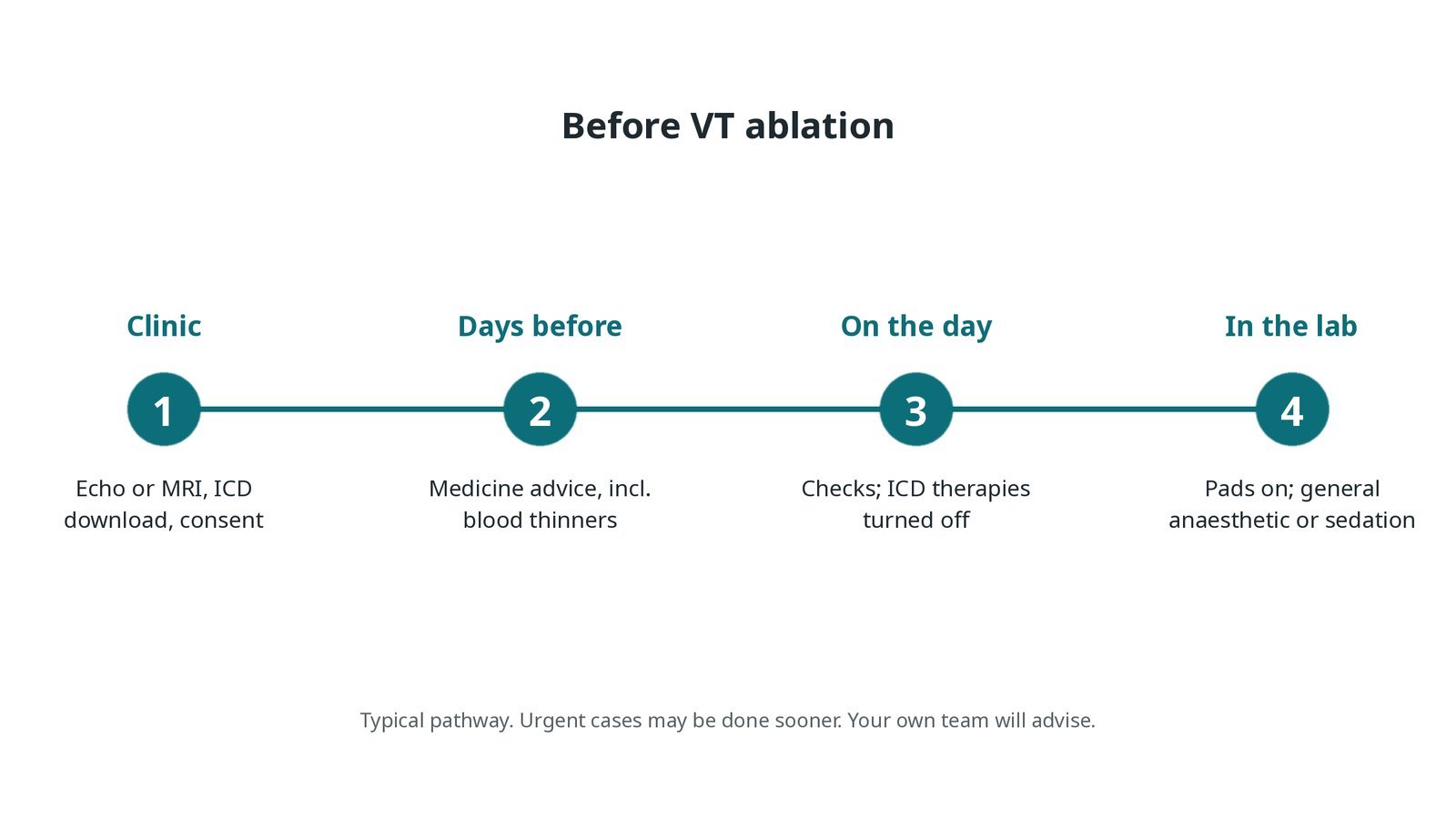
Before VT ablation: scans, ICD download and consent; medicine advice; ICD therapies turned off on the day; defibrillator pads and anaesthetic or sedation in the lab.
Step by step
1. Getting ready
External defibrillator pads go on first, and many patients then have a general anaesthetic; some have sedation, as per centre protocol. The device physiologist turns off the ICD's shock and pacing therapies for the case, so it does not treat the rhythms started during mapping. An arterial line monitors blood pressure.
Watch: VT with a falling blood pressure: tell the operator, and be ready to pace or shock through the pads as directed.
2. Access
Sheaths go in the femoral vein and usually the femoral artery, under ultrasound; sizes per device and operator. Heparin is given, as per centre protocol. The left ventricle is reached across the aortic valve (retrograde) or across the atrial septum (transseptal). Some cases also need epicardial access through a needle under the breastbone. In unstable patients a heart pump or ECMO may be placed first to support the circulation.
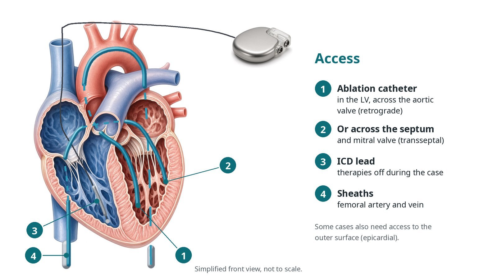
Access: (1) ablation catheter in the LV across the aortic valve (retrograde); (2) or across the septum and mitral valve (transseptal); (3) ICD lead, therapies off; (4) femoral artery and vein sheaths.
3. Mapping and ablation
A. Map. A mapping catheter builds a 3D voltage map of the ventricle, showing healthy muscle and scar.
B. Find the circuit. If the VT is tolerated, it is started and its circuit followed (activation mapping). If not, abnormal signals in the scar and pace mapping guide the targets (substrate mapping).
C. Ablate. Radiofrequency energy is delivered across the channels and abnormal areas, per operator. The catheter tip is cooled with saline (irrigated), so fluid balance is watched.
D. Test. Pacing tries to start the VT again. Not being able to start it is a good sign.
Watch: A sudden pop on the recording, or the blood pressure falling with a widening heart shadow: think steam pop or tamponade. Tell the operator and get the echo and pericardiocentesis kit ready.
4. Finishing
Catheters and sheaths are removed. Artery sites are closed with a closure device or pressure, per operator. If epicardial access was used, a drain may stay in for a short time. The ICD therapies are turned back on and checked before you leave the lab.
Watch: ICD therapies still off at handover: do not leave the lab until the device has been reprogrammed and confirmed.
5. Recovery
You are monitored in recovery, on a cardiac ward or in the coronary care unit, depending on how the case went. Groin, blood pressure, rhythm and fluid balance are checked, and an echo may look for fluid around the heart.
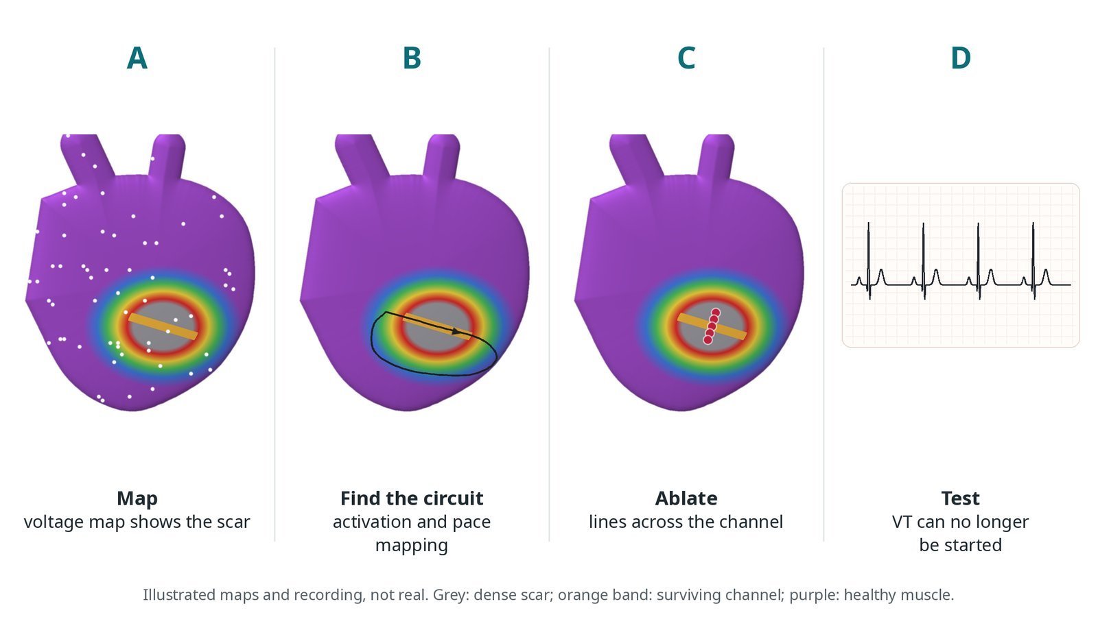
A: a voltage map shows the scar. B: activation and pace mapping find the circuit. C: ablation lines across the channel. D: test that VT can no longer be started.
Recovery and aftercare
First hours: bed rest with checks of the groin, blood pressure, heart rhythm and fluid balance, as per centre protocol.
Going home: usually after a few days, once the rhythm is stable and the ICD has been checked.
ICD: therapies are switched back on before you leave the lab and checked again before home.
Medicines: rhythm medicines may be changed, reduced or kept; take them exactly as advised.
Driving: DVLA rules for ICDs and VT apply; ask your team how long you must wait.
Follow-up: arrhythmia and device clinic, with remote monitoring of the ICD.
Call 999 if you have chest pain, fainting, breathlessness, more than one shock, or signs of a stroke.
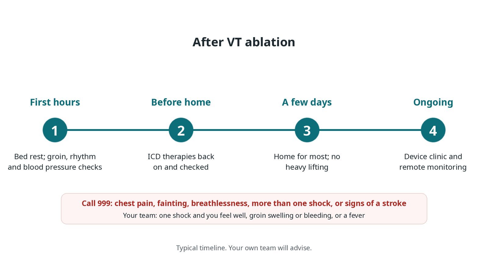
After VT ablation: (1) bed rest and checks; (2) ICD therapies back on and checked; (3) home for most after a few days; (4) device clinic and remote monitoring.
Risks and complications
Your team will explain the risks that apply to you. Possible problems include:
Bleeding, bruising or vessel damage at the groin
Fluid or bleeding around the heart (tamponade) (uncommon): May need a drain.
Stroke (uncommon): Higher when the left ventricle is reached through the aorta.
Steam pop: a small tissue rupture during heating (uncommon)
Damage to a heart valve, the conduction system or a coronary artery (rare)
Worsening heart failure or a severe drop in blood pressure (uncommon): Support devices may be needed in very unwell patients.
The VT coming back: Some people need a repeat procedure.
Cath lab kit list (printable)
Barts example kit list
An example from one UK centre, written generically. Every lab has its own protocol, and sizes and doses vary.
femoral venous and arterial sheaths, sizes per device and operator
ultrasound for vascular access
irrigated ablation catheter and radiofrequency generator, per operator
high-density mapping catheter, per operator
3D mapping system and skin patches
transseptal needle and steerable sheath, if the transseptal route is used
external defibrillator pads and defibrillator
ICD programmer and device physiologist
arterial line and anaesthetic set-up, as per centre protocol
epicardial access needle and pericardiocentesis kit, available
heart pump or ECMO, if planned, per operator
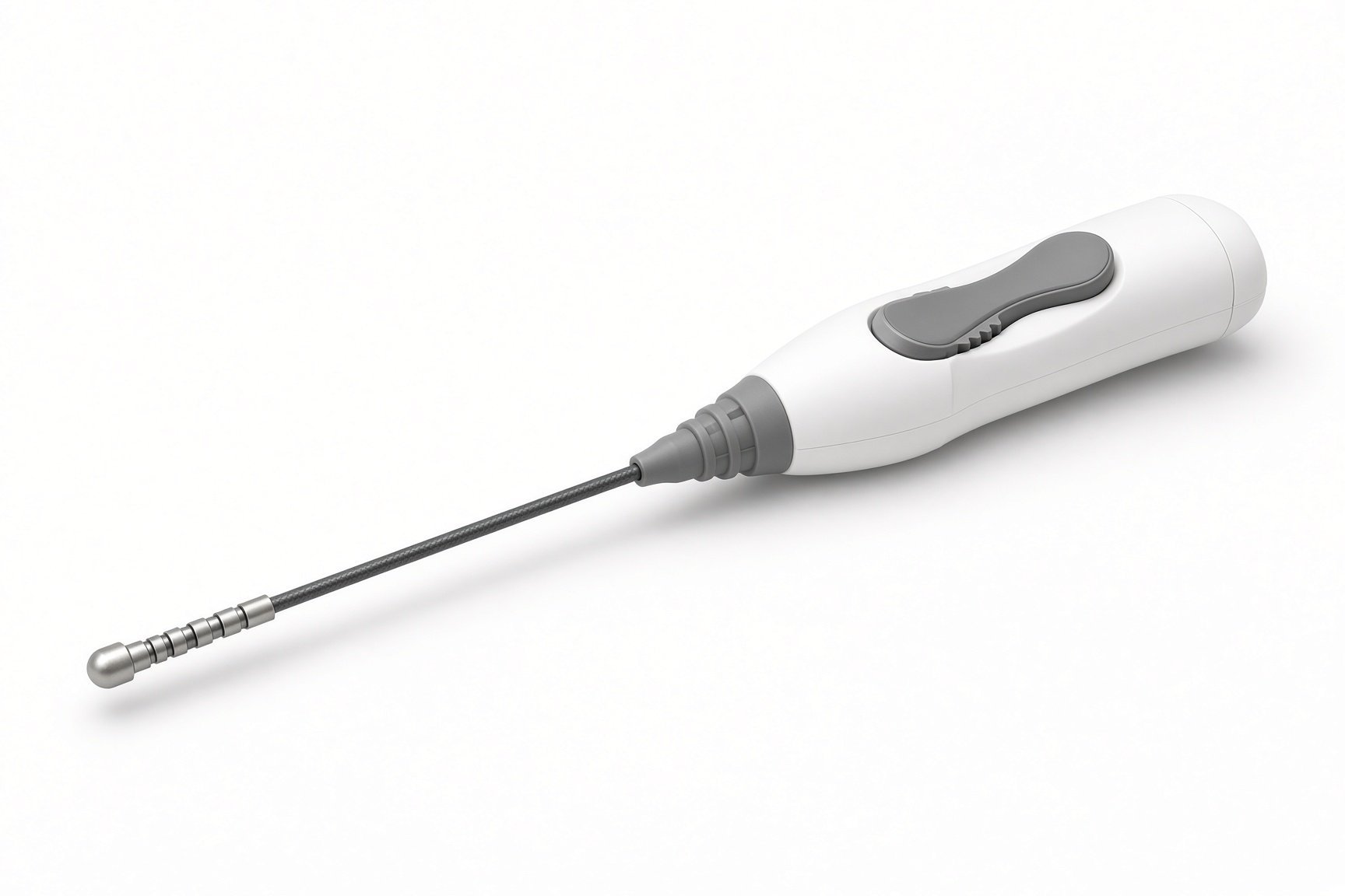
Irrigated ablation catheter.
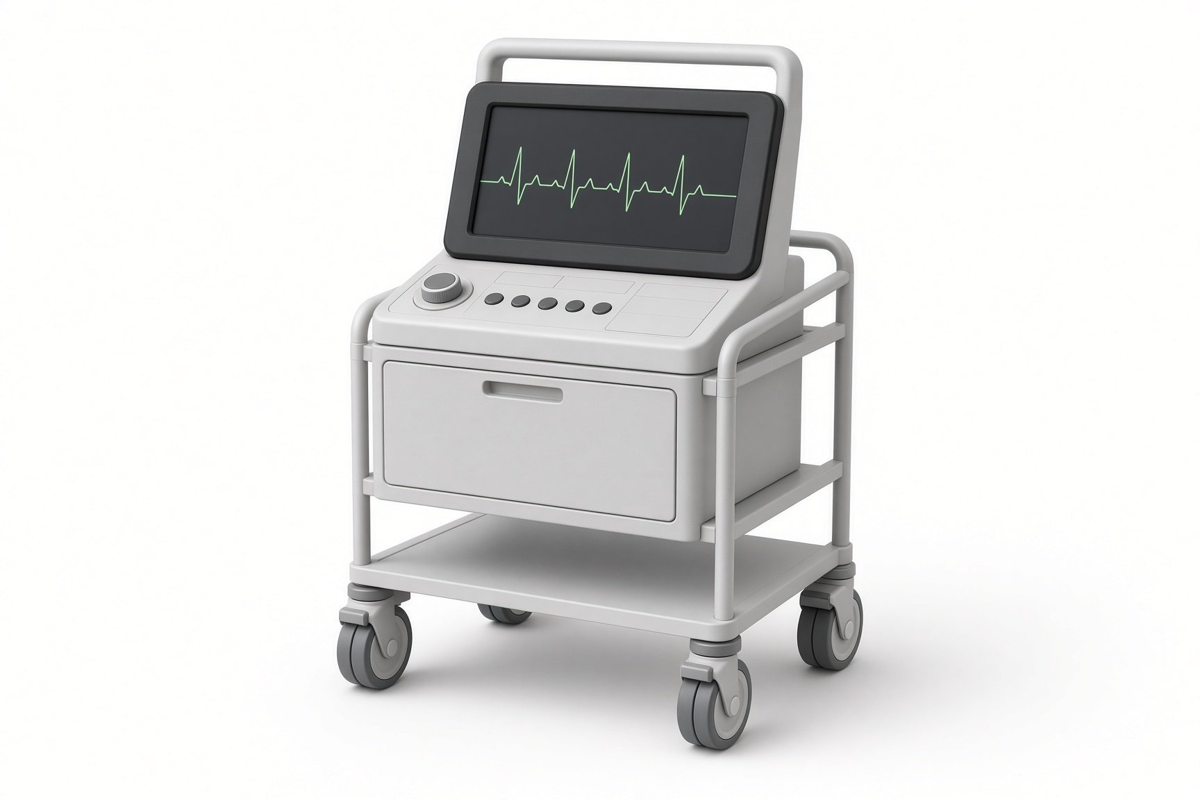
3D mapping system.
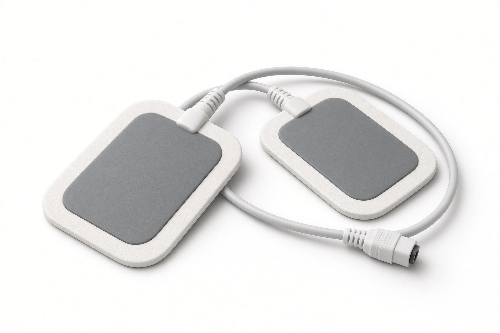
External defibrillator pads.
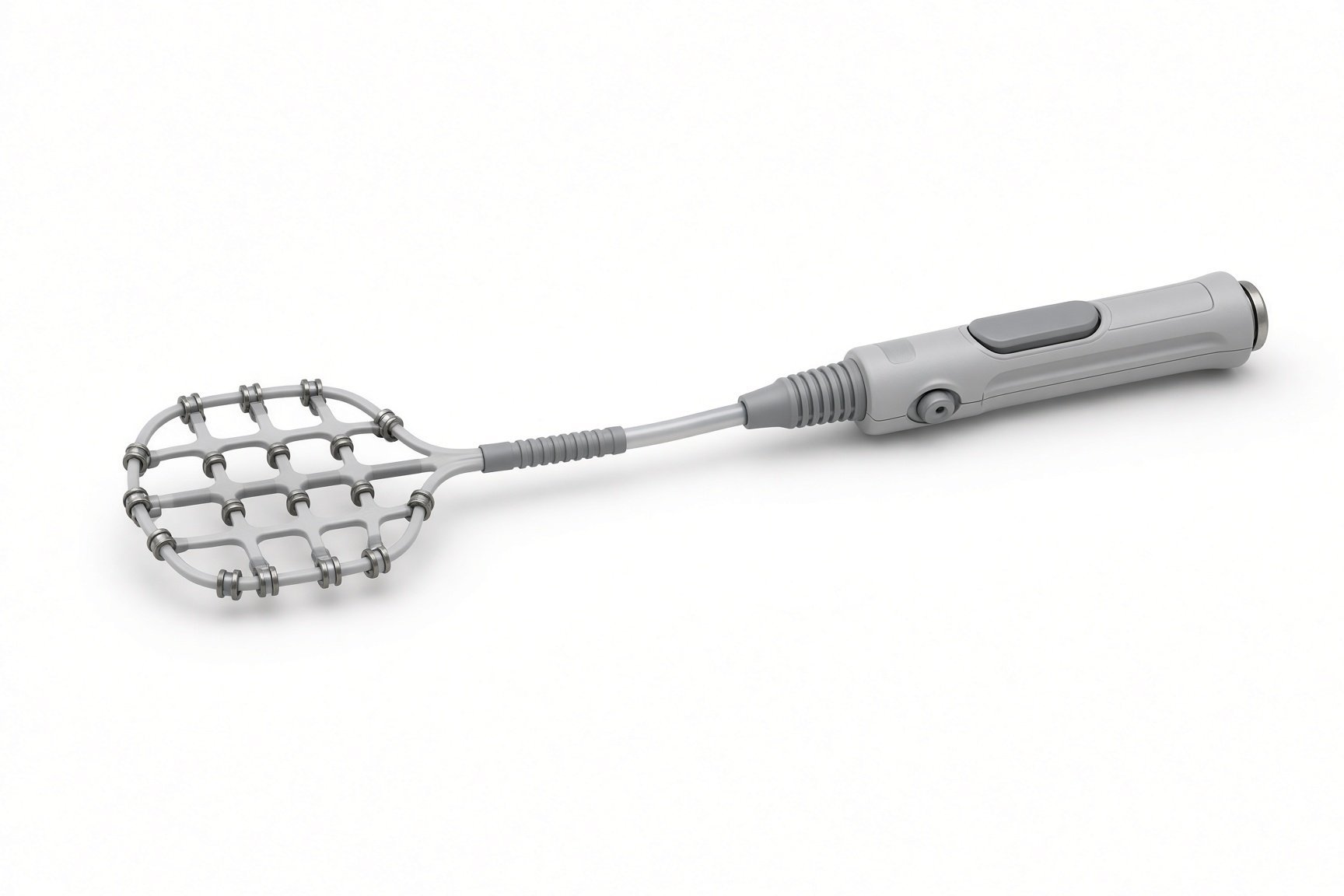
High-density mapping catheter.
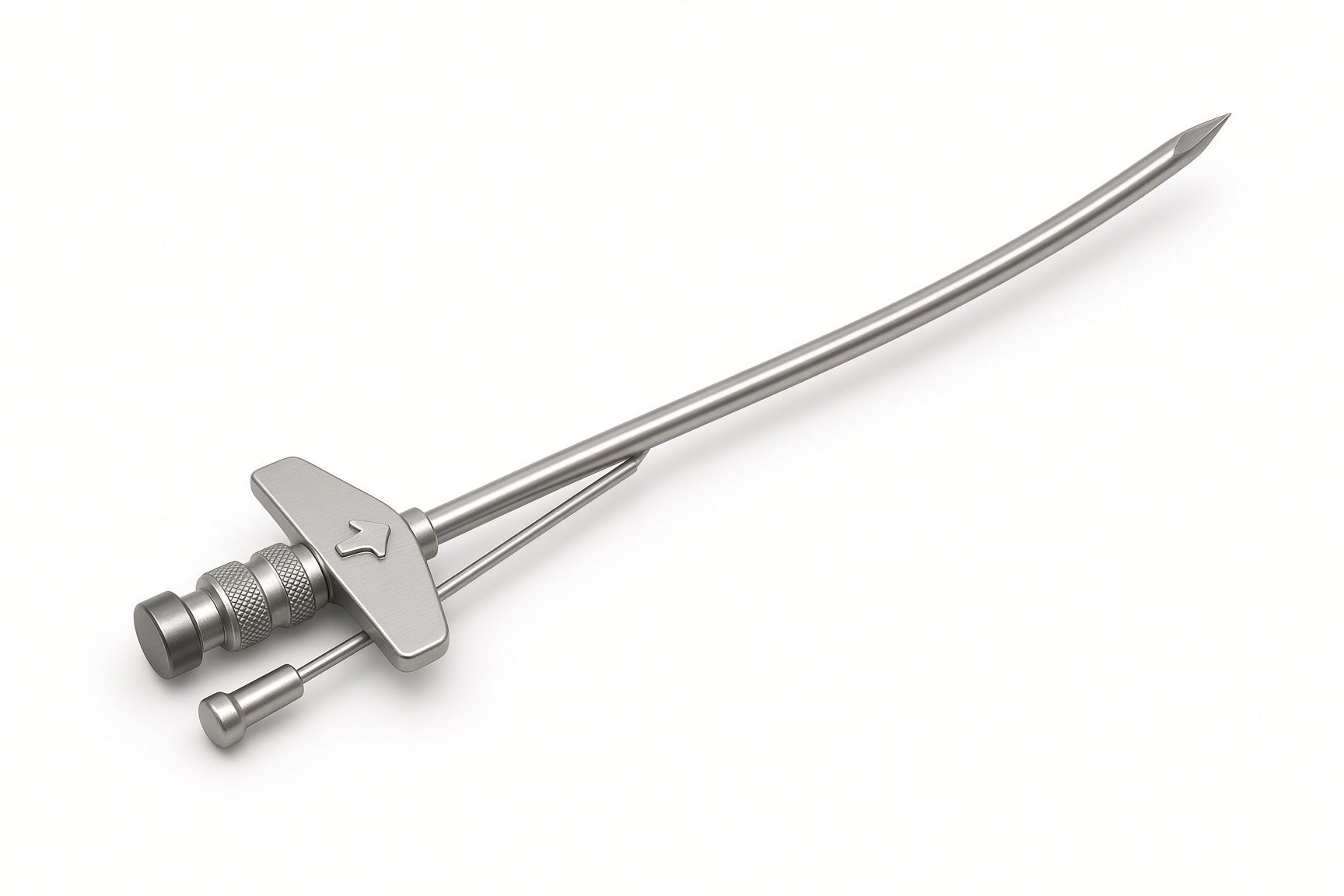
Transseptal needle.
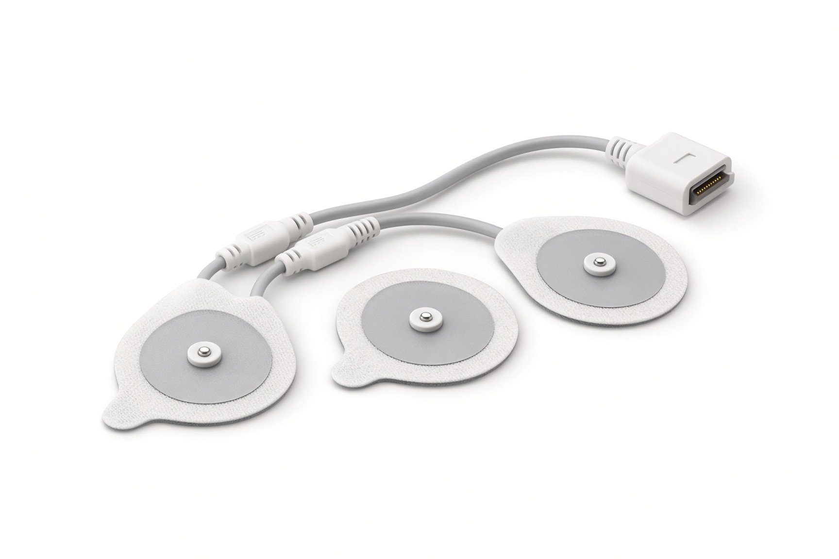
Mapping skin patches.
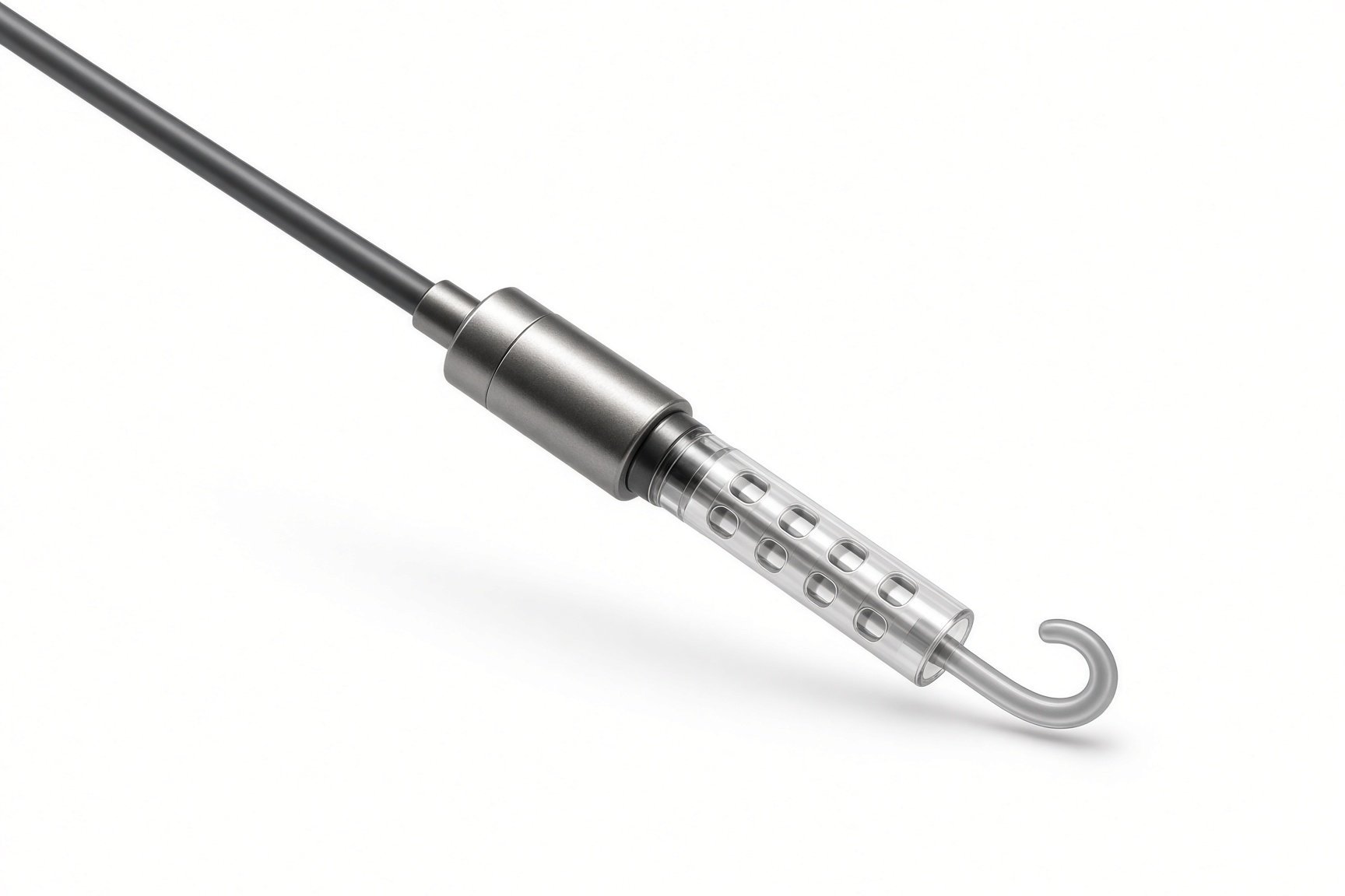
Heart pump, if needed.
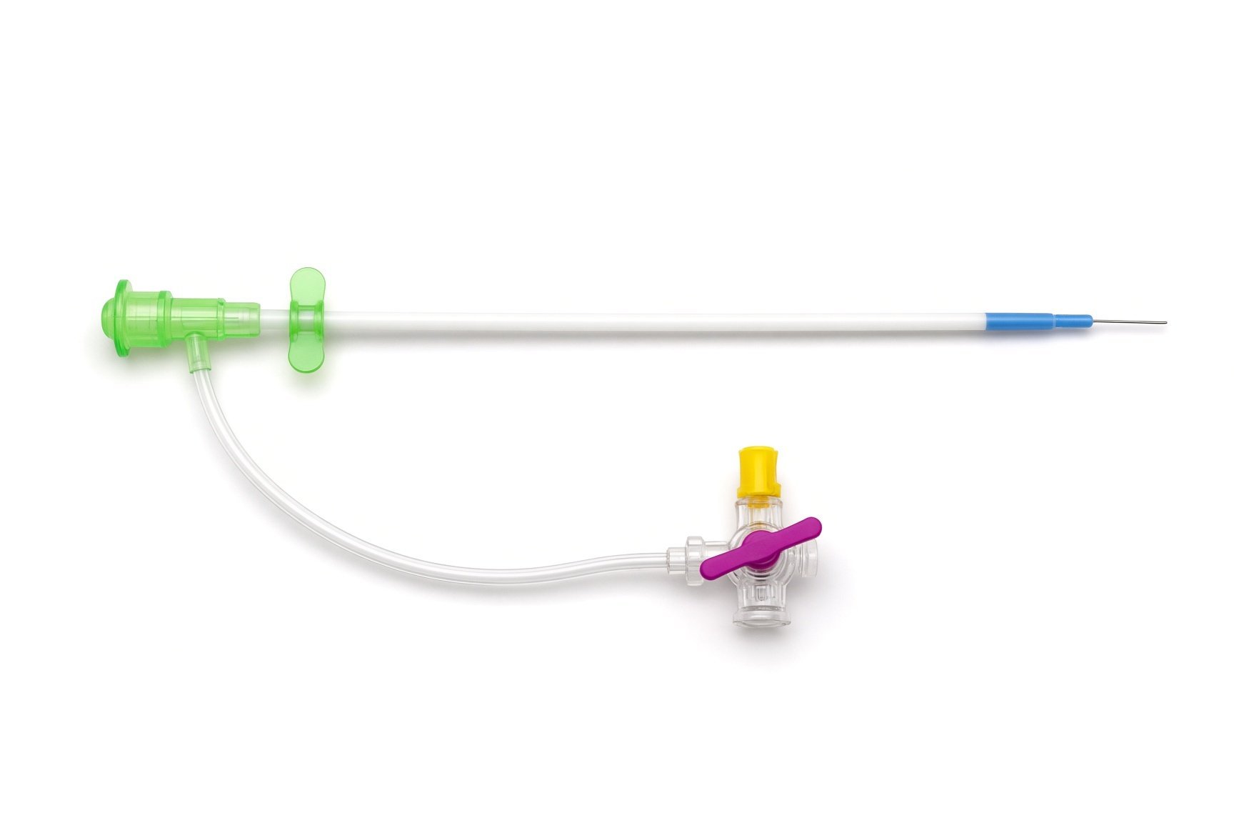
Femoral sheaths.
Heart pump if needed.
Procedural steps (Barts example)
Getting ready
Access
Mapping and ablation
Finishing
Recovery
First time in the cath lab?
Pads before programming. External pads go on and the defibrillator is checked before the ICD is switched off.
Know the fluid count. Irrigated catheters add a lot of saline over a long case. Keep the running total and know who you report it to.
Have the rescue kit in reach. Echo and the pericardiocentesis set should be close for every VT case.
Close the loop on the ICD. The ICD therapies must be back on before the patient leaves. Say it out loud at the end.
Pads on and the defibrillator ready before the ICD therapies are turned off.
Note the time the ICD therapies went off. They must be back on before the patient leaves the lab.
Keep a running total of irrigation fluid and tell the team as it builds up, as per centre protocol.
FAQ
Will I still need my ICD?
Yes. Ablation reduces VT and shocks, but the ICD stays as a safety net.
Will I be asleep?
Often, under general anaesthetic. Some centres use sedation, which can make the VT easier to start.
Why does it take so long?
Building a detailed map of the ventricle, ablating and testing the result all take time.
Can the VT come back?
It can. Some people need a repeat procedure, or medicines as well.
Related Cath Lab 101 guides
Sources
All checked on 12 November 2026. Each page's own date is shown where it gives one.
Zeppenfeld K, et al. 2022 ESC Guidelines for the management of patients with ventricular arrhythmias and the prevention of sudden cardiac death. Eur Heart J 2022.
Cronin EM, et al. 2019 HRS/EHRA/APHRS/LAHRS expert consensus statement on catheter ablation of ventricular arrhythmias.
StatPearls (NCBI Bookshelf), search: ventricular tachycardia. ncbi.nlm.nih.gov/books
Disclaimer
Education only. This page is for general education for healthcare students, cath lab staff and curious readers. It isn't medical advice and doesn't replace advice from your own doctor, heart team or local unit protocols. Equipment and medicines are described generically. No product or manufacturer is recommended. If you think someone is having a heart emergency, call 999.
Disclosure: I work for a company that makes a device used in this procedure. This page is independent education, isn't sponsored or reviewed by any company, and doesn't recommend any product.
All diagrams on this page are original illustrations created for rodnieoro.com.