SVT and atrial flutter ablation: a Cath Lab 101 guide
Reviewed by Rodnie Oro, cath lab charge nurse · Last reviewed: November 2026 · Education only, not medical advice
SVT and atrial flutter ablation in one minute
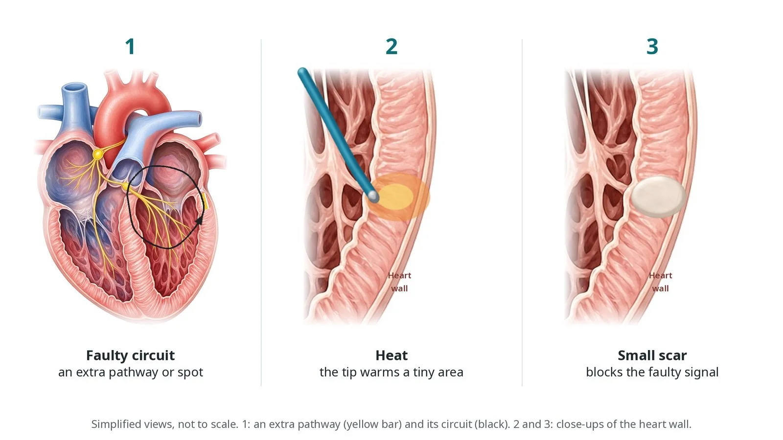
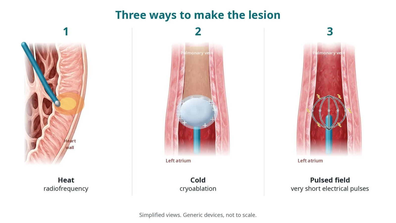
What it is: Catheters from the groin vein record the heart's electrical signals (an EP study) and find the cause of a fast rhythm. A small area is then ablated, usually with heat (radiofrequency), sometimes with cold (cryo), to stop it.
Who it is for: People with supraventricular tachycardia (SVT), such as AVNRT or an accessory pathway (including WPW), or with typical atrial flutter, who want a cure or whose medicines do not help.
Main risks: Bruising at the groin; rarely, damage to the heart's normal wiring needing a pacemaker (mainly with AVNRT and pathways near the AV node), fluid around the heart, or the rhythm coming back.
Usually: Local anaesthetic with light sedation, sometimes a general anaesthetic, about 1 to 3 hours, often home the same day.
On this page: What is SVT and atrial flutter ablation? · Who is it for? · When is it done? · Step by step · Recovery · Risks · Kit list · First time in the lab? · FAQ
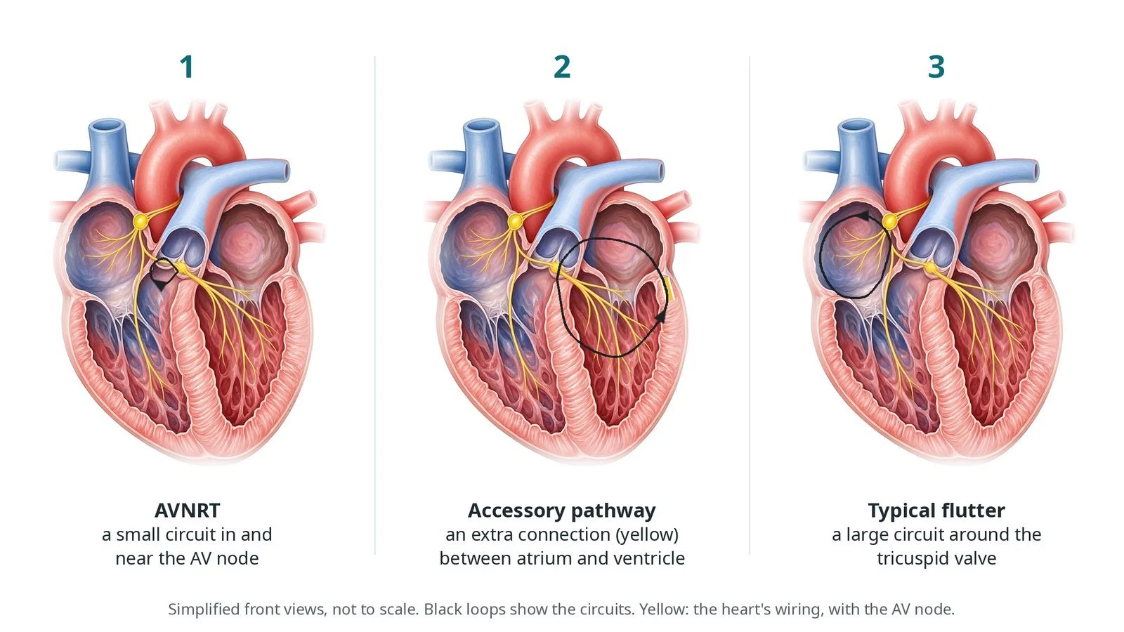
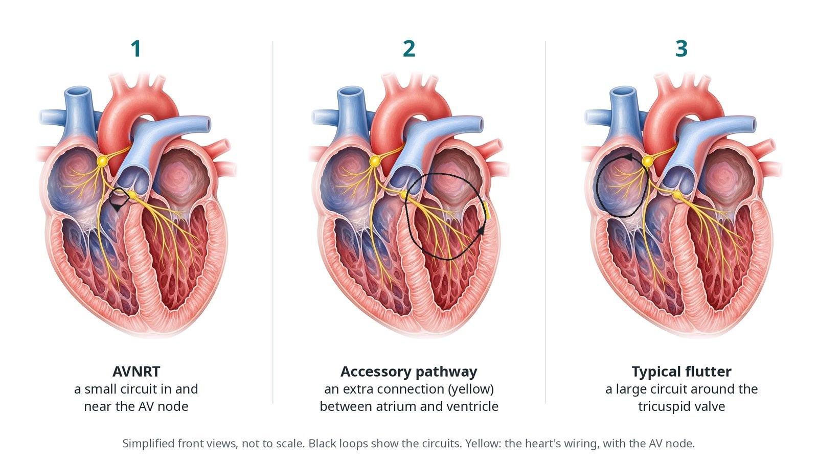
(1) AVNRT: a small circuit in and near the AV node. (2) Accessory pathway: an extra connection between atrium and ventricle. (3) Typical flutter: a large circuit around the tricuspid valve.
What is SVT and atrial flutter ablation?
Supraventricular tachycardia (SVT) means a fast rhythm that starts above the ventricles. Most SVT treated by ablation is caused by a circuit: a loop the signal goes round and round.
AVNRT (AV nodal re-entrant tachycardia) is the most common. The AV node has two inputs, a fast pathway and a slow pathway, and the signal can loop between them. The slow pathway lies low in the right atrium, between the opening of the coronary sinus and the tricuspid valve. Ablating it stops the loop and leaves the normal fast pathway working.
An accessory pathway is an extra strand of muscle across a valve ring, linking an atrium directly to a ventricle. When it shows on the resting ECG as a delta wave, this is the Wolff-Parkinson-White (WPW) pattern. The pathway is found by mapping the earliest signal along the valve ring and ablated there. Left-sided pathways are reached across the atrial septum or backwards through the aorta.
Typical atrial flutter is a large circuit round the tricuspid valve in the right atrium. It must pass through a narrow strip between the tricuspid valve and the inferior vena cava: the cavotricuspid isthmus (CTI). A line of ablation across the isthmus breaks the circuit. Success is shown when signals can no longer cross the line in either direction (bidirectional block).
New to the acronyms? See the Cath Lab Abbreviations A–Z.
Who is it for?
It may be considered for people with:
AVNRT or an accessory pathway causing symptoms; ablation is often offered as a first choice
WPW with a pathway that could be dangerous, or after fast AF with WPW
typical atrial flutter, especially if it keeps coming back
The ESC SVT guidelines support catheter ablation as first-line treatment for many of these rhythms. The decision is shared with you.
The decision is made by your heart team, who will talk through the reasons, the alternatives and the risks with you.
When is it done?
It is usually planned.
Before it, you usually have:
ECGs, ideally one recorded during the fast rhythm
an echo in some cases
advice on stopping rhythm medicines a few days before, if the team needs to start the rhythm in the lab
advice on blood thinners, especially for flutter, as per centre protocol
gives you a chance to ask questions and give consent
arranges blood tests, including kidney function and a blood count, and an ECG
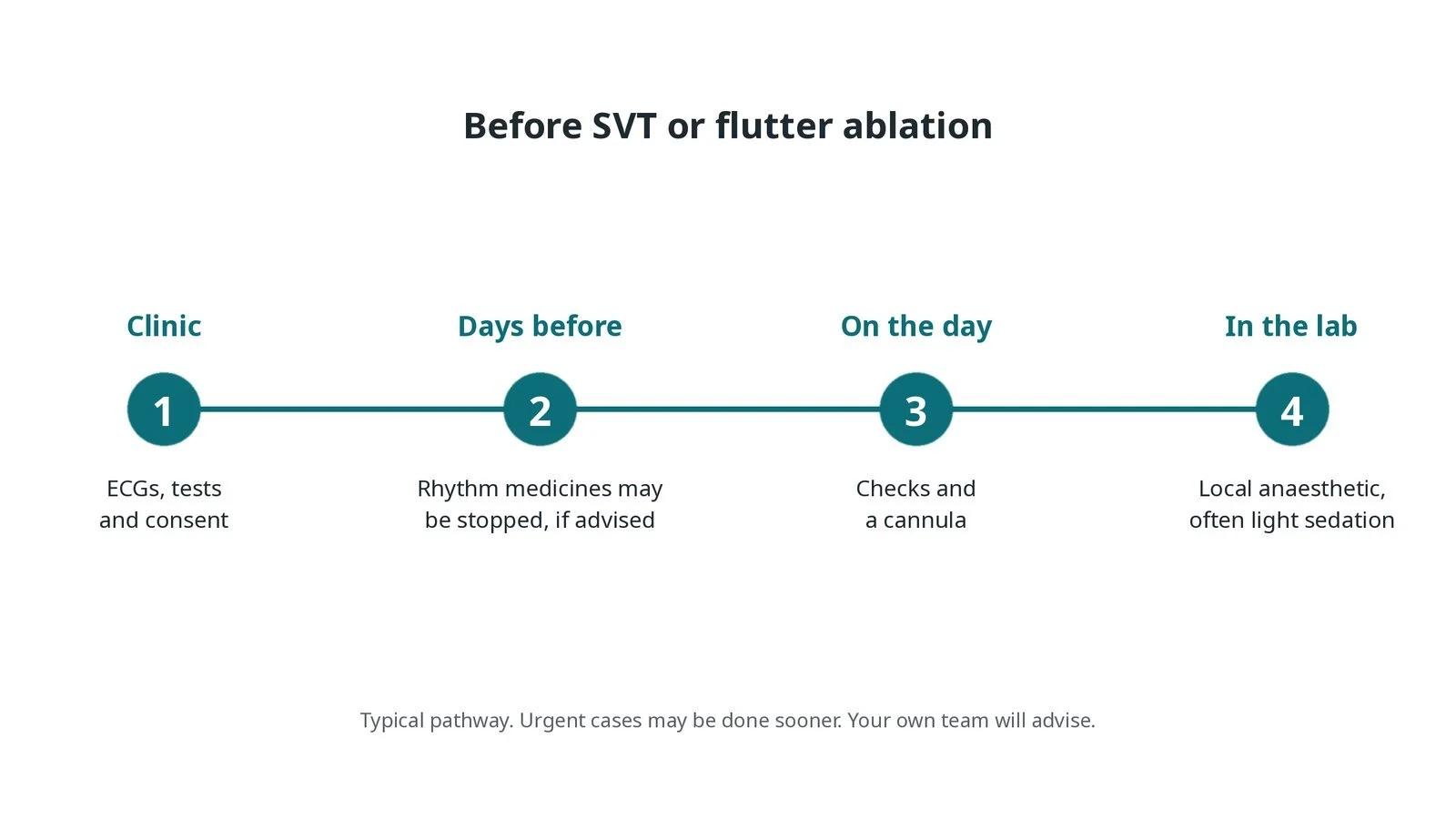
Before SVT or flutter ablation: ECGs, tests and consent; rhythm medicines may be stopped if advised; checks on the day; local anaesthetic and often light sedation.
Step by step
1. Getting ready
You lie on the X-ray table with ECG, blood pressure and oxygen monitoring, and defibrillator pads, as per centre protocol. Local anaesthetic is used in the groin, often with light sedation, because deep sedation can stop the rhythm from being started. A general anaesthetic is used in some centres and for children.
2. Access
Two to four sheaths go into the femoral vein under ultrasound; sizes per device and operator. Diagnostic catheters are placed in the high right atrium, at the His bundle, in the right ventricle and in the coronary sinus. For a left-sided pathway the catheter crosses the atrial septum (transseptal) or goes backwards through the aorta from the femoral artery.
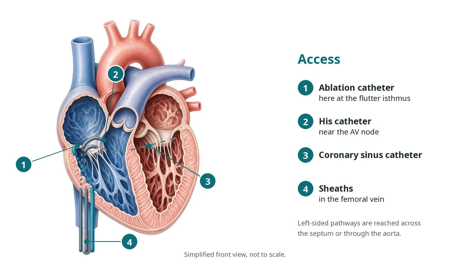
Access: (1) ablation catheter, here at the flutter isthmus; (2) His catheter near the AV node; (3) coronary sinus catheter; (4) sheaths in the femoral vein.
3. EP study and ablation
A. EP study. Pacing and timed extra beats start the fast rhythm, and its signals show which kind of SVT it is. A drug such as isoprenaline may help, per operator.
B. Target. For AVNRT, the slow pathway, low in the right atrium near the coronary sinus opening. For an accessory pathway, the point on the valve ring with the earliest signal. For typical flutter, the cavotricuspid isthmus.
C. Ablate. Radiofrequency energy, or cryo for some targets close to the AV node, is delivered, per operator. For flutter, a line of lesions runs from the tricuspid valve to the inferior vena cava.
D. Check. Pacing tries to start the rhythm again. For flutter, pacing on each side of the line shows bidirectional block. The team often waits and re-tests before finishing.
Watch: PR prolonging or AV block during energy near the AV node: say "stop" clearly; the operator stops energy straight away.
4. Finishing
The catheters and sheaths are removed. Pressure is held on the groin, or a stitch is used, per operator. You rest in bed for a short time.
5. Before going home
An ECG checks the rhythm and, after AVNRT or pathway ablation, that normal conduction is intact. Most people go home the same day.
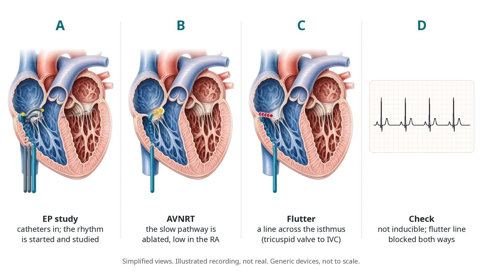
A: EP study: the rhythm is started and studied. B: AVNRT: the slow pathway is ablated low in the right atrium. C: flutter: a line across the isthmus. D: check: not inducible, flutter line blocked both ways.
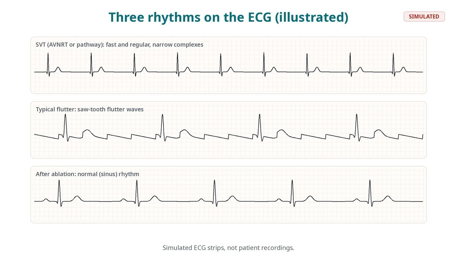
Simulated ECG strips, not patient recordings: SVT, fast and regular; typical flutter with saw-tooth waves; normal rhythm after ablation.
Recovery and aftercare
First hours: bed rest while the groin settles; checks of the groin, rhythm and blood pressure, as per centre protocol.
Going home: usually the same day; if you had sedation, someone should take you home and stay with you overnight.
Activity: avoid heavy lifting and strenuous exercise for about a week.
Medicines: your team will tell you whether to stop rhythm medicines; after flutter ablation, blood thinners may continue for a time.
Driving: DVLA rules depend on the rhythm and your symptoms; ask your team, and do not drive for at least 24 hours after sedation.
Follow-up: clinic review or a phone call, sometimes with an ECG or heart monitor.
Call 999 if you have chest pain, fainting, breathlessness, signs of a stroke, or heavy bleeding or swelling at the groin.
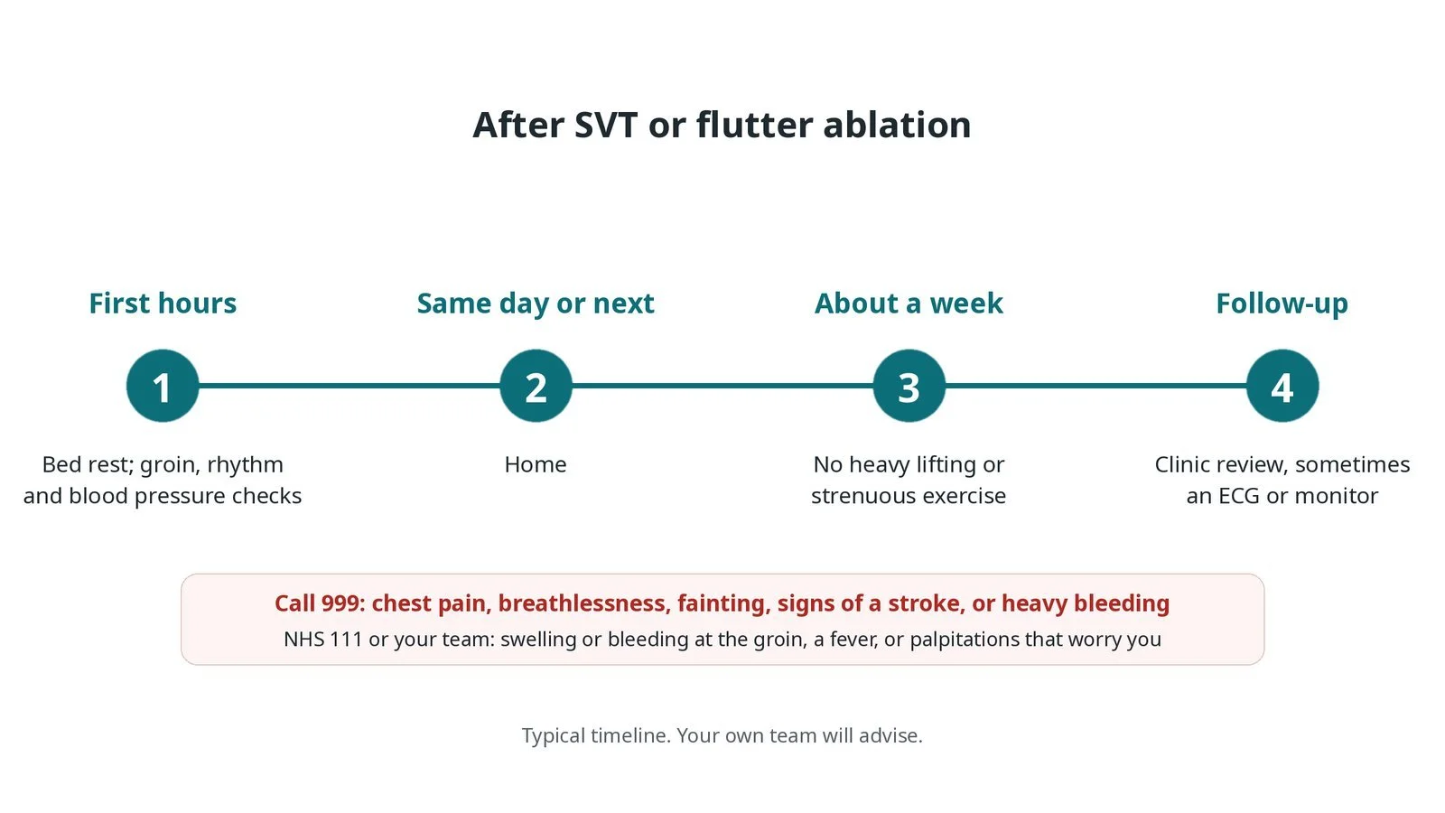
After SVT or flutter ablation: (1) bed rest and checks; (2) home the same day or next morning; (3) no heavy lifting for about a week; (4) clinic follow-up.
Risks and complications
Your team will explain the risks that apply to you. Possible problems include:
Bruising or bleeding at the groin
Heart block needing a pacemaker (rare): Mainly with AVNRT or pathways close to the AV node.
Fluid or bleeding around the heart (tamponade) (rare)
Stroke or a clot (rare): Mainly with left-sided procedures.
Damage to a vein or artery in the groin (uncommon)
The rhythm coming back, sometimes needing a repeat procedure (uncommon): Atrial fibrillation can also develop later after flutter ablation.
Cath lab kit list (printable)
Barts example kit list
An example from one UK centre, written generically. Every lab has its own protocol, and sizes and doses vary.
femoral venous sheaths (two to four), sizes per device and operator
ultrasound for vein access
diagnostic EP catheters: high right atrium, His, right ventricle and coronary sinus
steerable ablation catheter (radiofrequency or cryo), per operator
radiofrequency or cryo console and grounding patch
recording system and stimulator
3D mapping system, if used, per operator
isoprenaline and other drugs to start the rhythm, as per centre protocol
transseptal kit, for left-sided pathways
defibrillator pads and defibrillator
Procedural steps (Barts example)
Getting ready
Access
EP study and ablation
Finishing
Before going home
First time in the cath lab?
Learn the catheter positions. High right atrium, His, right ventricle and coronary sinus: know which signal is which on the recording screen.
Keep sedation light. The rhythm often needs to be started on purpose, and deep sedation can prevent it.
Watch the PR interval near the AV node. Heart block is rare, but it is the main risk with AVNRT; speak up early.
Expect the rhythm. Starting the patient's SVT is planned. Warn them they may feel their usual palpitations.
Ask the operator how much sedation they want: too much can make the rhythm impossible to start.
During slow pathway ablation, watch the PR interval and the junctional beats. Call out any sign of heart block at once.
FAQ
Will I feel my palpitations in the lab?
You may. The team often starts the rhythm on purpose to study it, and stops it quickly.
How likely is a cure?
For AVNRT, accessory pathways and typical flutter, ablation usually works well. Your team will give you their own figures.
Will I need a pacemaker?
Very rarely. The risk is mainly with AVNRT and pathways near the AV node, and the team watches closely for it.
Can I stop my tablets?
Often, after a successful ablation, but only when your team says so.
Related Cath Lab 101 guides
AV node ablation (ablate and pace) (coming soon)
Sources
All checked on 12 November 2026. Each page's own date is shown where it gives one.
Brugada J, et al. 2019 ESC Guidelines for the management of patients with supraventricular tachycardia. Eur Heart J 2020.
StatPearls (NCBI Bookshelf), search: atrioventricular nodal reentrant tachycardia. ncbi.nlm.nih.gov/books
StatPearls (NCBI Bookshelf), search: atrial flutter. ncbi.nlm.nih.gov/books
Disclaimer
Education only. This page is for general education for healthcare students, cath lab staff and curious readers. It isn't medical advice and doesn't replace advice from your own doctor, heart team or local unit protocols. Equipment and medicines are described generically. No product or manufacturer is recommended. If you think someone is having a heart emergency, call 999.
Disclosure: I work for a company that makes a device used in this procedure. This page is independent education, isn't sponsored or reviewed by any company, and doesn't recommend any product.
All diagrams on this page are original illustrations created for rodnieoro.com.