Transvenous lead extraction: a Cath Lab 101 guide
Reviewed by Rodnie Oro, cath lab charge nurse · Last reviewed: November 2026 · Education only, not medical advice
Transvenous lead extraction in one minute
What it is: Lead extraction removes pacemaker or ICD leads through the veins they sit in. Leads that have been in place for a while are held by scar tissue, so special tools are used: locking stylets to grip the lead, and laser or rotating mechanical sheaths that free the scar tissue as they are passed over the lead. Snares from the groin can help.
Who it is for: People with an infected device or lead, leads that have failed or are causing problems, blocked veins needing a new lead, and some other selected reasons, as decided by an extraction team.
Main risks: Serious complications are uncommon but can be life-threatening: a tear in the superior vena cava or heart causing major bleeding, needing emergency surgery. Other risks include bleeding, tamponade, a collapsed lung, pieces of lead left behind, and infection. Your team will explain your own risk.
Usually: Under general anaesthetic in a hybrid theatre or a lab with cardiac surgery ready, about 2 to 4 hours, with a hospital stay of a few days or longer if there is infection.
On this page: What is transvenous lead extraction? · Who is it for? · When is it done? · Step by step · Recovery · Risks · Kit list · First time in the lab? · FAQ
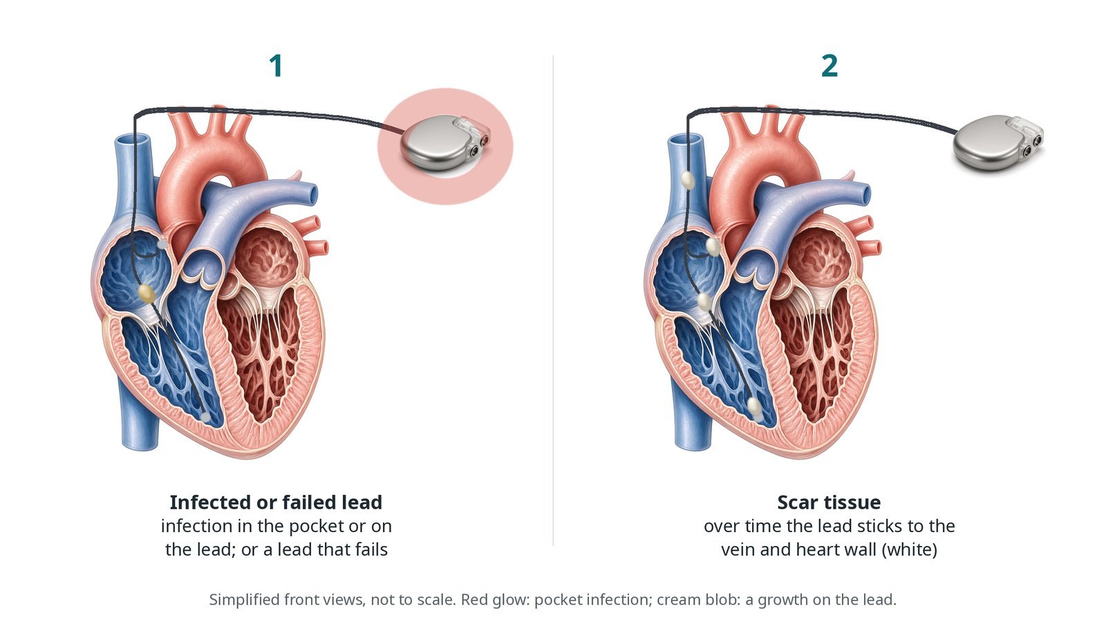
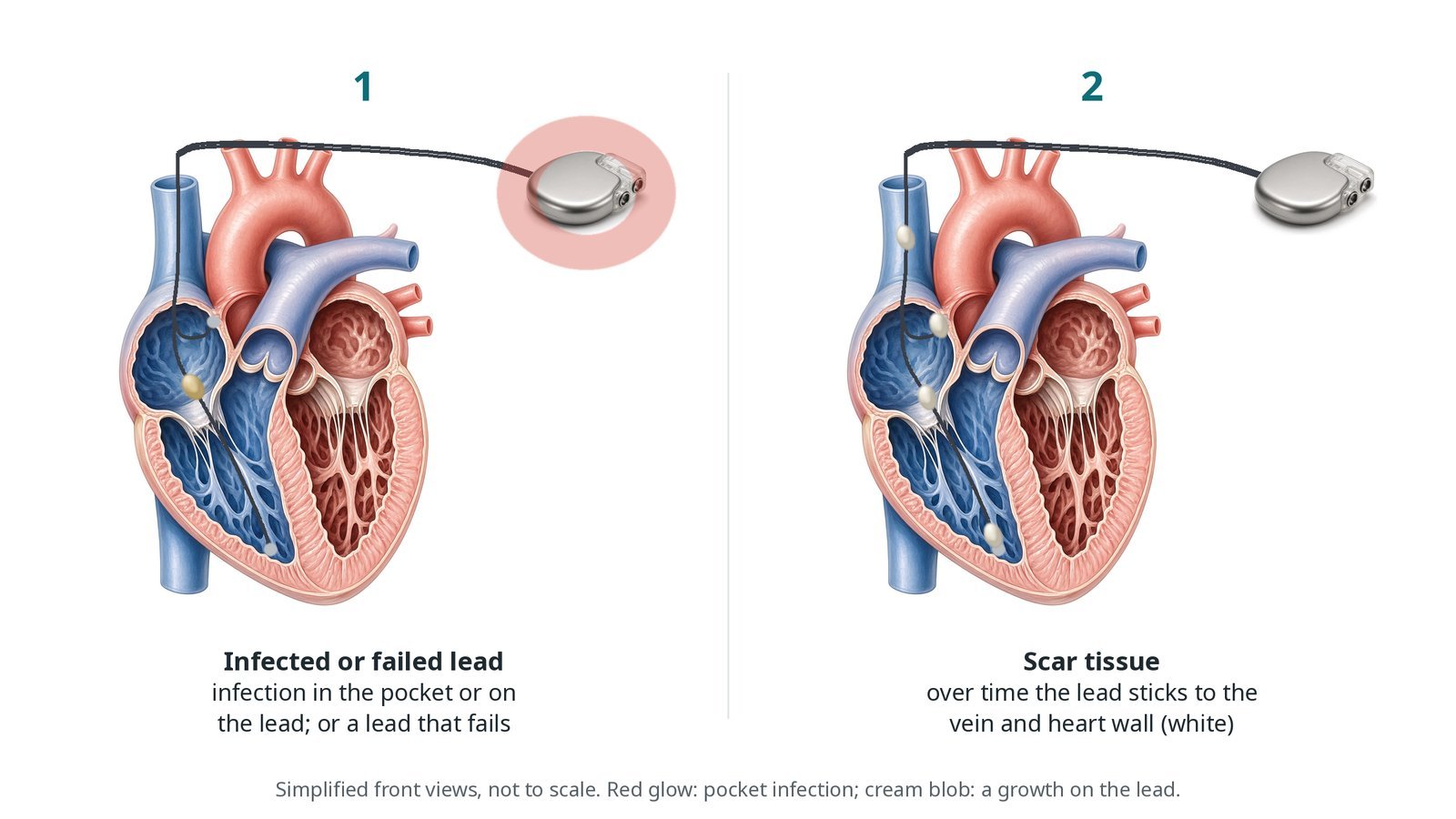
(1) Infected or failed lead: infection in the pocket or on the lead, or a lead that fails. (2) Scar tissue: over time the lead sticks to the vein and heart wall.
What is transvenous lead extraction?
Leads go from the generator pocket into a vein below the collarbone, through the brachiocephalic vein and superior vena cava (SVC), into the right atrium and right ventricle. Over months and years, scar tissue (fibrosis) grows around them, most often where the lead enters the vein, in the SVC, at the tricuspid valve and at the tip in the heart wall.
A new lead can often be pulled out with simple traction. Older leads usually need a locking stylet passed down the inside of the lead to grip it along its length, and a sheath passed over the lead to cut or laser through the scar tissue. Near the heart, the sheath is held against the wall while the lead is pulled back (counter-traction), so the heart wall is not pulled inwards.
The SVC is the most dangerous area: a tear here bleeds into the chest quickly. Teams prepare for this with surgical standby, blood cross-matched, and often a compliant balloon that can be inflated in the SVC to slow bleeding while the surgeon opens the chest.
When extraction is for infection, all the hardware is removed and antibiotics are given. A new device is placed later, on the other side or with a different system, once the infection has cleared.
New to the acronyms? See the Cath Lab Abbreviations A–Z.
Who is it for?
Decided by an extraction team, using EHRA and HRS consensus. Common reasons:
infection of the pocket, the leads or the blood (including infection on a heart valve or lead)
leads that have failed, are under advisory, or are causing problems
blocked veins when a new lead is needed
selected other reasons, such as too many leads or to allow an MRI scan
Who it may not suit: Some people who are very frail, or whose leads carry very high risk, may be managed differently, for example with long-term antibiotics. This is a careful shared decision.
The decision is made by your heart team, who will talk through the reasons, the alternatives and the risks with you.
When is it done?
Planned, or more urgently for infection.
Before it you usually have:
blood cultures and a TOE to look for infection on the leads or valves
imaging such as a chest X-ray, CT or venogram
blood tests and blood cross-matched
a plan for pacing during and after, especially if pacing-dependent
a heart team discussion and consent, including the risk of emergency surgery
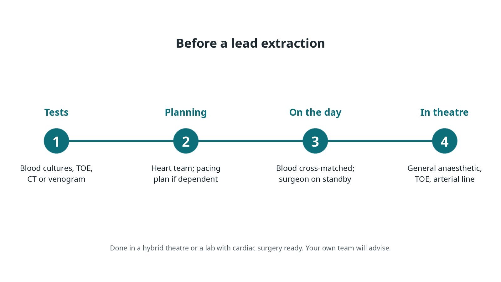
Before a lead extraction: blood cultures, TOE and imaging; heart team planning and a pacing plan; blood cross-matched and a surgeon on standby; general anaesthetic, TOE and arterial line.
Step by step
1. Getting ready
You have a general anaesthetic. An arterial line, large drips, a urinary catheter and a TOE probe are placed. Defibrillator pads go on, and the chest and groins are prepared so surgery can start immediately if needed. A temporary pacing wire is placed if you are pacing-dependent.
2. Access
The generator pocket is opened, the box removed, and the leads freed back to where they enter the vein. A femoral venous sheath is often placed in the groin for snares, a temporary wire or a guidewire for the SVC balloon.
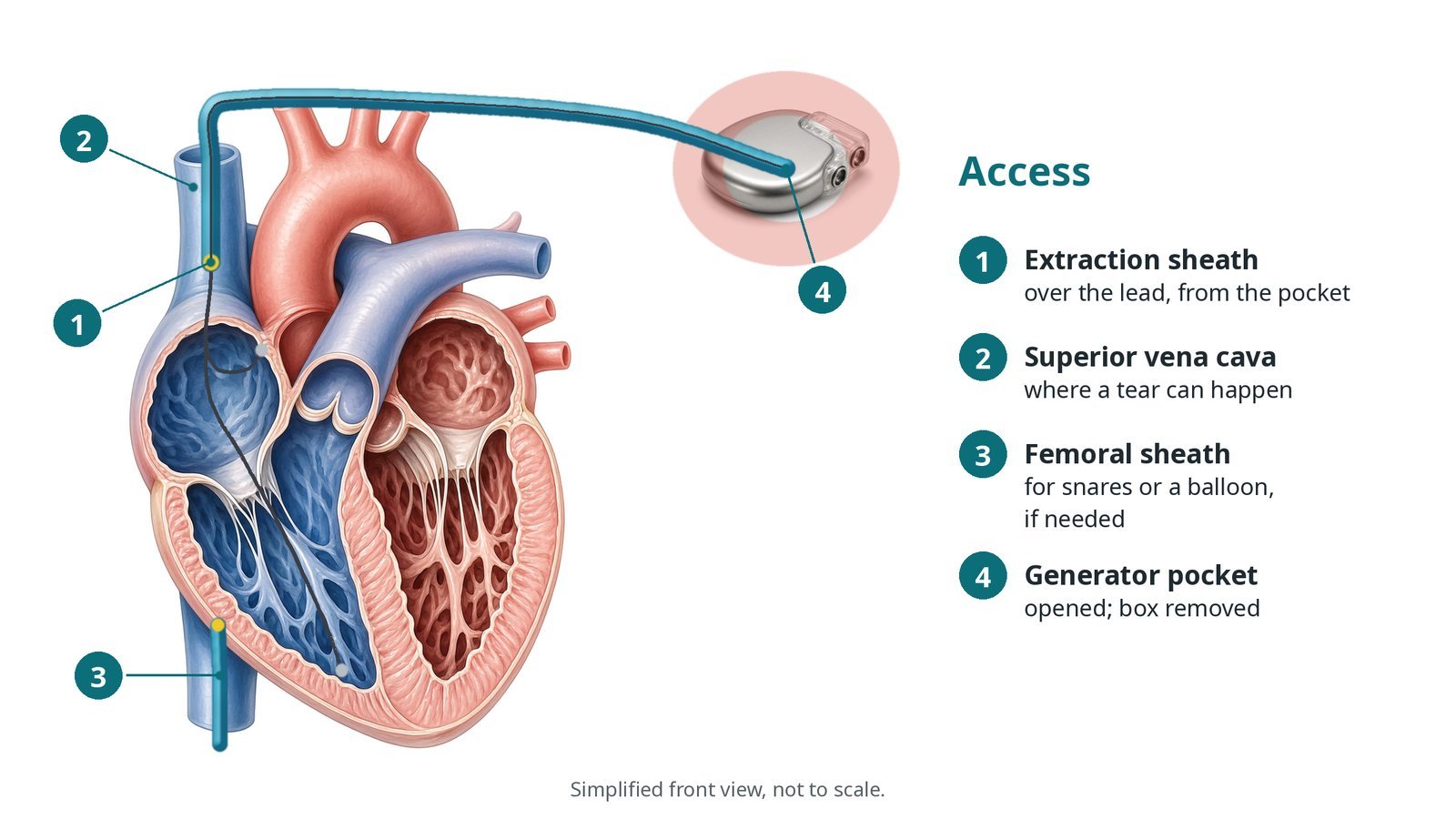
Access: (1) extraction sheath over the lead, from the pocket; (2) superior vena cava, where a tear can happen; (3) femoral sheath for snares or a balloon if needed; (4) generator pocket opened.
3. Removing the lead
A. Lock. The lead is cut, a locking stylet is passed down its inside and locked, and a tie is often added to the outside. Gentle traction is tried.
B. Sheath. A laser or rotating mechanical sheath is passed over the lead, freeing scar tissue as it advances along the vein and SVC.
C. Counter-traction. At the heart, the sheath is held against the wall while the lead is pulled back, so the heart wall is not pulled in.
D. Out. The lead and sheath are removed, and TOE checks for fluid around the heart. If needed, snares from the groin catch the lead or pieces of it.
Watch: A sudden fall in blood pressure, a new effusion on TOE or blood in the chest drain means a possible SVC or heart tear. Call it out at once: the SVC balloon is inflated, fluids and blood are given, and the surgeon opens the chest.
4. Finishing
The pocket is cleaned, and infected tissue removed. It is closed, sometimes with a drain. If new leads are needed and there is no infection, they may be placed at the same time; if there is infection, a new device is usually delayed.
5. After the extraction
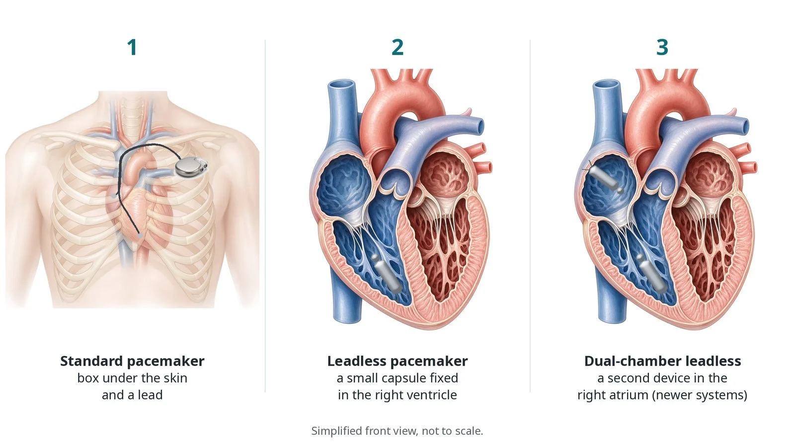
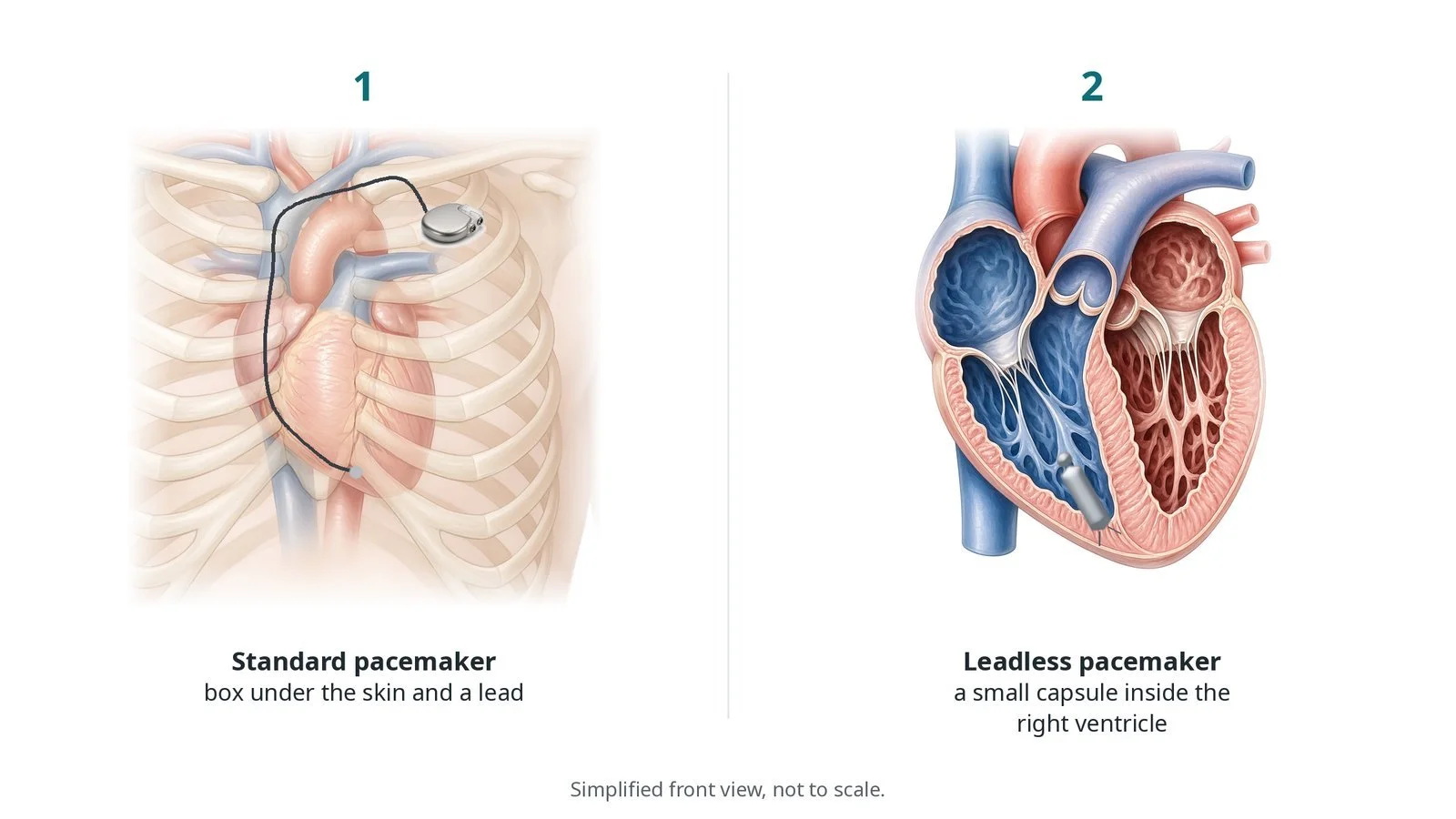
You go to a recovery area, ward or intensive care. Echo and a chest X-ray are done. If extraction was for infection, antibiotics continue, and a new device is placed once blood cultures are clear for long enough, usually on the other side or with a leadless or subcutaneous device, as decided by the team.
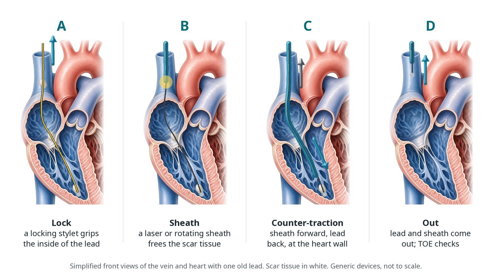
A: a locking stylet grips the inside of the lead. B: a laser or rotating sheath frees the scar tissue. C: counter-traction at the heart wall. D: the lead and sheath come out and TOE checks for fluid.
Recovery and aftercare
First hours: recovery or intensive care with checks of blood pressure, rhythm, chest drain if present, and wounds.
Hospital stay: a few days, longer if antibiotics are needed for infection.
Medicines: antibiotics if there was infection, for the time advised by the infection team; blood thinners restart as advised.
New device: if one is needed, it is usually placed once infection has cleared, timed by the team.
Driving: rules depend on your device and why it was removed or replaced. Your team will advise on DVLA rules.
Follow-up: device clinic, wound checks and, after infection, a review with the infection team.
Call 999 if you have chest pain, sudden breathlessness, fainting or coughing up blood.
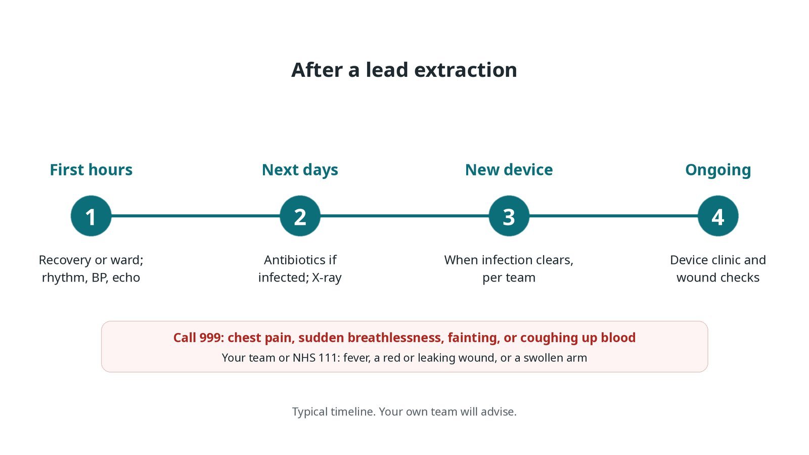
After a lead extraction: (1) recovery with rhythm, blood pressure and echo checks; (2) antibiotics if infected and a chest X-ray; (3) a new device when infection clears, timed by the team; (4) device clinic and wound checks.
Risks and complications
Your team will explain the risks that apply to you. Possible problems include:
Tear of the superior vena cava or heart with major bleeding (rare): May need emergency surgery; can be life-threatening.
Bleeding around the heart (tamponade) (uncommon)
Bleeding or a bruise at the pocket
Collapsed lung (pneumothorax) (uncommon)
Part of the lead left behind (uncommon)
Damage to the tricuspid valve (uncommon)
Need for blood transfusion
Cath lab kit list (printable)
Barts example kit list
An example from one UK centre, written generically. Every lab has its own protocol, and sizes and doses vary.
general anaesthetic, arterial line and TOE probe
locking stylets and lead extension or ties
laser sheaths and laser console, or mechanical rotational sheaths, per operator
outer telescoping sheaths
femoral sheath, snares and retrieval tools
compliant balloon for the superior vena cava, with its guidewire, prepared
temporary pacing wire and box
pericardiocentesis kit and chest drain
cardiac surgery set, perfusion and cross-matched blood immediately available
cell salvage, as per centre protocol
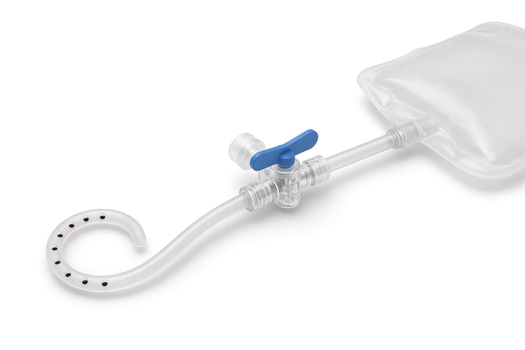
Pericardiocentesis kit.
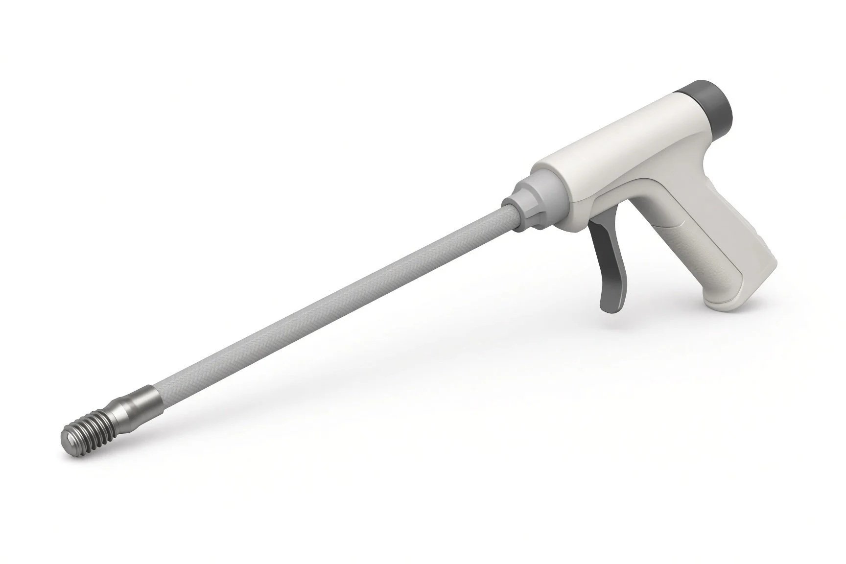
Mechanical rotational sheath.
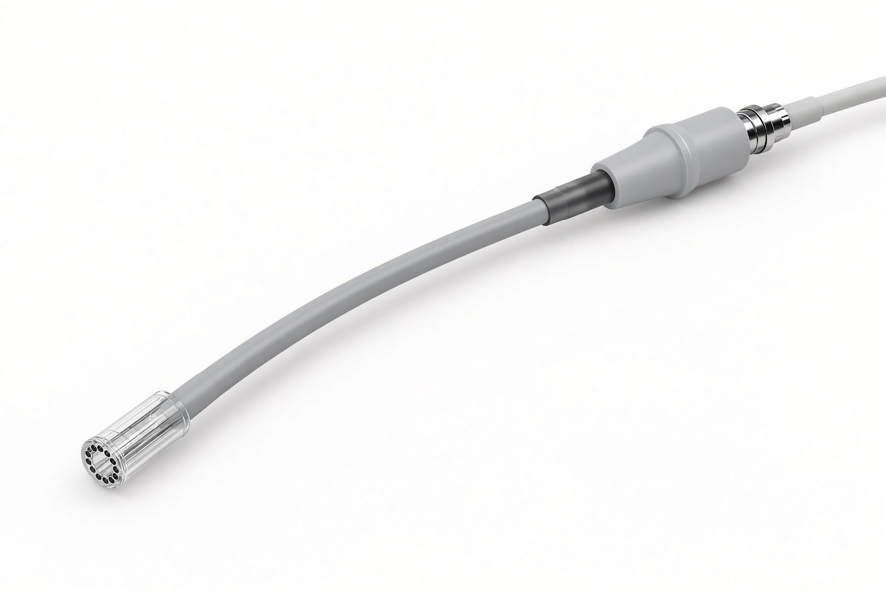
Laser extraction sheath.
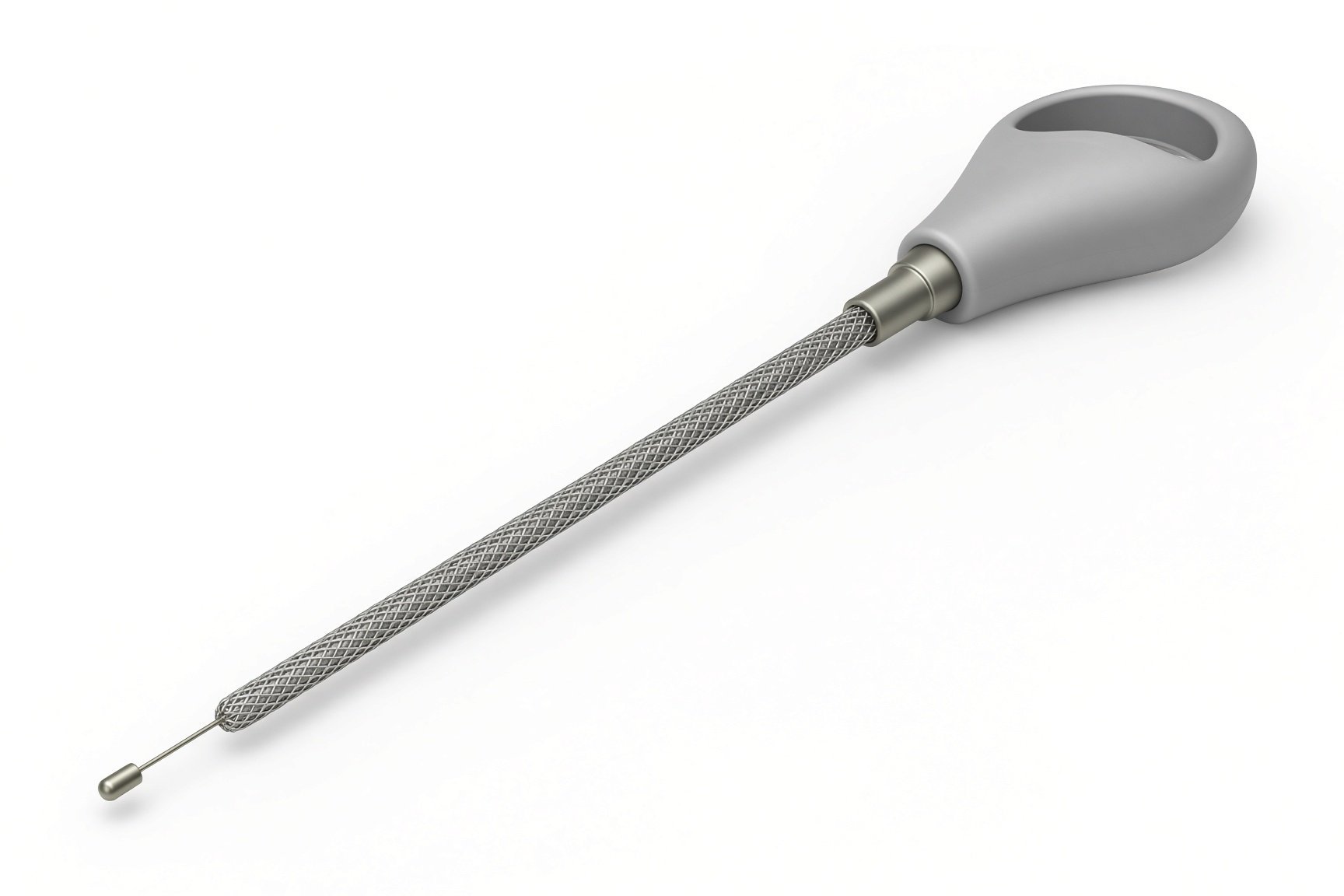
Locking stylets.
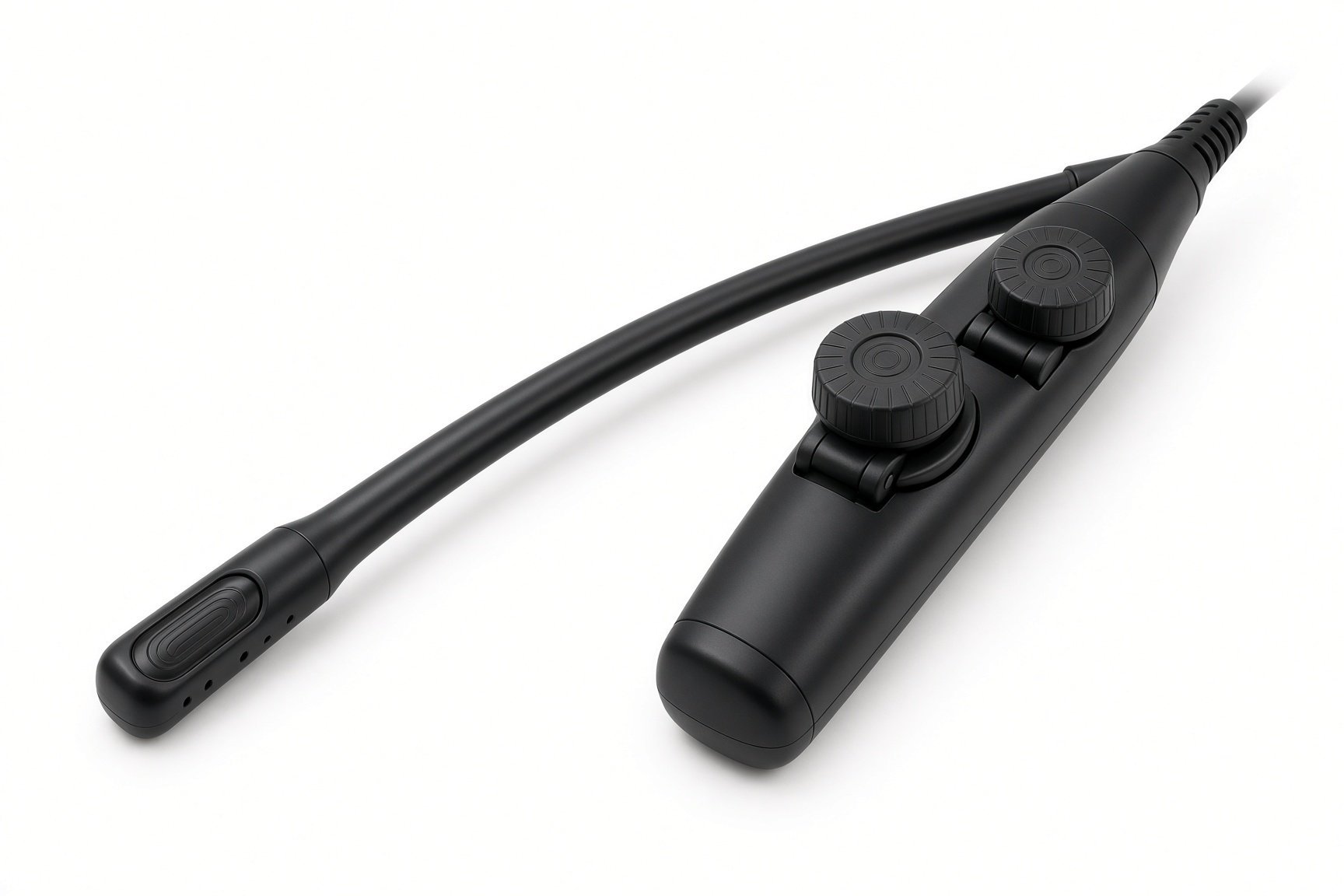
TOE probe.
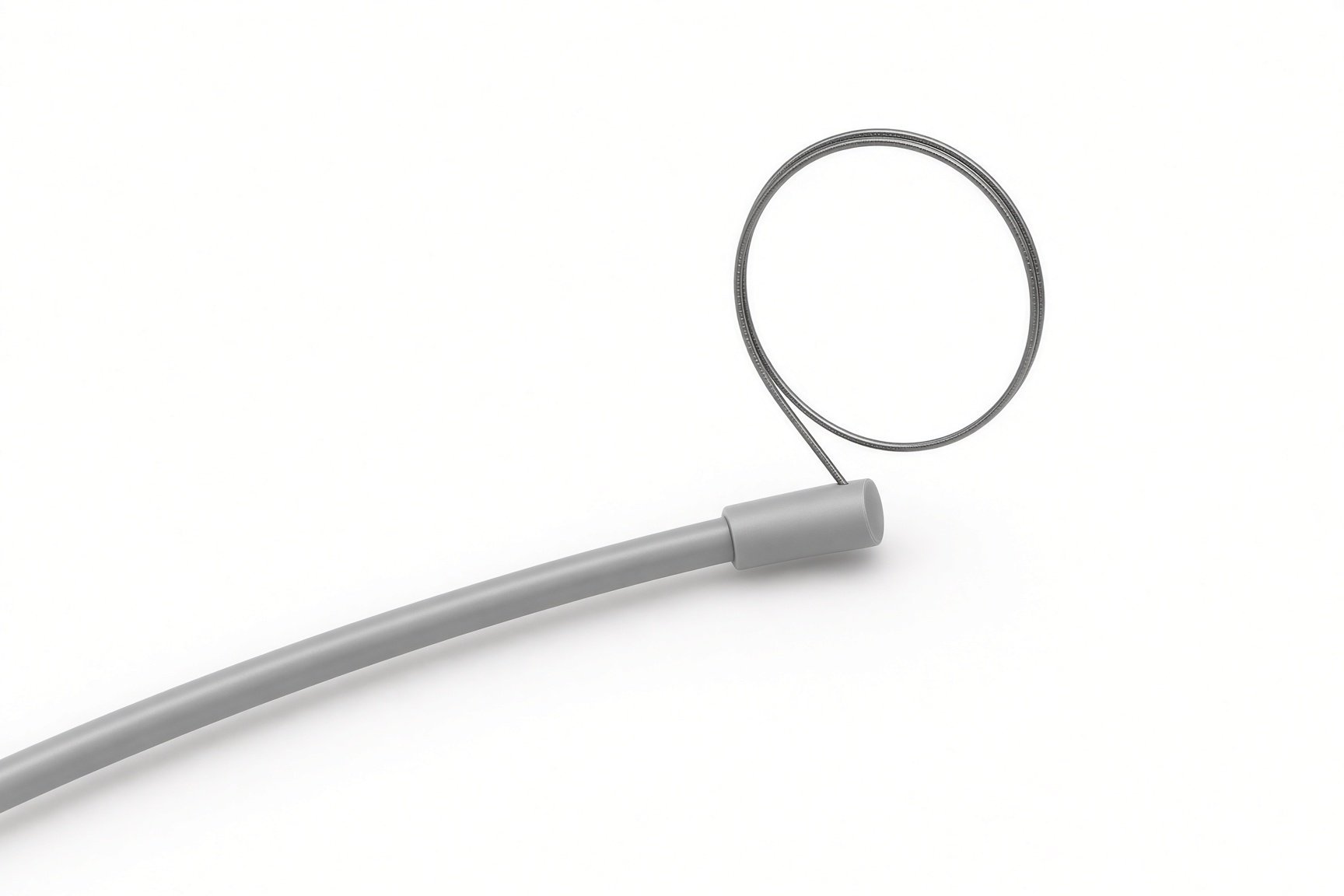
Femoral snares.
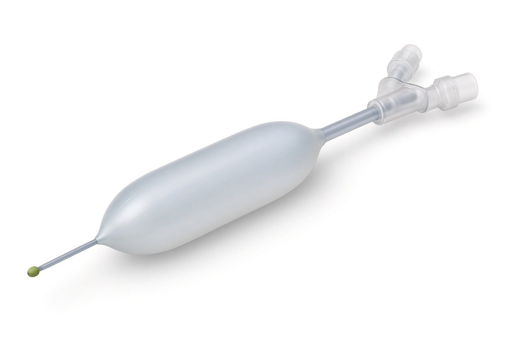
SVC occlusion balloon.
Pericardiocentesis kit.
Procedural steps (Barts example)
Getting ready
Access
Removing the lead
Finishing
After the extraction
First time in the cath lab?
Everyone knows the emergency plan. An SVC tear is rare but fast. Know where the balloon is and what your role is.
Watch the arterial line. The first sign of trouble is usually a falling pressure. Say it out loud.
Track every piece. Count lead parts, stylets and tools out of the body; fragments matter.
Infection cases are different. The pocket may be messy, and specimens for the lab are needed. Ask what to send.
Do a team brief that covers the SVC tear plan: who calls it, where the balloon is, and who opens the chest.
Check blood is in the theatre fridge or immediately available, as per centre protocol.
Call out blood pressure trends every few minutes during sheath advancement.
Keep laser safety rules (eye protection, signage) if a laser is used.
FAQ
Why can't the leads just be pulled out?
Scar tissue holds older leads to the vein and heart. Pulling hard could tear them, so special tools free the lead safely.
Why do I need surgery on standby?
In rare cases a vein or the heart tears and bleeds. A surgeon must be ready to repair it at once.
Will I be without a pacemaker?
If you need pacing, a temporary pacing system is used until a new device is fitted.
Why wait for a new device after an infection?
Putting a new device in before the infection has cleared could infect it too.
Related Cath Lab 101 guides
Pacemaker and ICD generator change and upgrade (coming soon)
Leadless pacemaker implant (coming soon)
Sources
All checked on 12 November 2026. Each page's own date is shown where it gives one.
Bongiorni MG, et al. 2018 EHRA expert consensus statement on lead extraction. Europace 2018;20(7):1217
Kusumoto FM, et al. 2017 HRS expert consensus statement on cardiovascular implantable electronic device lead management and extraction. Heart Rhythm 2017;14(12):e503–e551
Blomström-Lundqvist C, et al. European Heart Rhythm Association (EHRA) international consensus document on how to prevent, diagnose, and treat cardiac implantable electronic device infections. Europace 2020;22(4):515–549
StatPearls (NCBI Bookshelf), search: pacemaker lead extraction. ncbi.nlm.nih.gov/books
Disclaimer
Education only. This page is for general education for healthcare students, cath lab staff and curious readers. It isn't medical advice and doesn't replace advice from your own doctor, heart team or local unit protocols. Equipment and medicines are described generically. No product or manufacturer is recommended. If you think someone is having a heart emergency, call 999.
Disclosure: I work in the medical device industry. This page is independent education and isn't sponsored or reviewed by any company.
All diagrams on this page are original illustrations created for rodnieoro.com.