Left ventricular (endomyocardial) biopsy: a Cath Lab 101 guide
Reviewed by Rodnie Oro, cath lab charge nurse · Last reviewed: October 2026 · Education only, not medical advice
LV biopsy in one minute
What it is: an endomyocardial biopsy takes a few tiny pieces of heart muscle (each a few millimetres) from inside the heart using a catheter with small jaws (a bioptome). In an LV biopsy, samples come from the left ventricle, reached through an artery in the wrist or groin.
Who it is for: people with unexplained heart muscle disease, where the result could change treatment, for example suspected myocarditis, some cardiomyopathies, infiltrative conditions or heart transplant monitoring, as decided by a specialist.
Main risks: fluid around the heart from a small perforation (tamponade), heart rhythm problems, bleeding at the access site and, rarely, stroke or valve damage. Your team will explain your own risk.
Usually: under local anaesthetic with or without light sedation, about 1 hour, often home the same or next day.
On this page: What is an LV biopsy? · Who is it for? · When is it done? · Step by step · Recovery · Risks · Kit list · First time in the lab? · FAQ
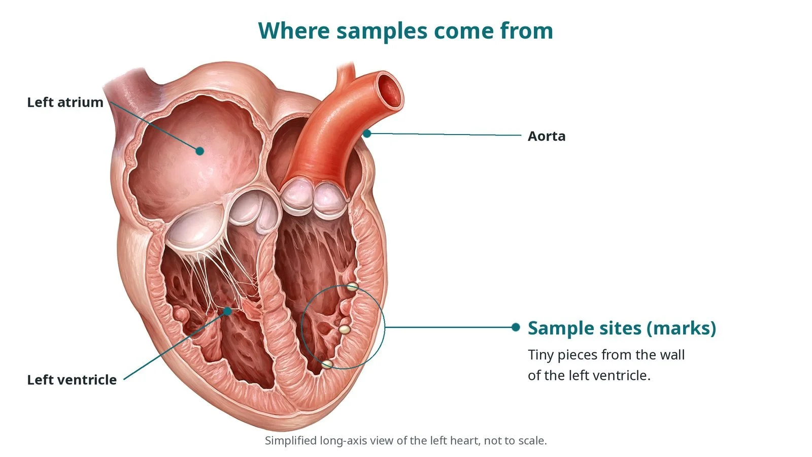
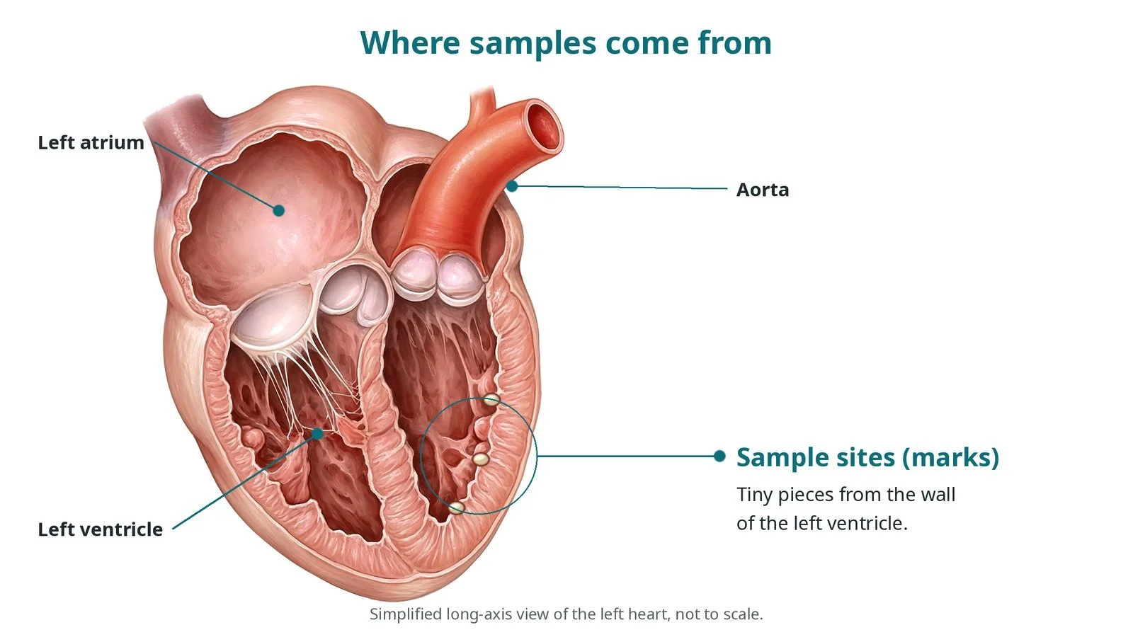
Samples are taken from the wall of the left ventricle (marks).
What is an LV biopsy?
Some heart muscle conditions cannot be diagnosed by scans and blood tests alone. Looking at the heart muscle under a microscope can show inflammation, abnormal deposits (such as amyloid), scarring, infection or transplant rejection.
An endomyocardial biopsy (EMB) takes several small samples from the inside lining and muscle of the heart. Biopsies can be taken from the right ventricle (through a vein) or the left ventricle (through an artery). The LV route is chosen when the disease mainly affects the left ventricle, guided by MRI findings or local practice.
Samples are sent for:
histology: looking at the tissue under the microscope, preserved in fixative
virology or other tests: looking for viruses or other causes, in a separate pot as per laboratory instructions
sometimes electron microscopy or special stains
New to the acronyms? See the Cath Lab Abbreviations A–Z.
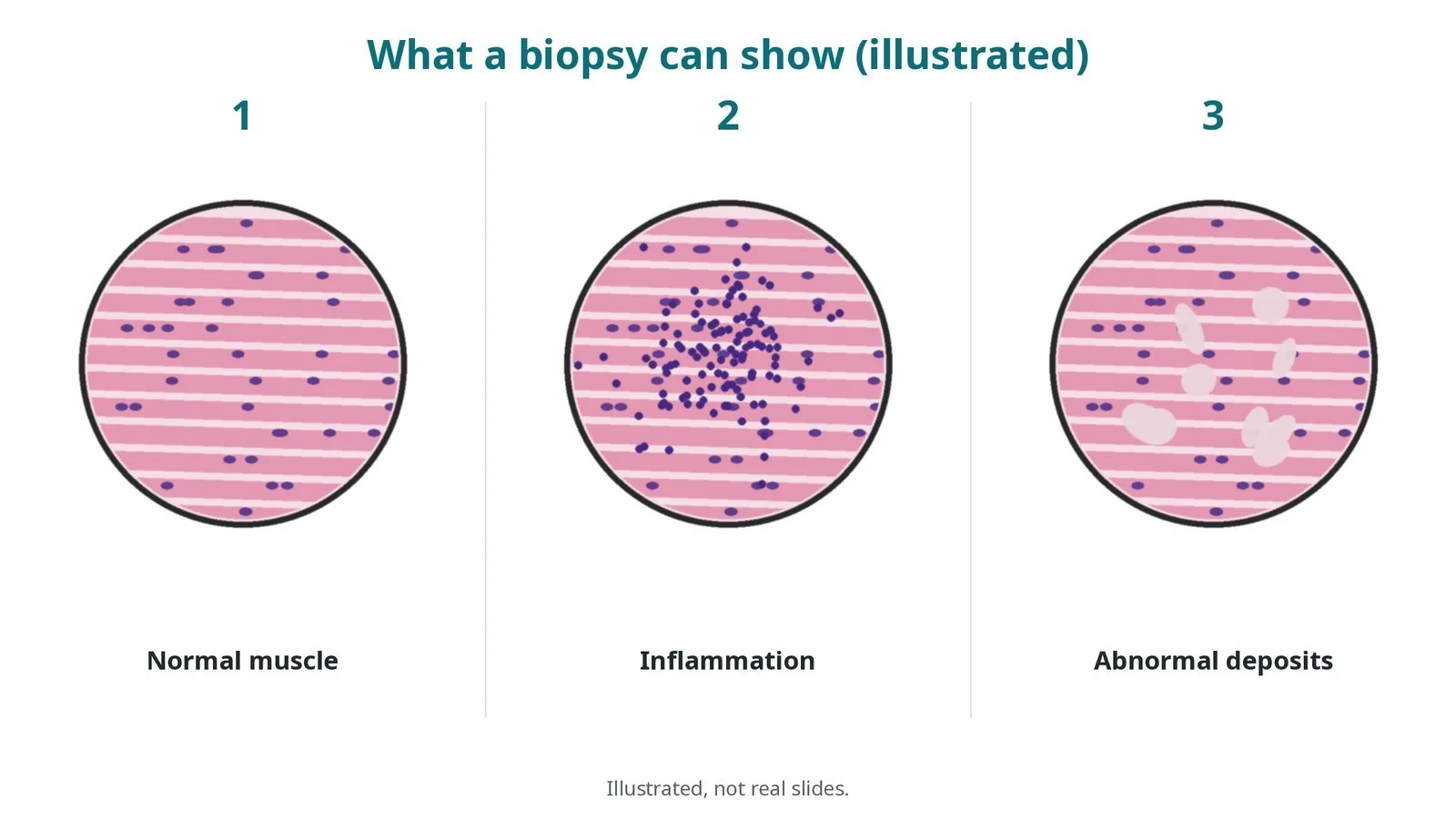
Illustrated, not real slides. A biopsy can show (1) normal muscle, (2) inflammation or (3) abnormal deposits.
Who is it for?
The ESC 2023 cardiomyopathy guidelines say endomyocardial biopsy should be considered when the result is likely to help diagnosis or treatment and other tests have not given the answer, for example in suspected myocarditis with rapid deterioration, suspected infiltrative or storage diseases, or unexplained cardiomyopathy. It is also used to monitor for rejection after heart transplant. Decisions are made by specialist teams.
When is it done?
It can be planned or urgent. Before it, the team usually arranges:
echo and often cardiac MRI, to target the area of disease
an ECG and blood tests, including clotting
a review of blood thinners, as per centre protocol
checks that the left ventricle has no clot
Step by step
1. Getting ready
You lie on the X-ray table. The procedure is done under local anaesthetic, with light sedation if needed. ECG, blood pressure and oxygen levels are monitored, and an echo machine is available in the room.
2. Access
A sheath is placed in the radial artery (wrist) or femoral artery (groin) under ultrasound; sizes per device and operator.
Heparin is given, dose as per centre protocol.
A pigtail catheter may be used to measure pressures and outline the left ventricle.
A guide catheter is passed across the aortic valve into the left ventricle to provide a channel for the bioptome. A radio-opaque marker on the chest may help with positioning.
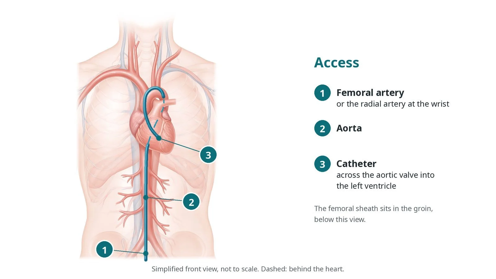
Access: (1) femoral artery, or the radial artery at the wrist; (2) aorta; (3) catheter across the aortic valve into the left ventricle.
3. Taking the samples
A. Guide in the LV. The guide catheter tip is positioned in the LV, away from the valve apparatus.
B. Bioptome in. The bioptome is passed through the guide to the wall, checked on X-ray (and sometimes echo).
C. Bite. The jaws are opened, pushed gently against the wall and closed to take a tiny piece of muscle.
D. Withdraw. The bioptome is withdrawn and the sample placed in the correct pot. This is repeated several times, per operator.
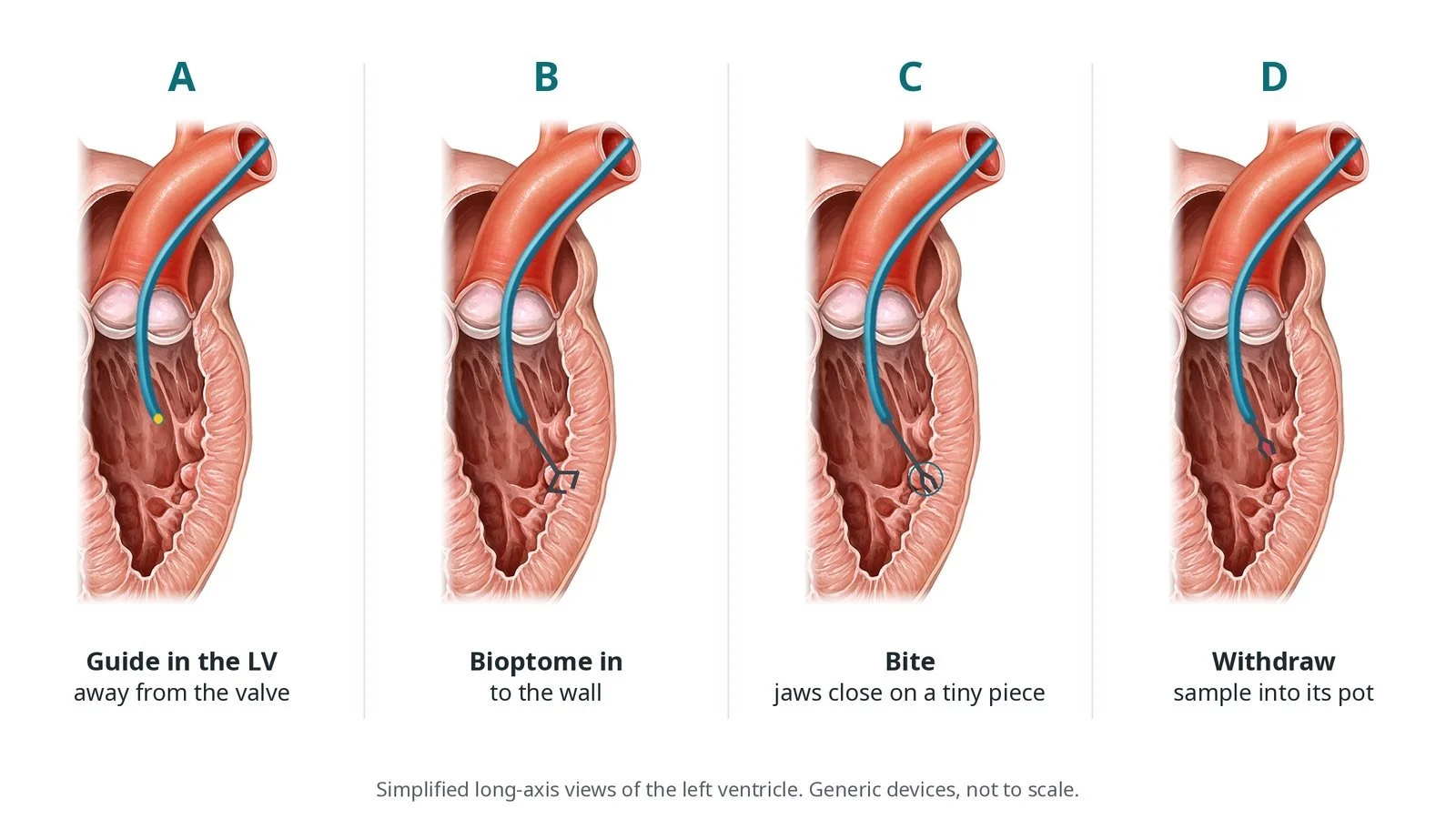
A: guide in the LV. B: bioptome in. C: bite. D: withdraw the sample.
4. Finishing
An echo checks for fluid around the heart. Catheters are removed and the artery closed with a wrist band, closure device or pressure, per operator.
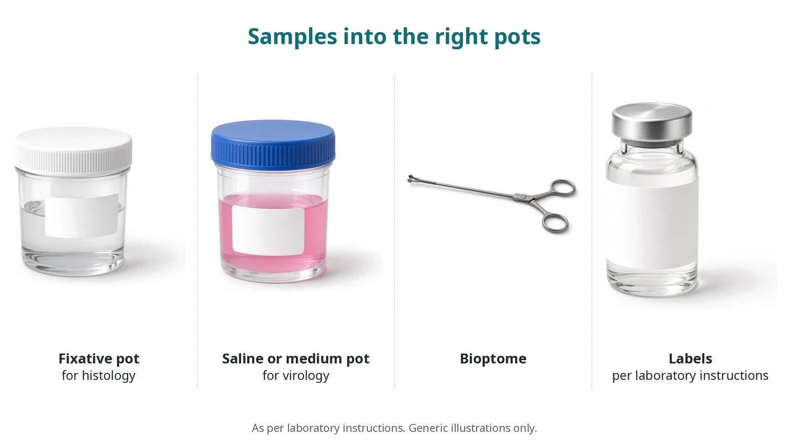
Samples go into the right pots: fixative for histology; saline or medium for virology, as per laboratory instructions.
Recovery and aftercare
First hours: bed rest with checks of the access site, heart rhythm and blood pressure, and sometimes a repeat echo, as per centre protocol.
Going home: often the same or next day.
Results: can take days to weeks, depending on the tests.
Activity: avoid heavy lifting and strenuous use of the arm or leg used for a few days, or as advised.
Driving: ask your team; driving rules depend on the underlying heart condition.
Call 999 if you have chest pain, sudden breathlessness, fainting, signs of a stroke, or heavy bleeding from the access site.
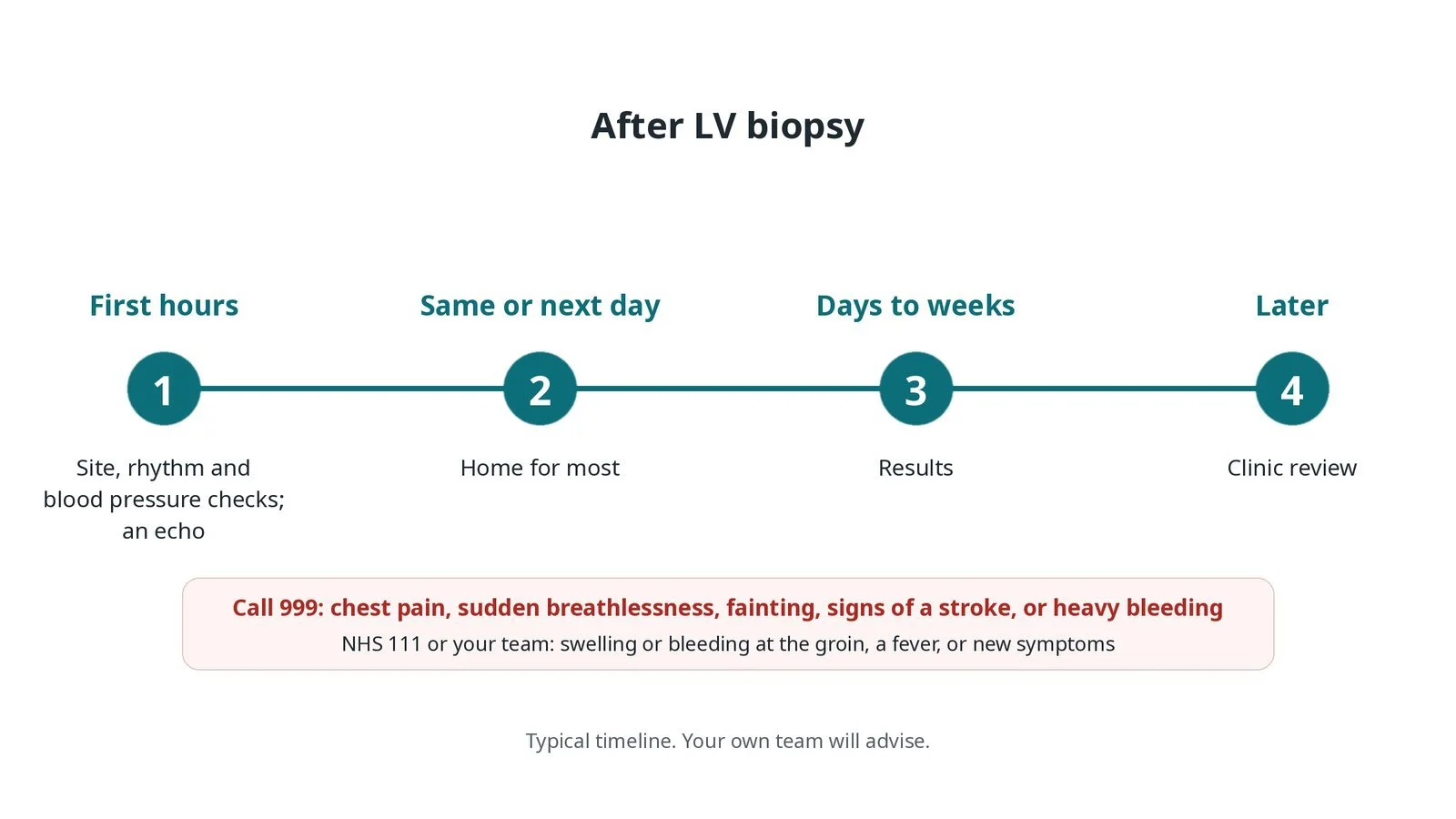
After LV biopsy: (1) checks and echo; (2) home for most; (3) results in days to weeks; (4) clinic review.
Risks and complications
Your team will discuss your own risks. Possible problems include:
a small hole in the heart wall causing fluid around the heart (tamponade), which may need draining
heart rhythm problems, usually brief
bleeding or artery injury at the access site
stroke or embolism (rare)
damage to the mitral valve apparatus (rare)
a non-diagnostic result, because disease can be patchy
Cath lab kit list (printable)
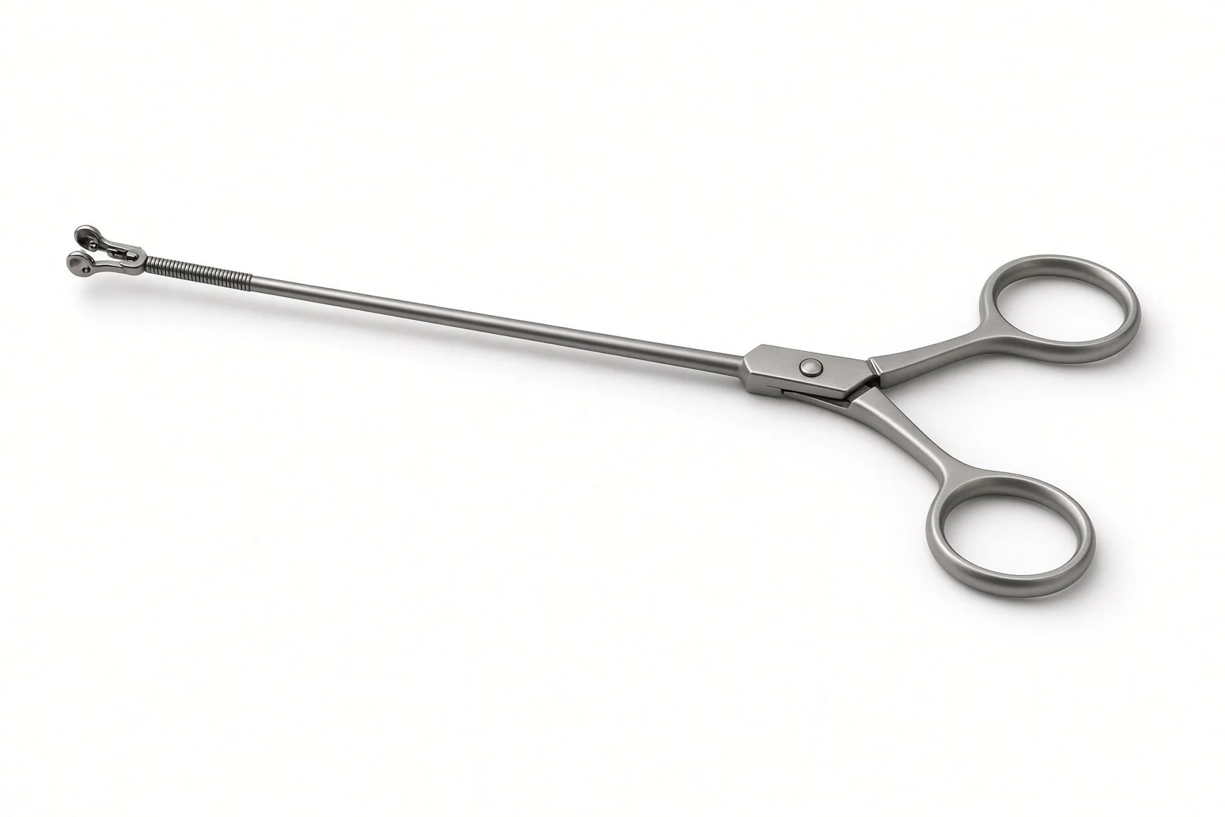
Bioptome
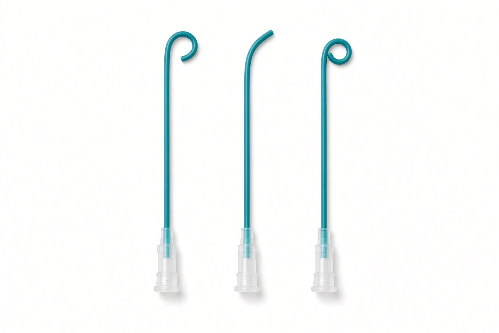
Pigtail diagnostic catheter (from the guide kit list; no published kit-page picture)
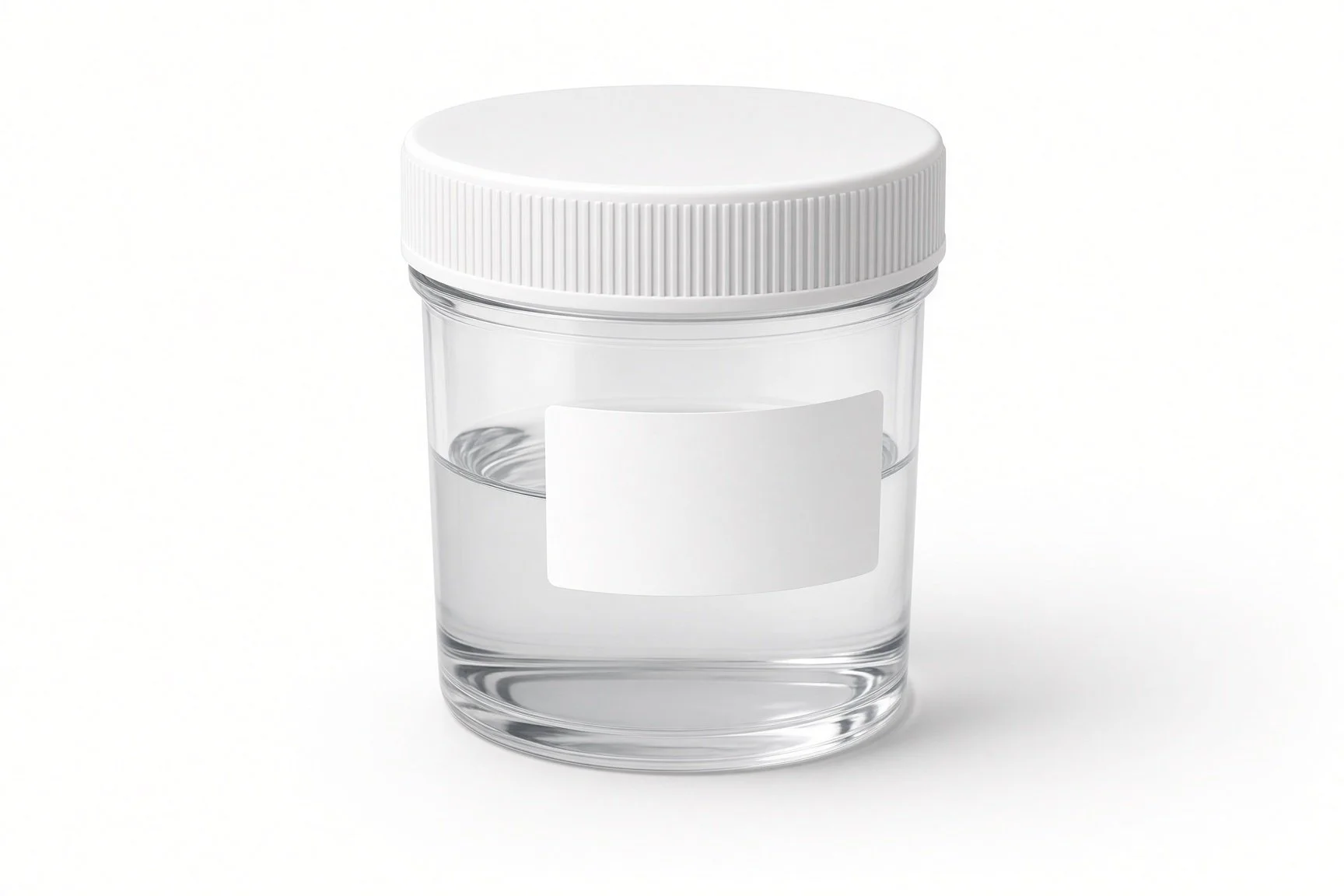
Fixative pot for histology
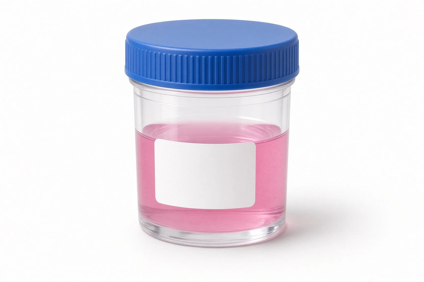
Saline or medium pot for virology
Labels per laboratory instructions
Example kit list
Written generically. Every lab has its own protocol, and sizes and doses vary.
echo machine in the room
radial and/or femoral arterial sheaths, sizes per device and operator
guide catheter suitable for the LV, per operator, with haemostasis set-up as per local practice
pigtail diagnostic catheter
bioptome, size and length per device and operator
radio-opaque skin marker, per operator
sample pots: fixative for histology; saline or medium for virology, as per laboratory instructions
heparin and local anaesthetic, as per centre protocol
bail-out: pericardiocentesis kit ready
Procedural steps (example)
Local anaesthetic; arterial access; heparin
Pigtail: pressures and LV angiogram if needed
Guide catheter across the aortic valve into the LV
Bioptome through the guide; several samples taken
Samples into labelled pots and sent as per laboratory instructions
Echo for effusion; sheath out; haemostasis
First time in the cath lab?
💡 First-time tip
Pots and labels first. Have the right pots labelled before the first sample; samples are tiny and precious. Handle samples gently. Use a fine needle or forceps; do not crush the tissue. Pericardiocentesis kit open-ready. Know where it is and how to set it up. Watch the rhythm. Extra beats are common when the bioptome touches the wall. Echo after. A post-procedure echo is routine.
FAQ
Is it painful?
You may feel the access site and some brief flutters, but the heart biopsy itself is not usually painful.
How many samples are taken?
Usually several, because disease can be patchy.
When will I get the results?
Some results come in days; others, such as special tests, can take a few weeks.
Will it damage my heart?
The samples are tiny and the heart heals. Serious complications are uncommon but possible; your team will discuss them.
Related Cath Lab 101 guides
Sources
All checked on 10 October 2026. Each page's own date is shown where it gives one.
Arbelo E, et al. 2023 ESC Guidelines for the management of cardiomyopathies. Eur Heart J 2023;44(37):3503–3626. PubMed 37622657
British Heart Foundation. Myocarditis. bhf.org.uk
British Heart Foundation. Cardiomyopathy. bhf.org.uk
DVLA / GOV.UK. Cardiovascular disorders: assessing fitness to drive. gov.uk
Disclaimer
Education only. This page is for general education for healthcare students, cath lab staff and curious readers. It isn't medical advice and doesn't replace advice from your own doctor, heart team or local unit protocols. Equipment and medicines are described generically. No product or manufacturer is recommended. If you think someone is having a heart emergency, call 999.
Disclosure: I work in the medical device industry. This page is independent education and isn't sponsored or reviewed by any company.
All diagrams on this page are original illustrations created for rodnieoro.com.