Ureteroscopy for kidney stones: an OR 101 guide
Reviewed by Rodnie Oro, cath lab charge nurse · Last reviewed: October 2026 · Education only, not medical advice
Ureteroscopy in one minute
What it is: a thin camera scope passed through the urethra and bladder into the ureter or kidney to break up or remove a stone.
Who it is for: people with a stone that will not pass, causes pain, infection or blockage.
Main risks: infection, bleeding, stent discomfort, injury to the ureter (rare), stone fragments left behind.
Usually: under general anaesthetic, 30–90 minutes, often a day case.
On this page: What is ureteroscopy? · Who is it for? · When is it done? · Step by step · Recovery · Risks · Kit list · First time in theatre? · FAQ
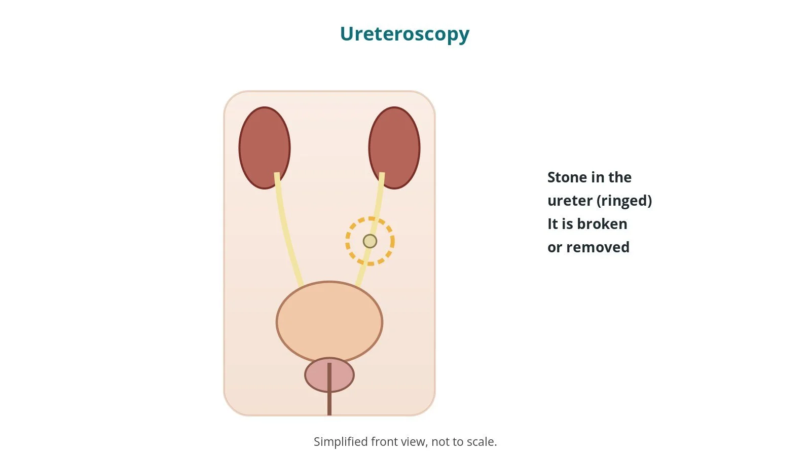
What is ureteroscopy?
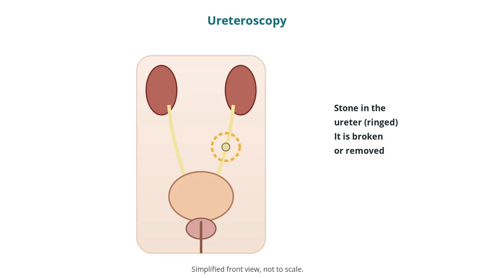
The ureters carry urine from the kidneys to the bladder. A kidney stone can block a ureter and cause severe pain or infection.
In ureteroscopy (URS) the surgeon:
- passes a thin semi-rigid or flexible ureteroscope up the urethra and bladder into the ureter
- uses a guidewire and X-ray to guide the way
- breaks the stone with a laser fibre, and may remove pieces with a small basket
- often leaves a temporary stent to keep urine draining
A blocked, infected kidney is an emergency and is drained first, as per national guidance.
New to the acronyms? See the Cath Lab Abbreviations A–Z.
Who is it for?
It may be considered for people with:
- a stone that has not passed or is unlikely to pass
- pain, blockage or infection from a stone
- some kidney stones, as an alternative to shock wave lithotripsy
The decision is made by your urology team, who will talk through the reasons, the alternatives (waiting, shock wave lithotripsy, keyhole surgery) and the risks with you.
When is it done?
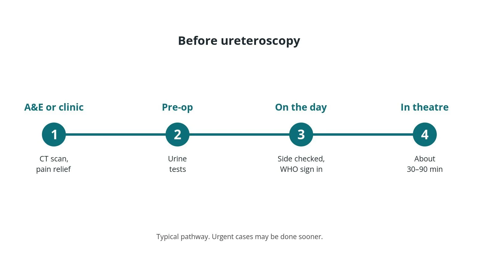
Most operations are planned; some are urgent. Before it, you usually have:
- a CT scan to find the stone
- blood and urine tests, with infection treated first
- a review of medicines, as per centre protocol
- a chance to ask questions and give consent
Step by step
1. Getting ready
Team brief and WHO checklist first, including the side and site where relevant. The anaesthetic team and ODP give the anaesthetic (usually general, as per centre protocol); antibiotics are given as per centre protocol. The circulating practitioner checks positioning in stirrups (lithotomy), pressure areas, warming, the camera stack and the irrigation fluid. The scrub practitioner sets up the endoscopy trolley and does the first count. Laser safety is set up as per centre protocol (signs, eye protection, a trained operator), and the X-ray and radiation protection are checked; the side is confirmed.
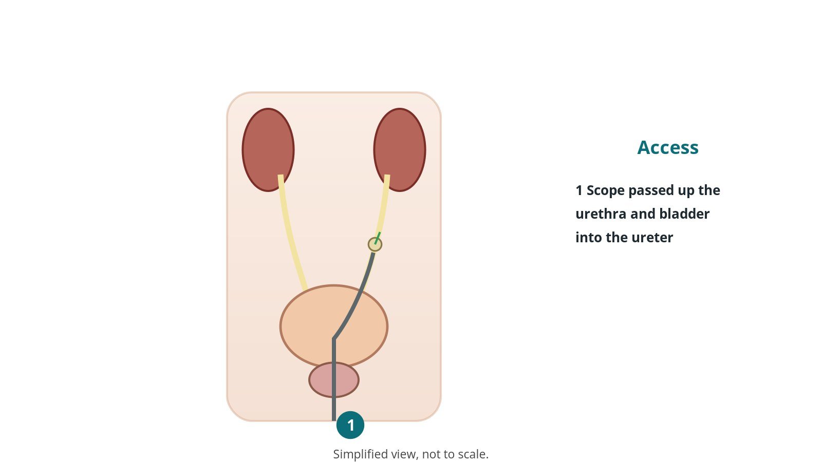
2. Access
No skin cuts: the scope is passed up the urethra, through the bladder and into the ureter, over a guidewire.
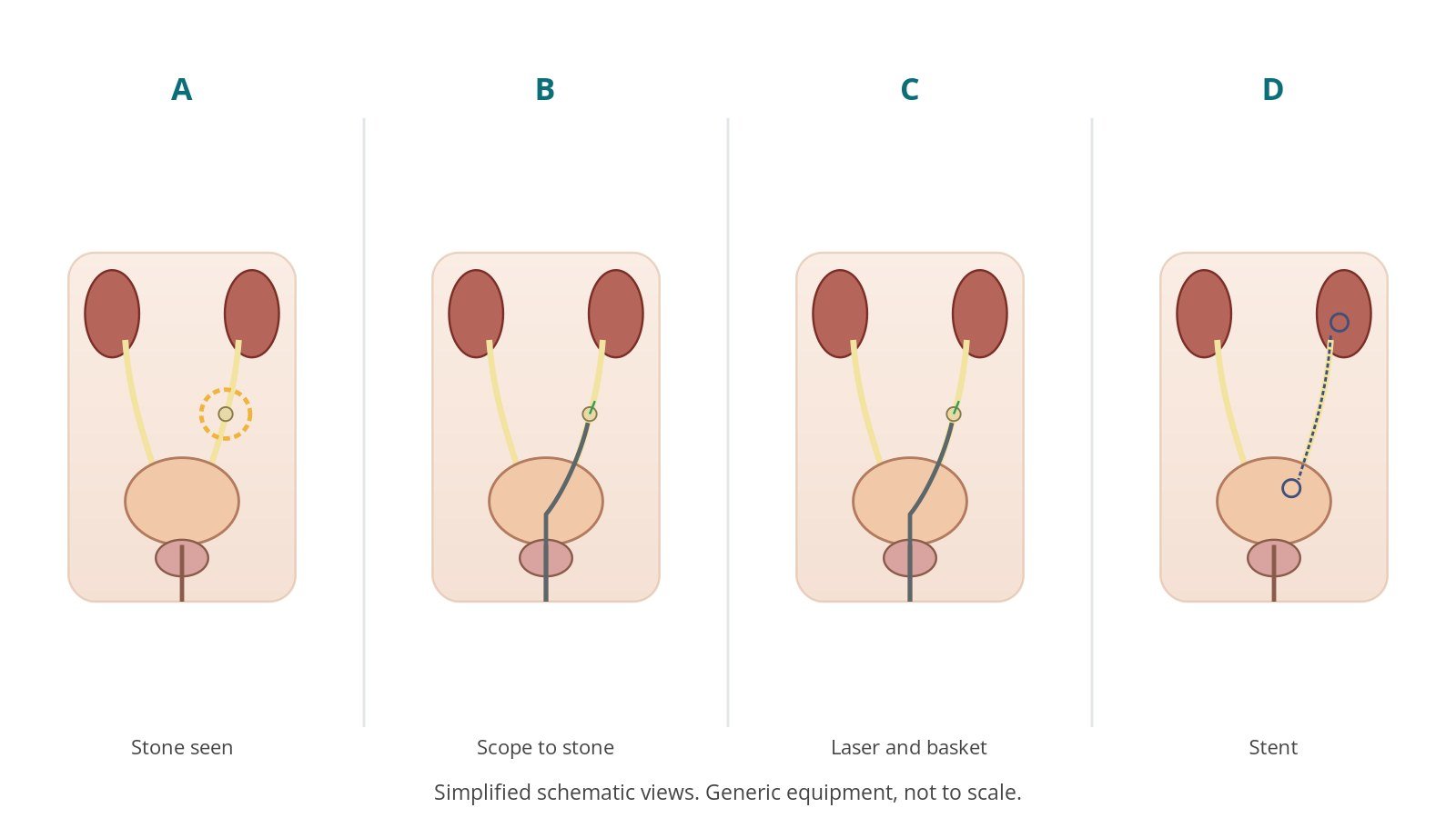
3. Treating the stone
- A. Stone seen. The stone is found on X-ray and with the scope.
- B. Scope to stone. A guidewire is placed and the scope passed up the ureter.
- C. Laser and basket. The stone is broken with laser and fragments removed.
- D. Stent. A temporary stent may be left to help drainage.
The team calls out X-ray screening, and only the trained operator fires the laser.
4. Finishing
Fragments are sent for analysis. A stent and sometimes a catheter are placed, per surgeon. Final counts are confirmed and the patient goes to recovery with a full handover, including stent details and removal plan.
Recovery and aftercare
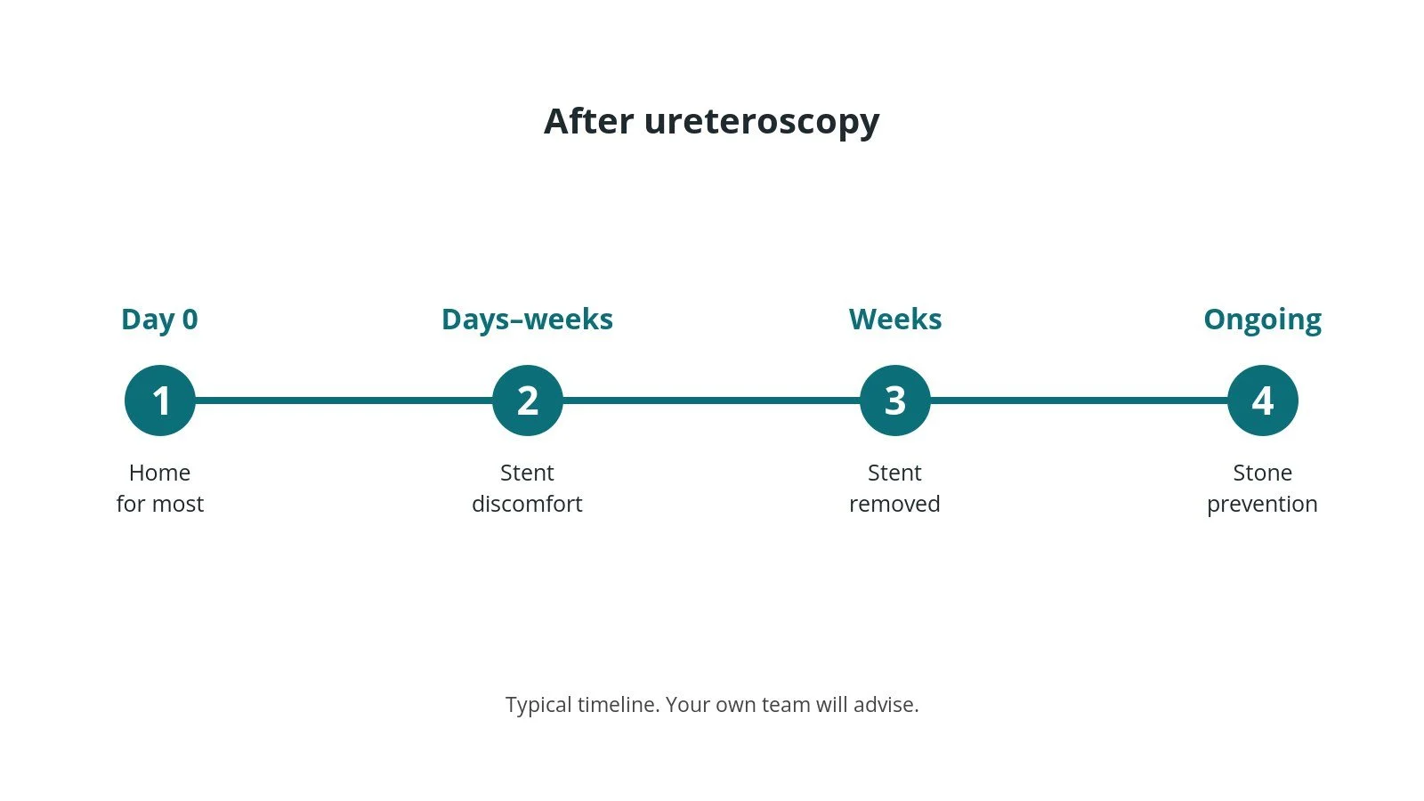
- Same day: often home the same day.
- Stent: may cause discomfort, frequent urination and blood in the urine; it must be removed or changed as planned.
- Drink plenty of fluids.
- Pain: take pain relief as advised.
- Stent removal: a date is arranged; do not miss it.
- Prevention: diet and fluid advice to reduce future stones.
Call 999 if you have chest pain, sudden breathlessness or collapse. Contact your team urgently if you cannot pass urine, pass large clots, have a high temperature or shivering, or have severe pain.
Risks and complications
Your team will explain the risks that apply to you. Possible problems include:
- urine infection, sometimes serious
- blood in the urine
- stent discomfort
- fragments left behind needing further treatment
- injury to the ureter (rare)
- narrowing of the ureter later (rare)
Theatre kit list (printable)
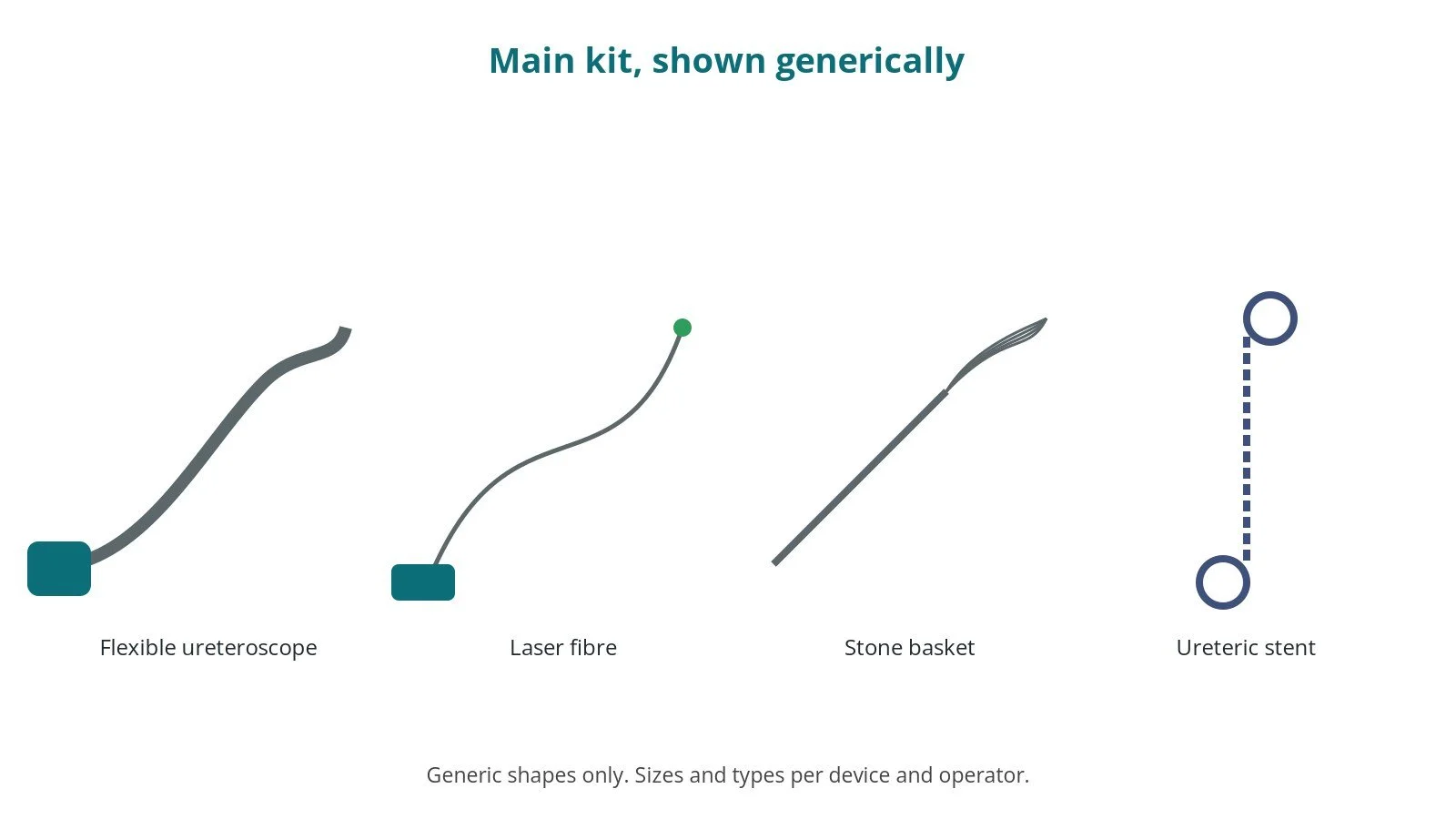
Example kit list
Every theatre has its own protocol, and sizes and doses vary.
- endoscopy stack: camera, light source, monitor
- semi-rigid and flexible ureteroscopes, per device and operator
- guidewires and access sheath, per surgeon
- laser and fibres, settings per device and operator
- stone basket, per surgeon
- ureteric stent, size per device and operator
- X-ray, contrast and radiation protection
- irrigation fluid
Procedural steps
- Team brief, WHO sign in; side confirmed; laser and X-ray safety; anaesthesia
- Lithotomy positioning; prep and drape; count
- Cystoscopy; guidewire under X-ray
- Ureteroscope to stone
- Laser and basket; fragments collected
- Stent if used; counts; stent logged; sign out and handover
First time in theatre?
💡 First-time tip
- Know your role. Scrub, circulating, anaesthetic and ODP each have their own checks; ask at the team brief.
- Counts matter. Swabs, needles and instruments are counted before, during and at closure; speak up if one is missing.
- Specimens. Label every specimen with site and side, and check it with the scrub practitioner before it leaves theatre.
- Laser safety. Wear eye protection when the laser is on and keep the door signs up.
- Stent log. Record the stent and removal plan so it is never forgotten.
FAQ
Will there be cuts?
No; it is done through the urethra.
Why do I need a stent?
To keep urine draining while swelling settles.
Will the stone come back?
Stones can recur; fluid and diet advice helps.
Related guides
Sources
All checked on 14 October 2026. Each page's own date is shown where it gives one.
- NICE. Renal and ureteric stones: assessment and management (NG118). nice.org.uk/guidance/ng118
- NHS. Kidney stones. nhs.uk/conditions/kidney-stones
- StatPearls (NCBI Bookshelf), search: ureteroscopy. ncbi.nlm.nih.gov/books
- British Association of Urological Surgeons. Patient information leaflets. baus.org.uk
- World Health Organization. Surgical Safety Checklist. who.int
Disclaimer
Education only. This page is for general education for healthcare students, theatre staff and curious readers. It isn't medical advice and doesn't replace advice from your own doctor, surgical team or local unit protocols. Equipment and medicines are described generically. No product or manufacturer is recommended. If you think someone is having a medical emergency, call 999.
Disclosure: I work in the medical device industry. This page is independent education and isn't sponsored or reviewed by any company.
All diagrams on this page are original illustrations created for rodnieoro.com.