Laparoscopic cholecystectomy (lap chole): an OR 101 guide
Reviewed by Rodnie Oro, cath lab charge nurse · Last reviewed: October 2026 · Education only, not medical advice
Lap chole in one minute
What it is: keyhole surgery to remove the gallbladder, usually because of gallstones.
Who it is for: people with painful gallstones, gallbladder inflammation, or problems caused by stones such as pancreatitis.
Main risks: bleeding, infection, bile leak, injury to the bile duct or bowel (rare), conversion to open surgery.
Usually: under general anaesthetic, about 1 hour, often home the same day.
On this page: What is a lap chole? · Who is it for? · When is it done? · Step by step · Recovery · Risks · Kit list · First time in theatre? · FAQ
What is a lap chole?
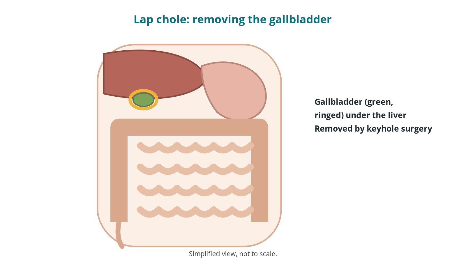
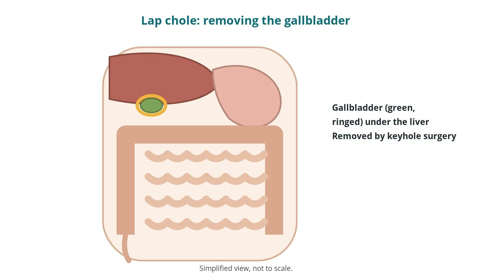
The gallbladder is a small pouch under the liver that stores bile. Gallstones can cause pain (biliary colic), inflammation (cholecystitis), jaundice or pancreatitis.
In a laparoscopic cholecystectomy the surgeon removes the gallbladder through small cuts, using a camera and long instruments:
- the abdomen is inflated with gas to make space
- the gallbladder's duct and artery are identified clearly, then clipped and divided
- the gallbladder is freed from the liver and removed in a bag
Bile then drains straight from the liver into the gut; most people live normally without a gallbladder. A few operations need conversion to open surgery.
New to the acronyms? See the Cath Lab Abbreviations A–Z.
Who is it for?
It may be considered for people with:
- repeated episodes of gallstone pain
- inflammation of the gallbladder (cholecystitis)
- pancreatitis or jaundice caused by gallstones (after treatment of the duct)
- gallbladder polyps in some cases
The decision is made by your surgical team, who will talk through the reasons, the alternatives and the risks with you.
When is it done?
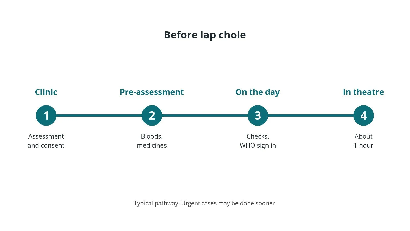
Most operations are planned; some are urgent. Before it, you usually have:
- an ultrasound scan, and sometimes an MRI scan of the bile ducts (MRCP)
- blood tests, including liver function
- a review of your medicines, especially blood thinners, as per centre protocol
- fasting instructions
- a chance to ask questions and give consent
Step by step
1. Getting ready
Team brief and WHO checklist first. The anaesthetic team and ODP give a general anaesthetic and insert a breathing tube; antibiotics and blood-clot prevention are given as per centre protocol. The circulating practitioner checks positioning, pressure areas, the diathermy plate, warming, and that the camera stack and gas insufflator work. The scrub practitioner sets up the laparoscopic trolley and an open set in case of conversion, and does the first count.
2. Access
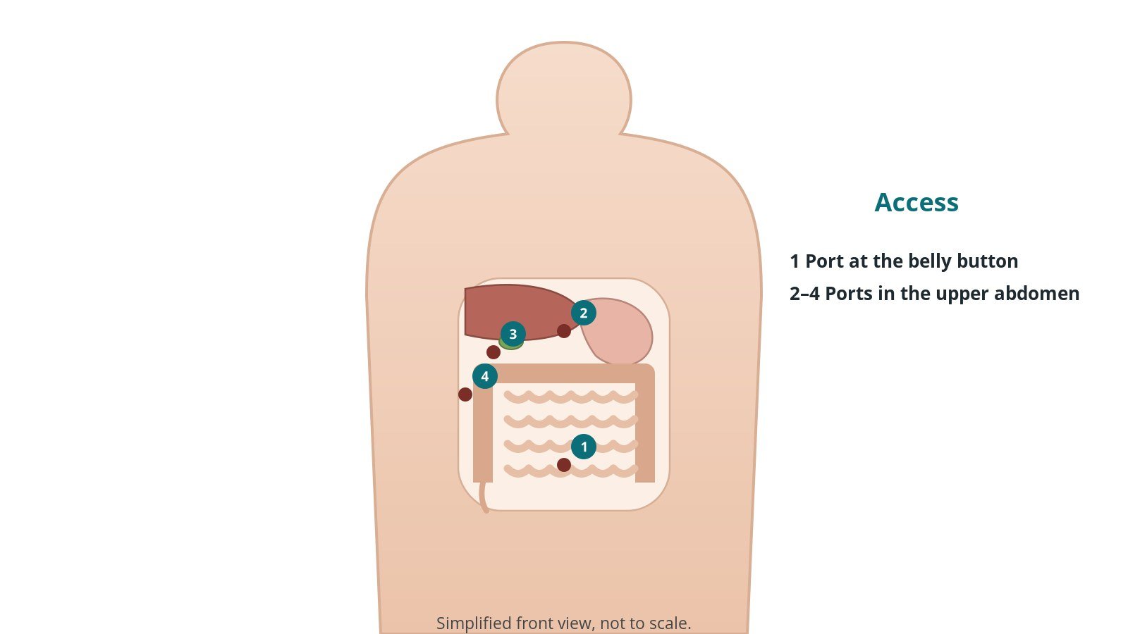
The patient lies on their back. A first port is placed, often at the belly button, using an open technique or entry needle, per surgeon, and the abdomen is filled with carbon dioxide gas. Usually three or four ports are used, sizes per device and operator. Some centres use X-ray of the ducts during surgery (cholangiogram).
3. Removing the gallbladder
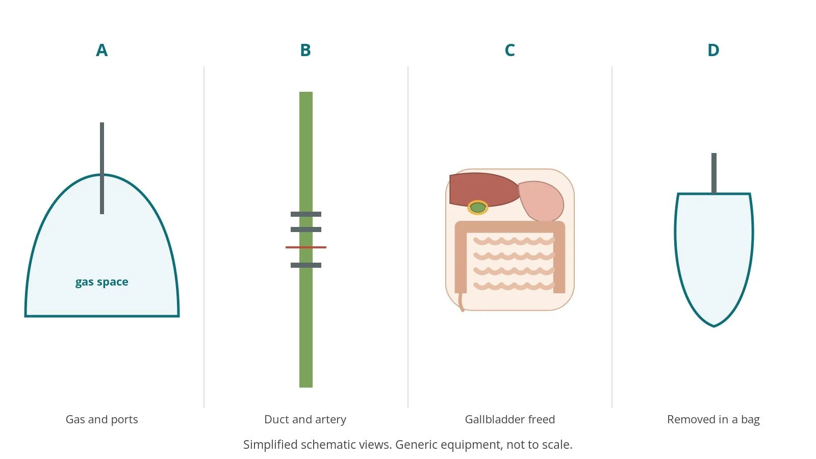
- A. Gas and ports. The abdomen is inflated and the ports and camera placed.
- B. Duct and artery. The area is dissected until the duct and artery are clearly seen (the "critical view of safety"), then clipped and divided.
- C. Gallbladder freed. The gallbladder is separated from the liver with an energy device.
- D. Removed in a bag. It is placed in a retrieval bag and taken out through the belly button cut.
The team often pauses at the critical view of safety; the circulating practitioner may be asked to record it.
4. Finishing
The gas is let out, ports removed and the larger cuts closed (deep layer, per surgeon), then the skin with sutures or glue. Local anaesthetic is often injected into the wounds. Final counts are confirmed, specimens labelled and checked, and the patient goes to recovery with a full handover.
Recovery and aftercare
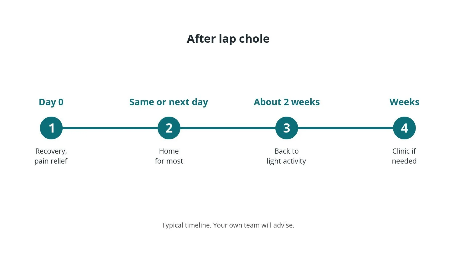
- First hours: in recovery, then a ward or day unit; pain relief and anti-sickness medicine as needed.
- Going home: often the same day or the next.
- Shoulder pain: common for a day or two from the gas.
- Activity: walking from the first day; avoid heavy lifting for about 2 weeks; ask your team.
- Eating: normal diet as tolerated.
- Work: often 1–2 weeks off, longer for manual jobs.
Call 999 if you have severe pain, heavy bleeding, breathlessness or collapse. Contact your team or GP if you have a fever, worsening tummy pain or swelling, vomiting, or a wound that becomes red, hot or leaky.
Risks and complications
Your team will explain the risks that apply to you. Possible problems include:
- bleeding
- wound infection or hernia at a port site
- bile leak
- injury to the bile duct (rare but serious)
- injury to the bowel or blood vessels (rare)
- stones left in the bile duct
- conversion to open surgery
- blood clots in the legs or lungs
Theatre kit list (printable)
Example kit list
Every theatre has its own protocol, and sizes and doses vary.
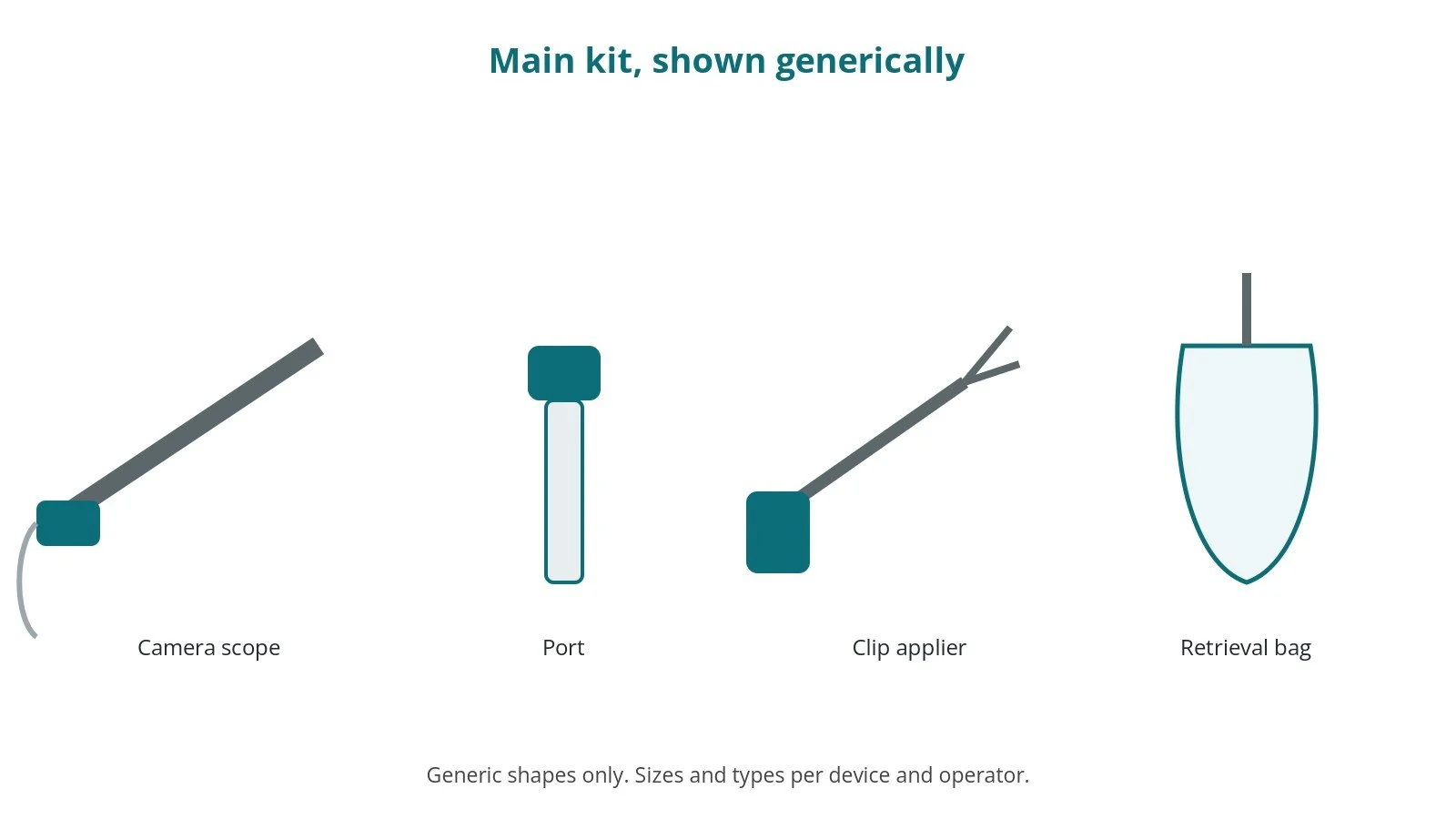
- laparoscopic stack: camera, scope, light source, gas insufflator and warmer
- ports, sizes per device and operator, and an entry needle or open-entry kit, per surgeon
- laparoscopic graspers, scissors, dissectors and suction-irrigation
- energy device, per surgeon
- clip applier and clips, sizes per device and operator
- specimen retrieval bag
- cholangiogram catheter and contrast, per surgeon and centre protocol
- closure sutures, local anaesthetic and dressings
Procedural steps
- Team brief, WHO sign in; anaesthesia; antibiotics as per centre protocol
- Supine position; prep and drape; first count
- First port; gas; camera; other ports
- Dissection; critical view of safety; clip and divide duct and artery
- Gallbladder off the liver; bag; removal; check for bleeding and bile
- Gas out; closure; local anaesthetic; counts; specimen check; sign out and handover
First time in theatre?
💡 First-time tip
- Know your role. Scrub, circulating, anaesthetic and ODP each have their own checks; ask at the team brief.
- Counts matter. Swabs, needles and instruments are counted before, during and at closure; speak up if one is missing.
- Specimens. Label every specimen with site and side, and check it with the scrub practitioner before it leaves theatre.
- Camera ready. Learn to white-balance the camera and warm the scope to stop fogging.
- Open set ready. An open tray stays available in case keyhole surgery has to convert to open.
FAQ
Can I live without a gallbladder?
Yes. Bile flows straight from the liver into the gut.
Will I need to change my diet?
Most people eat normally; some find fatty food upsets them for a while.
Why might it become open surgery?
If the anatomy is unclear or there is bleeding, the surgeon may convert for safety.
Related guides
Sources
All checked on 14 October 2026. Each page's own date is shown where it gives one.
- NHS. Gallbladder removal. nhs.uk/conditions/gallbladder-removal
- NICE. Gallstone disease: diagnosis and management (CG188). nice.org.uk/guidance/cg188
- StatPearls (NCBI Bookshelf), search: laparoscopic cholecystectomy. ncbi.nlm.nih.gov/books
- World Health Organization. Surgical Safety Checklist. who.int
Disclaimer
Education only. This page is for general education for healthcare students, theatre staff and curious readers. It isn't medical advice and doesn't replace advice from your own doctor, surgical team or local unit protocols. Equipment and medicines are described generically. No product or manufacturer is recommended. If you think someone is having a medical emergency, call 999.
Disclosure: I work in the medical device industry. This page is independent education and isn't sponsored or reviewed by any company.
All diagrams on this page are original illustrations created for rodnieoro.com.