Vitrectomy: an OR 101 guide
Reviewed by Rodnie Oro, cath lab charge nurse · Last reviewed: October 2026 · Education only, not medical advice
Vitrectomy in one minute
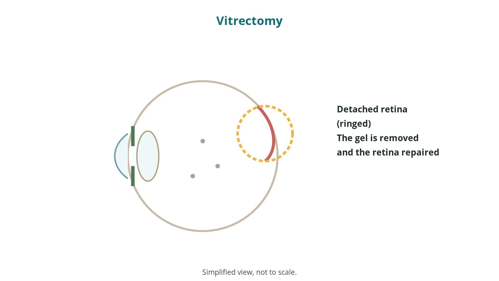
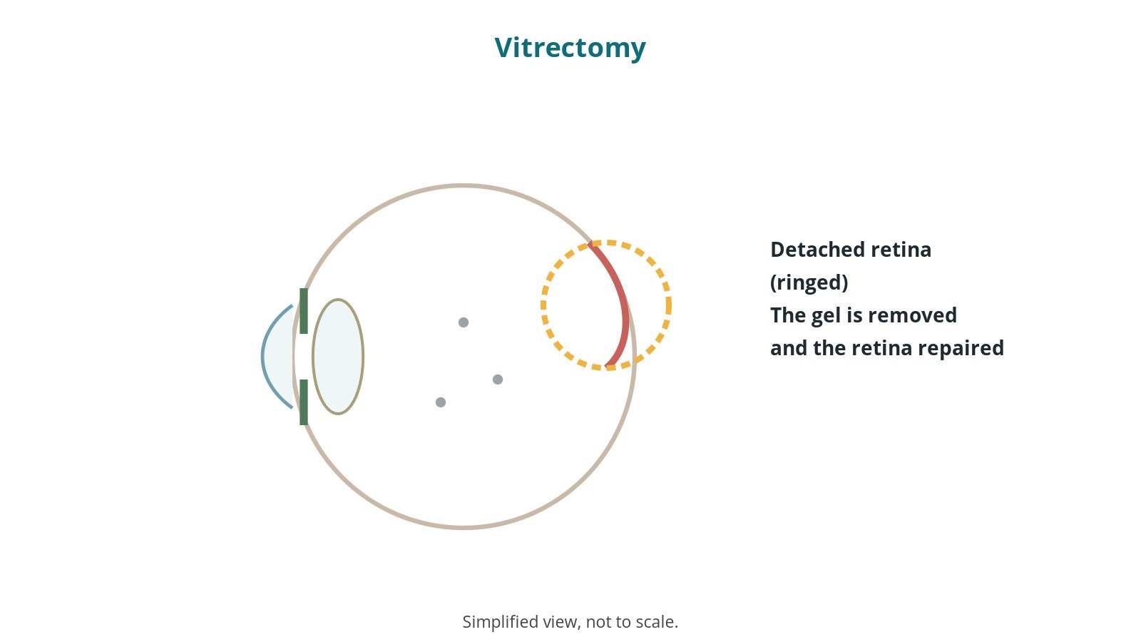
What it is: surgery to remove the vitreous gel inside the eye to treat problems of the retina.
Who it is for: people with retinal detachment, macular hole, bleeding in the eye or some diabetic eye disease.
Main risks: cataract, raised eye pressure, infection (rare), retinal detachment coming back, bleeding.
Usually: under local or general anaesthetic, 1–2 hours, often a day case.
On this page: What is a vitrectomy? · Who is it for? · When is it done? · Step by step · Recovery · Risks · Kit list · First time in theatre? · FAQ
What is a vitrectomy?
The vitreous is a clear gel filling the back of the eye. The retina lines the back of the eye and senses light.
In a vitrectomy the surgeon:
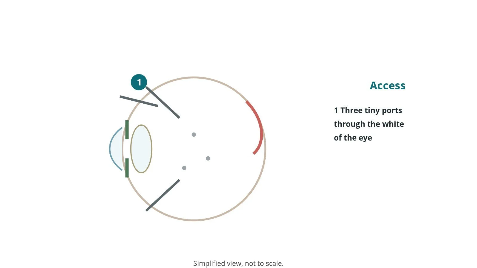
- places three tiny ports through the white of the eye
- removes the gel with a fine cutter under the microscope and a wide-angle viewing system
- repairs the retina, for example with laser or by peeling membranes
- may fill the eye with a gas bubble or silicone oil to hold the retina in place
After a gas bubble, the patient may need to keep their head in a set position (posturing) and must not fly.
New to the acronyms? See the Cath Lab Abbreviations A–Z.
Who is it for?
It may be considered for people with:
- retinal detachment
- macular hole or membranes on the retina
- bleeding inside the eye
- complications of diabetic eye disease
The decision is made by your eye team with you, talking through the reasons, the alternatives and the risks.
When is it done?
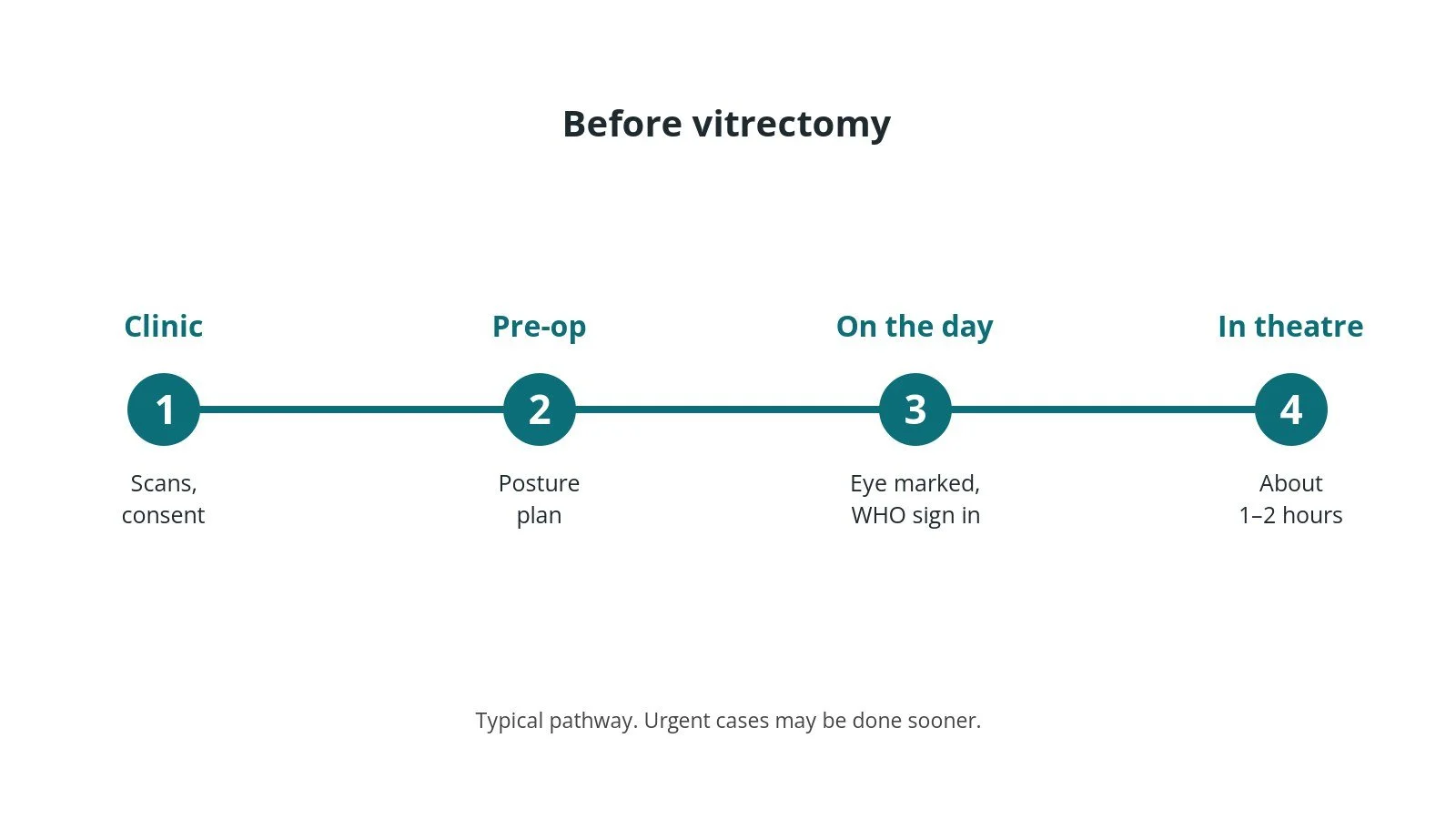
Most operations are planned; some are urgent. Before it, you usually have:
- an eye examination and scans
- a review of medicines, as per centre protocol
- a plan for posturing and travel after surgery
- a chance to ask questions and give consent
Step by step
1. Getting ready
Team brief and WHO checklist first, including the correct eye marked. The anaesthetic team and ODP give the anaesthetic (local injection around the eye, or general, as per centre protocol); eye drops and medicines are given as per centre protocol. The circulating practitioner checks positioning, pressure areas, the diathermy plate and warming. The scrub practitioner sets up the instrument trolley and does the first count. The vitrectomy machine, microscope, laser safety and any gas or oil are checked as per centre protocol.
2. Access
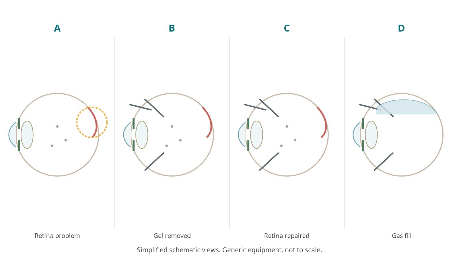
Three tiny ports are placed through the white of the eye.
3. Repairing the retina
- A. Retina problem. The detached retina is seen.
- B. Gel removed. Ports are placed and the gel removed with the cutter.
- C. Retina repaired. The retina is flattened and treated with laser.
- D. Gas fill. The eye is filled with gas or oil.
If gas is used, the team records it and the patient gets a wristband warning against nitrous oxide anaesthetic and flying, as per centre protocol.
4. Finishing
The ports are removed and checked for leaks, antibiotics given as per centre protocol, and a pad and shield applied. Gas or oil details are recorded and the patient goes to recovery with posturing instructions.
Recovery and aftercare
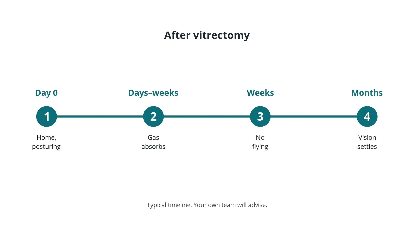
- Same day: often home the same day with a pad and shield.
- Posturing: keep the head in the position advised, sometimes for days.
- Gas bubble: blurred vision until it absorbs; no flying or high altitude.
- Drops: as prescribed.
- Oil: a second operation may be needed to remove it.
- Urgent help: for increasing pain or worse vision.
Contact the eye unit urgently if you have increasing pain, worsening vision, a red eye, flashing lights or a shadow in your vision.
Risks and complications
Your team will explain the risks that apply to you. Possible problems include:
- cataract developing or worsening
- raised eye pressure
- retinal detachment coming back
- infection inside the eye (rare)
- bleeding
- vision not improving as hoped
Theatre kit list (printable)
Example kit list
Every theatre has its own protocol, and sizes and doses vary.
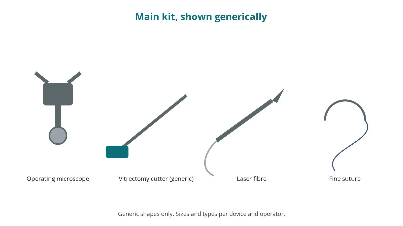
- operating microscope with wide-angle viewing system
- vitrectomy machine, cutter and light pipe, settings per device and operator
- trocars and ports, sizes per device and operator
- endolaser, with laser safety as per centre protocol
- gas or silicone oil, per surgeon
- fine eye instruments
- pad, shield and gas warning wristband
Procedural steps
- Team brief, WHO sign in; eye marked; laser and gas checks; anaesthetic
- Positioning; drape; count
- Ports placed
- Gel removed; retina repaired; laser
- Gas or oil fill
- Ports out; antibiotics; gas logged; wristband; sign out and posturing advice
First time in theatre?
💡 First-time tip
- Know your role. Scrub, circulating, anaesthetic and ODP each have their own checks; ask at the team brief.
- Counts matter. Swabs, needles and instruments are counted before, during and at closure; speak up if one is missing.
- Microscope. Drape the operating microscope sterile and balance it before the case.
- Gas warning. Make sure the gas wristband and record are done; nitrous oxide must be avoided.
- Laser safety. Wear eye protection and keep door signs up when the laser is used.
FAQ
Why do I need to posture?
So the bubble presses on the right part of the retina.
When can I fly?
Not until the gas has gone; your team will tell you.
Will my sight be blurry?
Yes, until the gas absorbs or oil is removed.
Related guides
Sources
All checked on 14 October 2026. Each page's own date is shown where it gives one.
- NHS. Retinal detachment. nhs.uk/conditions/retinal-detachment
- NHS. Macular hole. nhs.uk/conditions/macular-hole
- StatPearls (NCBI Bookshelf), search: vitrectomy. ncbi.nlm.nih.gov/books
- The Royal College of Ophthalmologists. www.rcophth.ac.uk
- World Health Organization. Surgical Safety Checklist. who.int
Disclaimer
Education only. This page is for general education for healthcare students, theatre staff and curious readers. It isn't medical advice and doesn't replace advice from your own doctor, surgical team or local unit protocols. Equipment and medicines are described generically. No product or manufacturer is recommended. If you think someone is having a medical emergency, call 999.
Disclosure: I work in the medical device industry. This page is independent education and isn't sponsored or reviewed by any company.
All diagrams on this page are original illustrations created for rodnieoro.com.