Mastoidectomy and tympanoplasty: an OR 101 guide
Reviewed by Rodnie Oro, cath lab charge nurse · Last reviewed: October 2026 · Education only, not medical advice
Mastoidectomy and tympanoplasty in one minute
What it is: mastoidectomy clears disease from the mastoid bone behind the ear; tympanoplasty repairs the eardrum.
Who it is for: people with cholesteatoma, long-term ear infection or a hole in the eardrum.
Main risks: hearing change, dizziness, taste change, facial nerve weakness (rare), disease coming back.
Usually: under general anaesthetic, 1–4 hours, day case or one night.
On this page: What are mastoidectomy and tympanoplasty? · Who is it for? · When is it done? · Step by step · Recovery · Risks · Kit list · First time in theatre? · FAQ
What are mastoidectomy and tympanoplasty?
The mastoid is the honeycomb bone behind the ear. Long-term infection or a skin cyst (cholesteatoma) can damage it and the middle ear.
- Mastoidectomy: the surgeon drills away diseased mastoid bone under an operating microscope.
- Tympanoplasty: the surgeon repairs a hole in the eardrum with a graft, often from tissue near the ear.
A facial nerve monitor is often used, per centre, because the nerve runs through this bone.
New to the acronyms? See the Cath Lab Abbreviations A–Z.
Who is it for?
It may be considered for people with:
- cholesteatoma
- chronic ear infection with discharge
- a hole in the eardrum that has not healed
The decision is made by your ENT team, who will talk through the reasons, the alternatives and the risks with you.
When is it done?
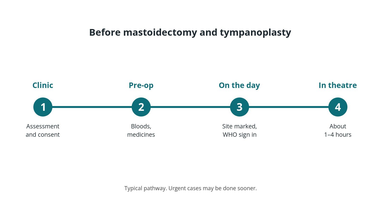
Most operations are planned; some are urgent. Before it, you usually have:
- a hearing test
- often a CT or MRI scan
- a review of medicines, as per centre protocol
- a chance to ask questions and give consent
Step by step
1. Getting ready
Team brief and WHO checklist first, including the side and site marking. The anaesthetic team and ODP give the anaesthetic (general, without long-acting muscle relaxant if the nerve monitor is used, as per centre protocol, as per centre protocol); antibiotics and blood-clot prevention are given as per centre protocol. The circulating practitioner checks positioning, pressure areas, the diathermy plate and warming. The scrub practitioner sets up the instrument trolley and does the first count. The microscope, drill and facial nerve monitor are checked; the head is turned with the ear up.
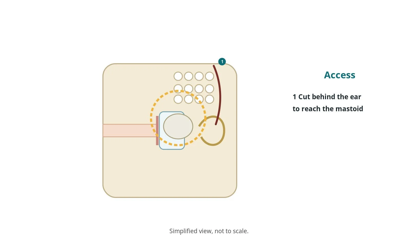
2. Access
A cut behind the ear, or through the ear canal, per surgeon.
3. Clearing disease and repairing the eardrum
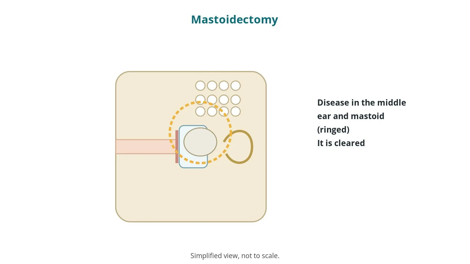
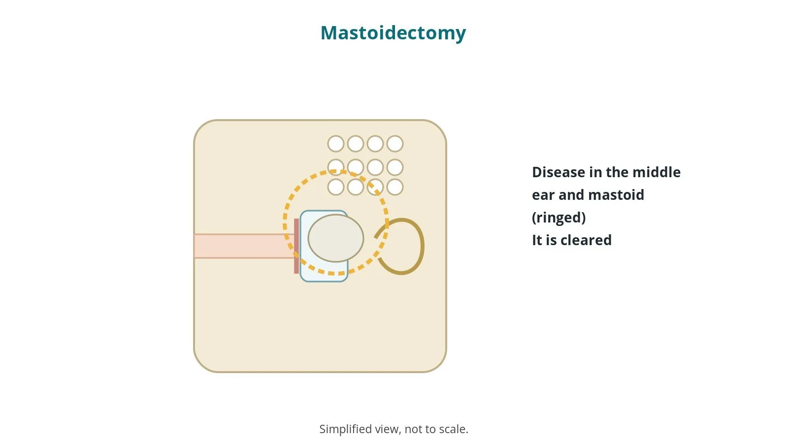
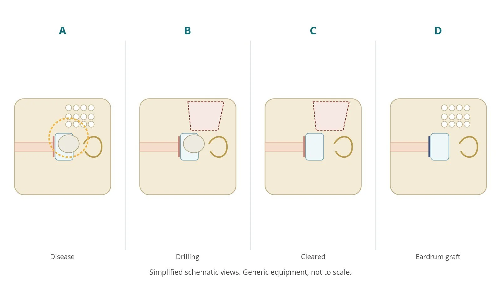
- A. Disease. The disease in the middle ear and mastoid is found.
- B. Drilling. Diseased bone is drilled away under the microscope.
- C. Cleared. The mastoid and middle ear are cleared.
- D. Eardrum graft. The eardrum is repaired with a graft.
The team keeps theatre still and quiet while the facial nerve monitor is in use.
4. Finishing
The ear canal is packed and the wound closed. A head bandage may be applied. Final counts are confirmed, specimens labelled, and the patient goes to recovery with a full handover including facial nerve checks.
Recovery and aftercare
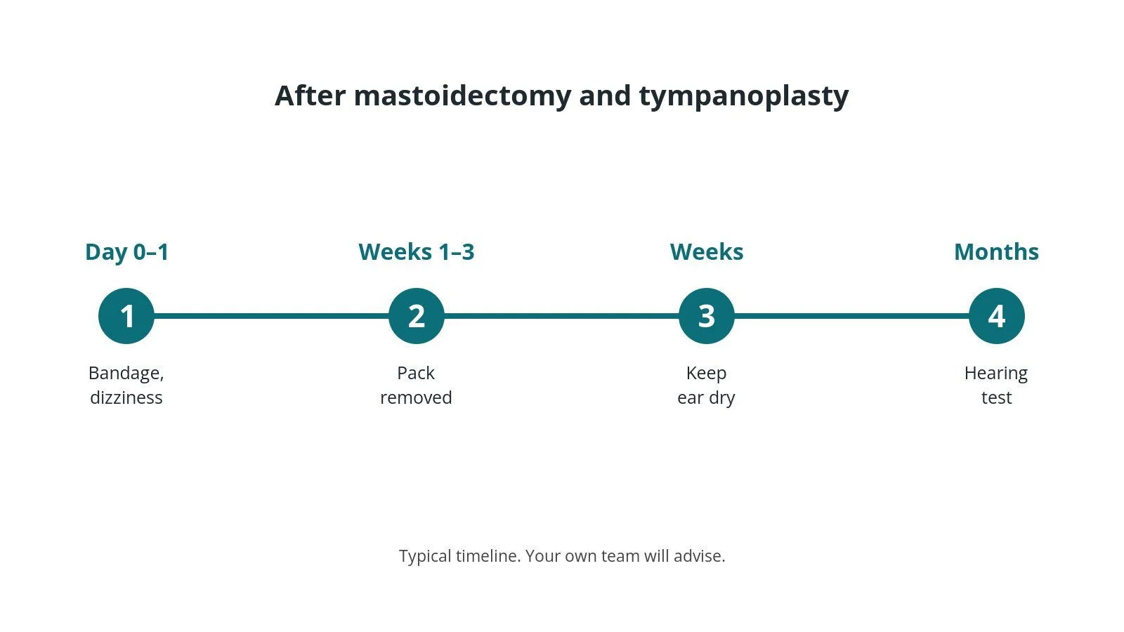
- First day: a head bandage and ear pack; some dizziness is common.
- Going home: often the same or next day.
- Keep dry: keep the ear dry until advised.
- Avoid: nose blowing and flying for a time, as advised.
- Pack removal: at clinic after 1–3 weeks.
- Hearing: checked at follow-up; disease checks may continue for years.
Call 999 if you are coughing or vomiting a lot of blood or have difficulty breathing. Contact your team if you have bleeding, a high temperature, increasing pain or are unable to eat or drink.
Risks and complications
Your team will explain the risks that apply to you. Possible problems include:
- hearing change, sometimes worse
- dizziness
- taste change on one side of the tongue
- tinnitus
- facial nerve weakness (rare)
- disease coming back
- graft not taking
Theatre kit list (printable)
Example kit list
Every theatre has its own protocol, and sizes and doses vary.
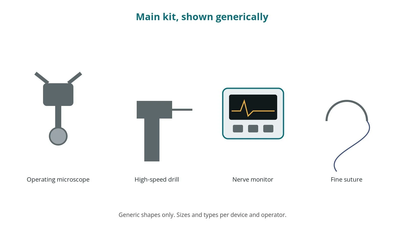
- operating microscope
- high-speed drill with burrs, per surgeon
- facial nerve monitor, per centre
- fine ear instruments
- graft materials, per surgeon
- ear packing, per surgeon
- head bandage
Procedural steps
- Team brief, WHO sign in; side marked; nerve monitor plan; anaesthesia
- Head turned; monitor set up; prep; count
- Cut behind the ear; graft harvested
- Mastoid drilled; disease cleared
- Eardrum graft placed; ear packed
- Closure; bandage; counts; sign out and handover with facial nerve check
First time in theatre?
💡 First-time tip
- Know your role. Scrub, circulating, anaesthetic and ODP each have their own checks; ask at the team brief.
- Counts matter. Swabs, needles and instruments are counted before, during and at closure; speak up if one is missing.
- Microscope. Drape the operating microscope sterile and balance it before the case.
- Nerve monitor. Make sure the monitor is connected and tested before drilling.
- Drill irrigation. Keep irrigation running when drilling to keep bone cool.
FAQ
Will my hearing improve?
Sometimes; the main aim is often to clear disease.
Will I be dizzy?
Some dizziness is common for a few days.
Can I get my ear wet?
Not until your team says so.
Related guides
Sources
All checked on 14 October 2026. Each page's own date is shown where it gives one.
- NHS. Cholesteatoma. nhs.uk/conditions/cholesteatoma
- NHS. Perforated eardrum. nhs.uk/conditions/perforated-eardrum
- StatPearls (NCBI Bookshelf), search: mastoidectomy. ncbi.nlm.nih.gov/books
- ENT UK. www.entuk.org
- World Health Organization. Surgical Safety Checklist. who.int
Disclaimer
Education only. This page is for general education for healthcare students, theatre staff and curious readers. It isn't medical advice and doesn't replace advice from your own doctor, surgical team or local unit protocols. Equipment and medicines are described generically. No product or manufacturer is recommended. If you think someone is having a medical emergency, call 999.
Disclosure: I work in the medical device industry. This page is independent education and isn't sponsored or reviewed by any company.
All diagrams on this page are original illustrations created for rodnieoro.com.