Craniotomy: an OR 101 guide
Reviewed by Rodnie Oro, cath lab charge nurse · Last reviewed: October 2026 · Education only, not medical advice
Craniotomy in one minute
What it is: surgery where a section of skull (bone flap) is lifted to reach the brain, then fixed back in place.
Who it is for: people with a brain tumour, bleeding, aneurysm, abscess or some types of epilepsy.
Main risks: bleeding, infection, seizures, stroke, weakness or speech problems, leak of brain fluid.
Usually: under general anaesthetic (sometimes awake for part), 3–6 hours, several days in hospital.
On this page: What is a craniotomy? · Who is it for? · When is it done? · Step by step · Recovery · Risks · Kit list · First time in theatre? · FAQ
What is a craniotomy?
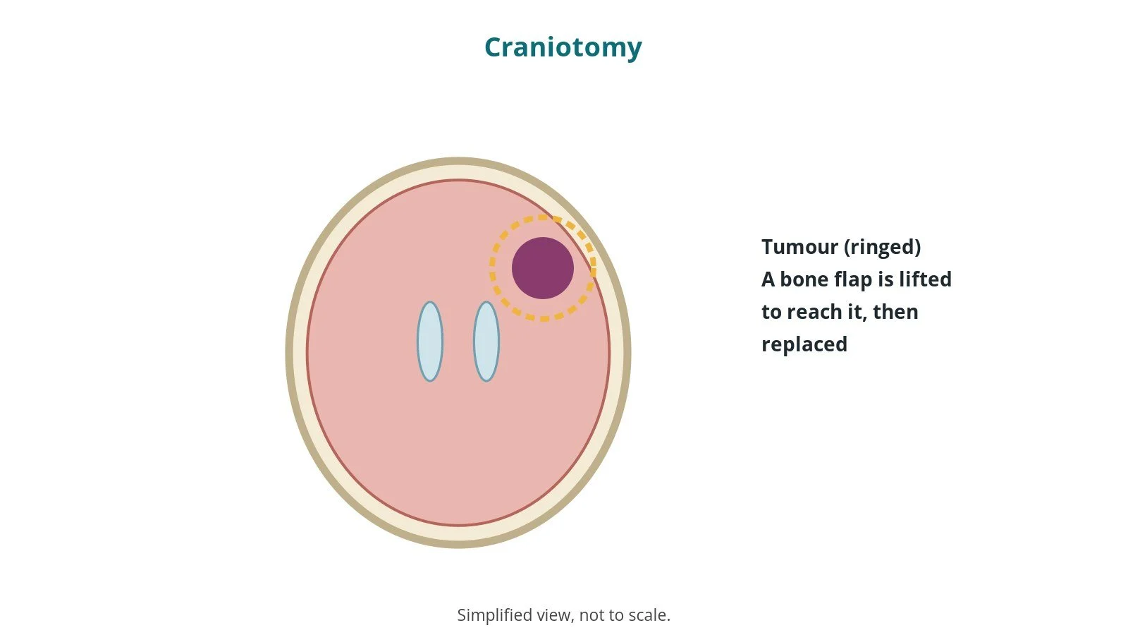
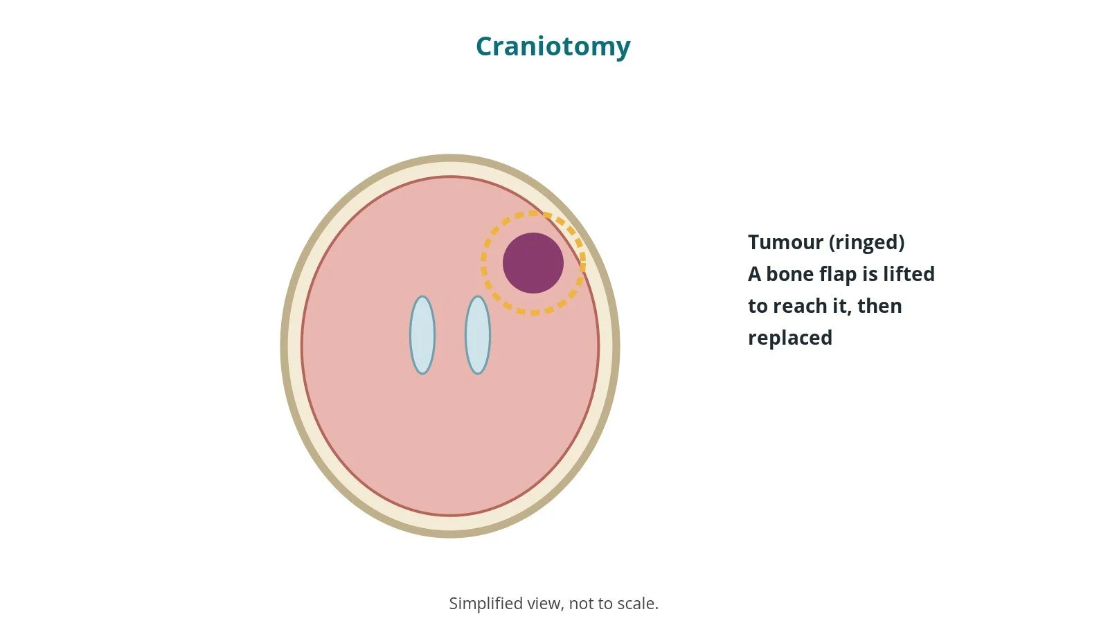
A craniotomy is an operation where the surgeon temporarily removes a piece of skull, the bone flap, to reach the brain.
The surgeon:
- holds the head still in a head clamp and plans the cut with image guidance (navigation), per centre
- makes a skin cut and drills small holes, then cuts the bone flap
- opens the brain's covering (the dura) and treats the problem, often using an operating microscope
- closes the dura and fixes the bone flap back with small plates and screws
Some operations are done with the patient awake for part of the surgery so speech or movement can be checked.
New to the acronyms? See the Cath Lab Abbreviations A–Z.
Who is it for?
It may be considered for people with:
- brain tumours
- bleeding in or around the brain
- aneurysms and abnormal blood vessels
- brain abscess
- some types of epilepsy
The decision is made by your neurosurgical team, often with a multidisciplinary team, who will talk through the reasons, the alternatives and the risks with you.
When is it done?
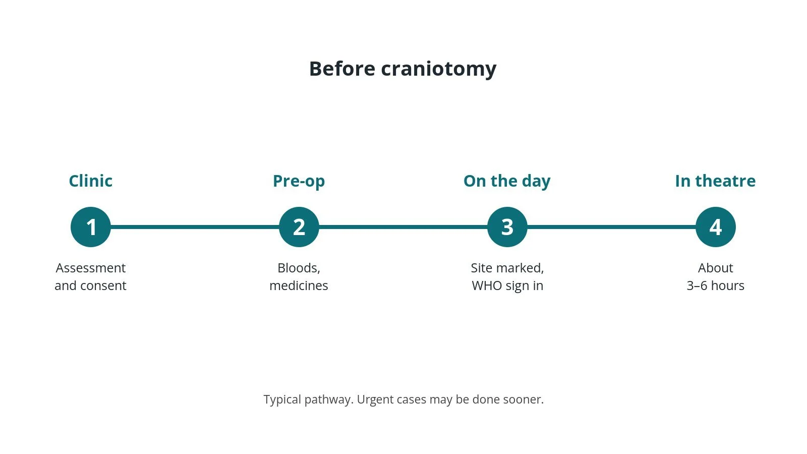
Most operations are planned; some are urgent. Before it, you usually have:
- an MRI or CT scan, often with planning scans for navigation
- blood tests and an ECG
- a review of medicines, especially blood thinners and seizure medicines, as per centre protocol
- a chance to ask questions and give consent
Step by step
1. Getting ready
Team brief and WHO checklist first, including the site and side marking. The anaesthetic team and ODP give the anaesthetic (general, or awake with sedation for part of the procedure, as per centre protocol); antibiotics and blood-clot prevention are given as per centre protocol. The circulating practitioner checks positioning, pressure areas, the diathermy plate and warming. The scrub practitioner sets up the instrument trolley and does the first count. The head clamp, navigation and microscope are checked; the head is positioned and pinned, and pressure areas protected.
2. Access
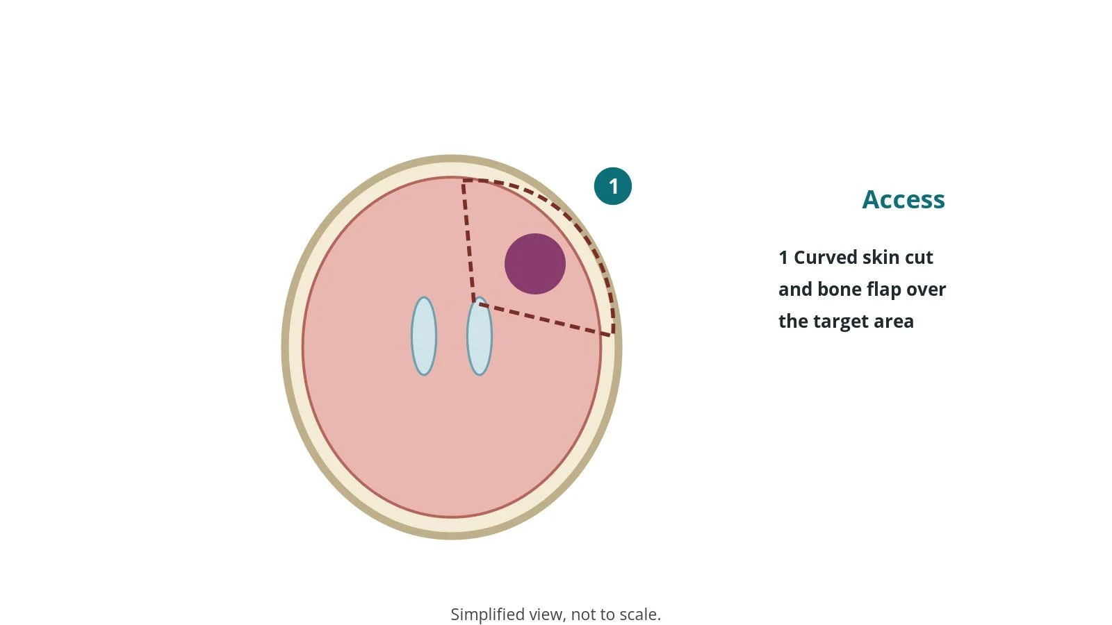
A curved skin cut is made over the area of the brain to be treated, often behind the hairline.
3. Treating the brain
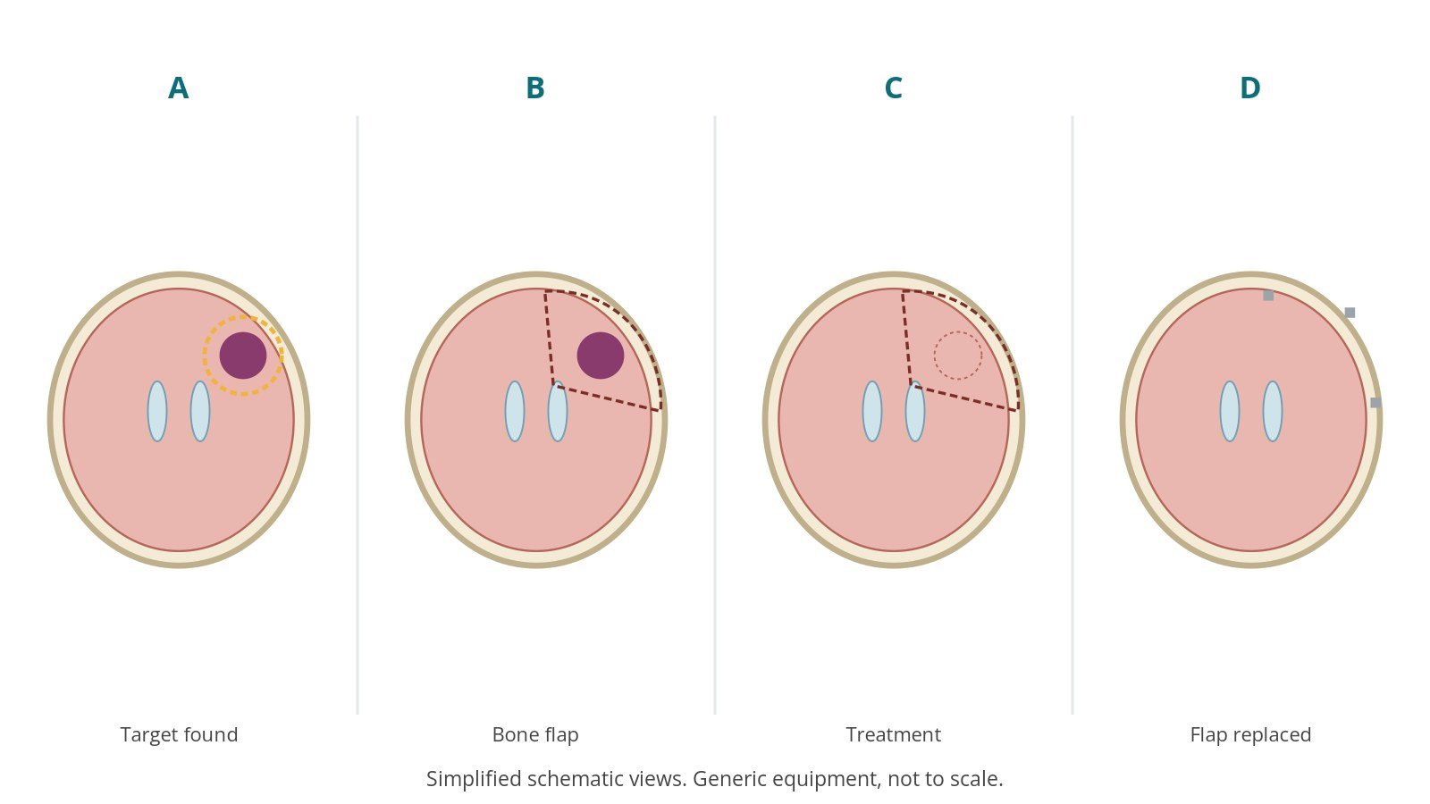
- A. Target found. The target is located with navigation.
- B. Bone flap. Holes are drilled and the bone flap lifted.
- C. Treatment. The dura is opened and the problem treated under the microscope.
- D. Flap replaced. The dura is closed and the flap fixed with small plates.
The anaesthetic team controls blood pressure and brain swelling as per centre protocol; in awake surgery the team checks speech and movement.
4. Finishing
The bone flap is fixed back, a drain may be placed, per surgeon, and the scalp closed. Final counts are confirmed, specimens labelled and checked, the head clamp removed, and the patient goes to recovery or intensive care with a full handover including neuro checks.
Recovery and aftercare
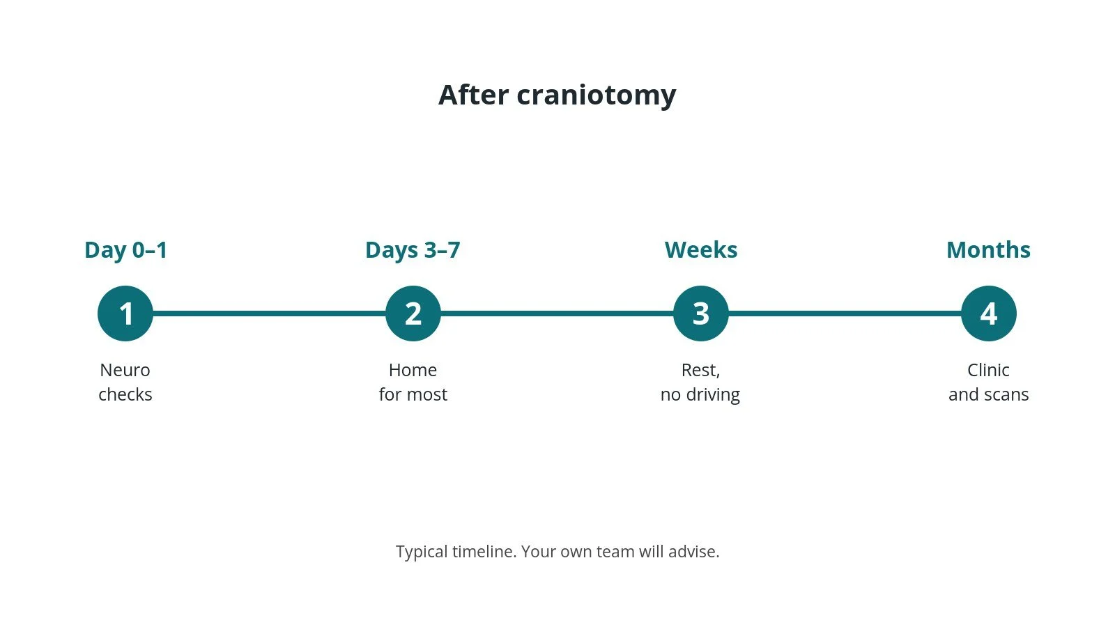
- First day: close neurological checks, often in high dependency or intensive care.
- Going home: often 3–7 days.
- Headache and swelling: common; pain relief is given.
- Driving: not allowed for a period; follow DVLA rules and your team.
- Wound: keep it clean and dry; stitches or clips removed as advised.
- Follow-up: clinic and scans; results discussed with the team.
Call 999 if you have a seizure, sudden severe headache, confusion, drowsiness, weakness or collapse. Contact your team if you have a fever, leaking wound, worsening headache or vomiting.
Risks and complications
Your team will explain the risks that apply to you. Possible problems include:
- bleeding in the brain
- infection of the wound, bone flap or brain
- seizures
- stroke, weakness, speech or vision problems
- leak of brain fluid
- blood clots in the legs or lungs
- swelling of the brain
Theatre kit list (printable)
Example kit list
Every theatre has its own protocol, and sizes and doses vary.
- head clamp with pins, per device and operator
- navigation system, per centre
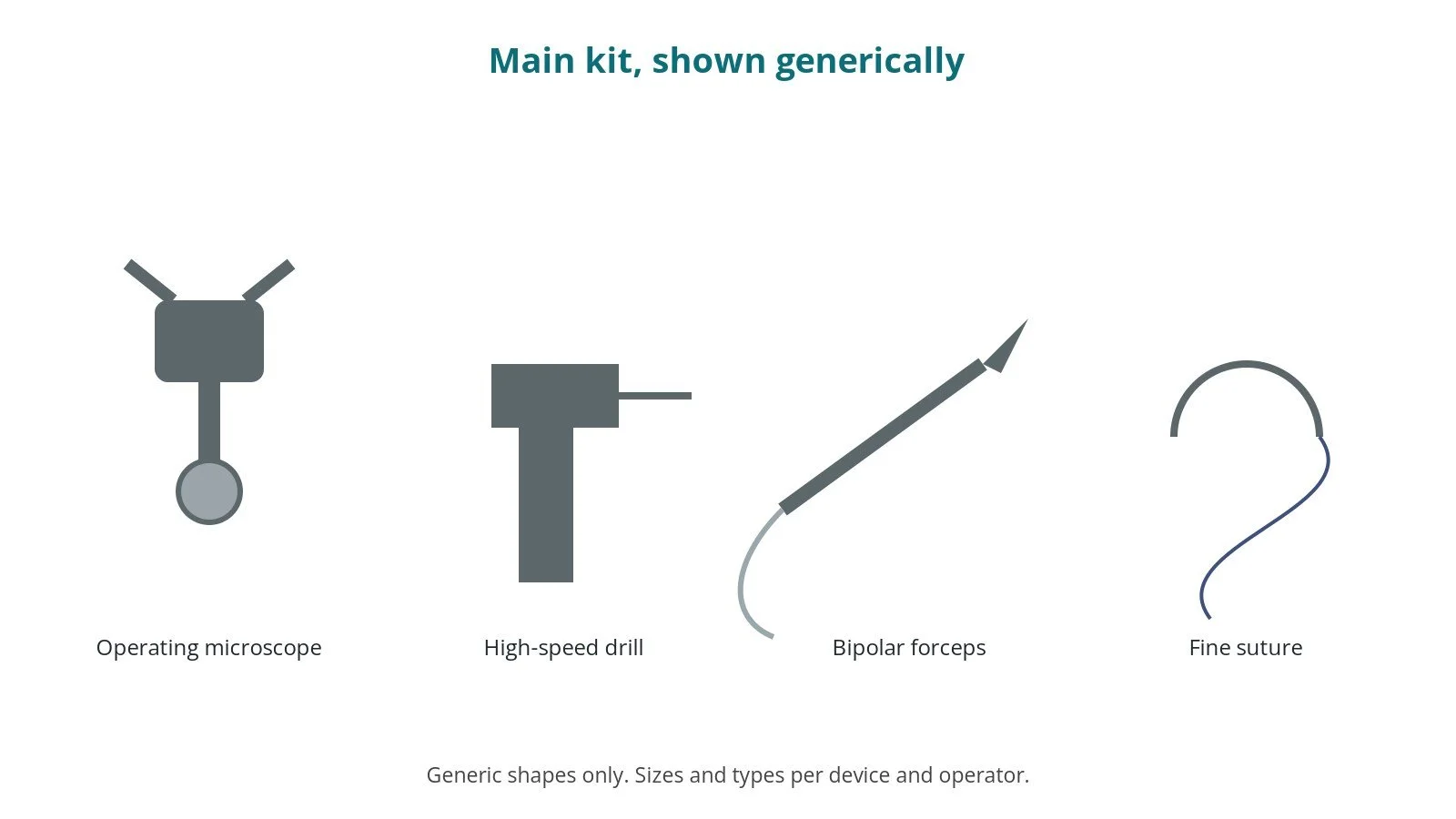
- high-speed drill and craniotome, per device and operator
- operating microscope
- bipolar diathermy and suction
- ultrasonic aspirator, per surgeon
- dural sealant or graft, per surgeon
- small plates and screws for the flap
- drain and closure sutures or clips
Procedural steps
- Team brief, WHO sign in; site and side marked; anaesthesia; lines
- Head clamp and positioning; navigation registered; count
- Skin cut; burr holes; bone flap
- Dura opened; microscope; lesion treated
- Haemostasis; dura closed; flap plated
- Closure; counts; specimens; clamp off; sign out and handover with neuro checks
First time in theatre?
💡 First-time tip
- Know your role. Scrub, circulating, anaesthetic and ODP each have their own checks; ask at the team brief.
- Counts matter. Swabs, needles and instruments are counted before, during and at closure; speak up if one is missing.
- Microscope. Drape the operating microscope sterile and balance it before the case.
- Bone flap. Keep the bone flap safe, moist and on the sterile field until it is replaced.
- Quiet theatre. Keep noise low, especially during awake surgery.
FAQ
Will my head be shaved?
Usually only a small strip along the cut.
Can I be awake?
For some operations, yes; your team will explain why.
When can I drive?
It depends on the reason for surgery; follow DVLA rules.
Related guides
Sources
All checked on 14 October 2026. Each page's own date is shown where it gives one.
- NICE. Brain tumours (primary) and brain metastases in over 16s (NG99). nice.org.uk/guidance/ng99
- NHS. Brain tumours. nhs.uk/conditions/brain-tumours
- StatPearls (NCBI Bookshelf), search: craniotomy. ncbi.nlm.nih.gov/books
- Society of British Neurological Surgeons. www.sbns.org.uk
- World Health Organization. Surgical Safety Checklist. who.int
Disclaimer
Education only. This page is for general education for healthcare students, theatre staff and curious readers. It isn't medical advice and doesn't replace advice from your own doctor, surgical team or local unit protocols. Equipment and medicines are described generically. No product or manufacturer is recommended. If you think someone is having a medical emergency, call 999.
Disclosure: I work in the medical device industry. This page is independent education and isn't sponsored or reviewed by any company.
All diagrams on this page are original illustrations created for rodnieoro.com.