Ventriculoperitoneal (VP) shunt: an OR 101 guide
Reviewed by Rodnie Oro, cath lab charge nurse · Last reviewed: October 2026 · Education only, not medical advice
VP shunt in one minute
What it is: surgery to place a thin tube with a valve that drains extra brain fluid from the brain into the abdomen.
Who it is for: people with hydrocephalus (a build-up of fluid in the brain).
Main risks: blockage, infection, over- or under-drainage, bleeding, needing further surgery.
Usually: under general anaesthetic, about 1 hour, a few days in hospital.
On this page: What is a VP shunt? · Who is it for? · When is it done? · Step by step · Recovery · Risks · Kit list · First time in theatre? · FAQ
What is a VP shunt?
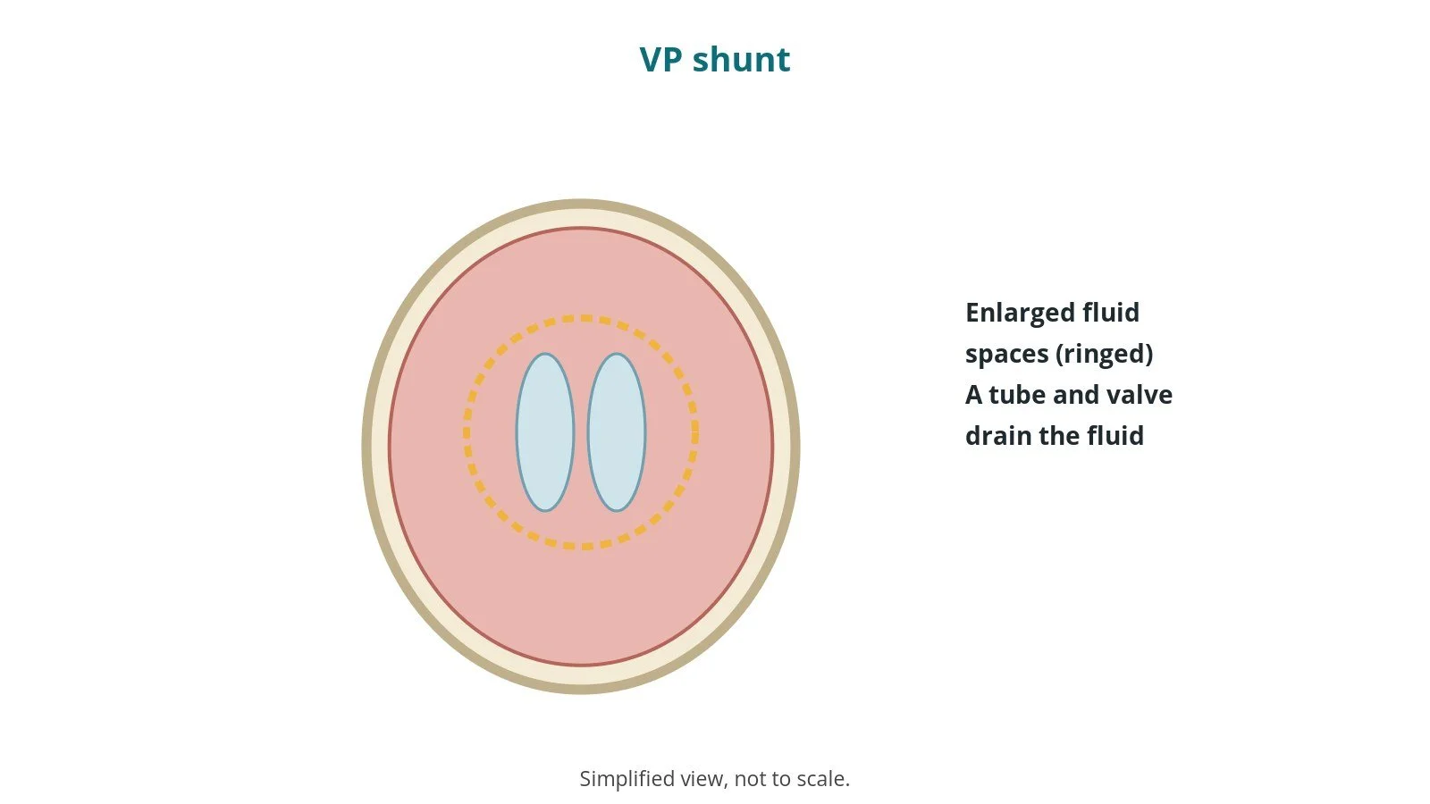
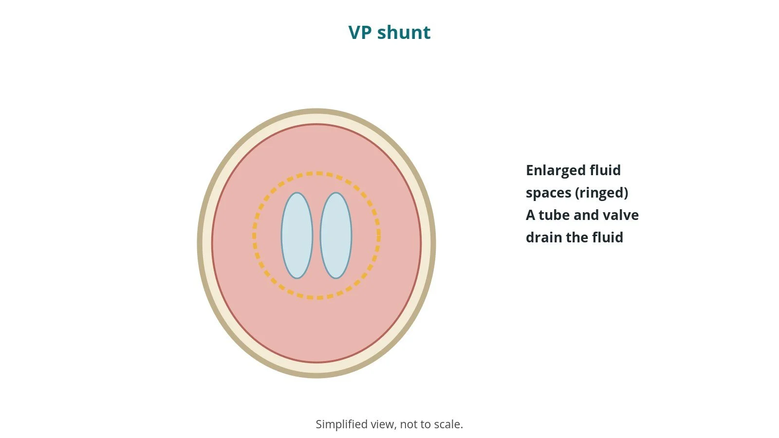
Cerebrospinal fluid (CSF) cushions the brain. In hydrocephalus it builds up in the brain's fluid spaces (ventricles) and raises pressure.
A ventriculoperitoneal (VP) shunt:
- has a tube in a ventricle, a valve under the scalp, and a tube tunnelled under the skin to the abdomen
- lets extra fluid drain to the abdomen, where it is absorbed
- has a valve setting chosen per device and operator; some can be adjusted with a magnet
An alternative for some people is endoscopic third ventriculostomy.
New to the acronyms? See the Cath Lab Abbreviations A–Z.
Who is it for?
It may be considered for people with:
- hydrocephalus in babies, children or adults
- normal pressure hydrocephalus in older adults, after assessment
The decision is made by your neurosurgical team, who will talk through the reasons, the alternatives and the risks with you.
When is it done?
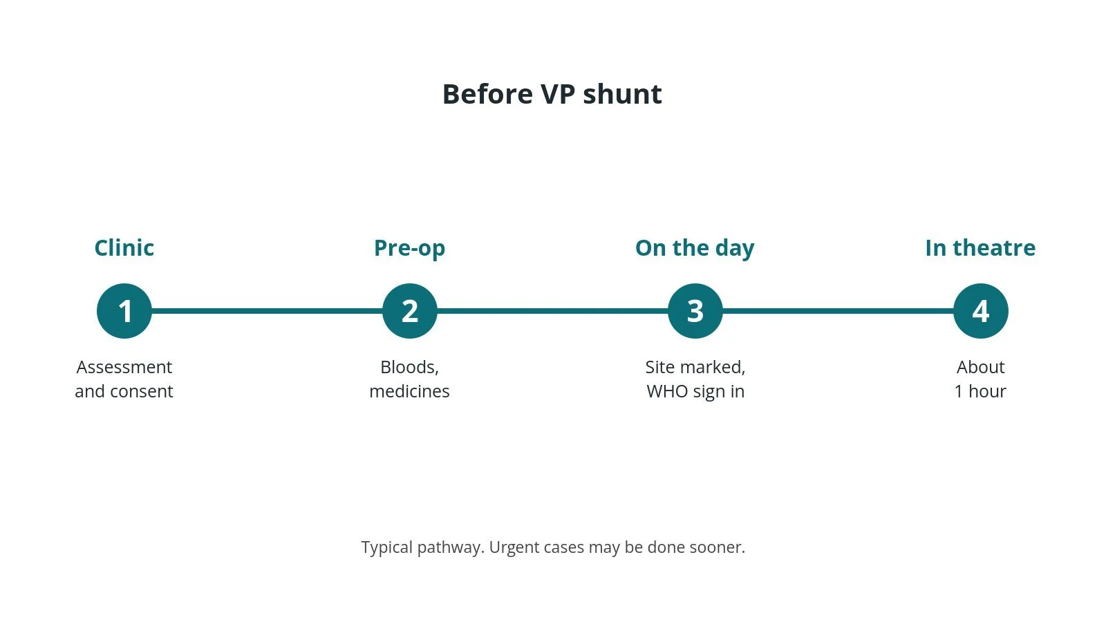
Most operations are planned; some are urgent. Before it, you usually have:
- a CT or MRI scan
- blood tests
- a review of medicines, as per centre protocol
- a chance to ask questions and give consent
Step by step
1. Getting ready
Team brief and WHO checklist first, including the site and side marking. The anaesthetic team and ODP give the anaesthetic (general, as per centre protocol); antibiotics and blood-clot prevention are given as per centre protocol. The circulating practitioner checks positioning, pressure areas, the diathermy plate and warming. The scrub practitioner sets up the instrument trolley and does the first count. Strict infection precautions are followed (limited theatre traffic, implant opened at the last moment), and the shunt check is done.
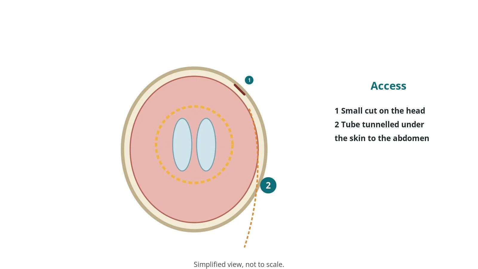
2. Access
A small cut on the head behind the ear or at the front, and a small cut on the abdomen; the tube is tunnelled under the skin between them.
3. Placing the shunt
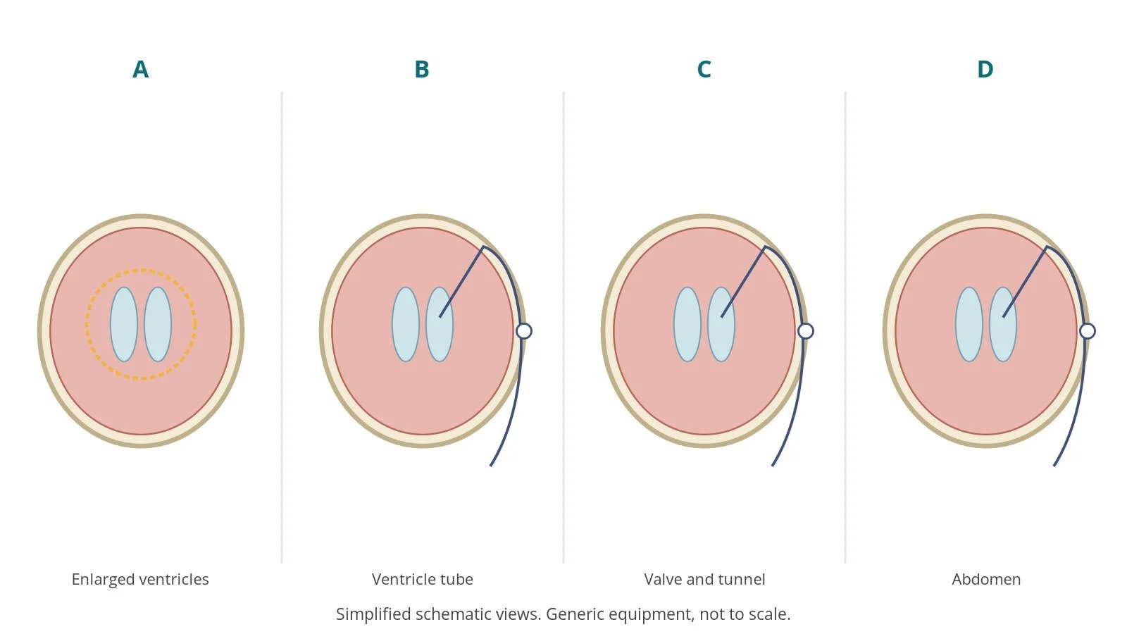
- A. Enlarged ventricles. The enlarged fluid spaces are confirmed.
- B. Ventricle tube. A burr hole is made and the tube passed into the ventricle.
- C. Valve and tunnel. The valve is connected and the tube tunnelled down.
- D. Abdomen. The end is placed in the abdomen and flow checked.
The shunt is handled as little as possible and gloves may be changed before touching it, as per centre protocol.
4. Finishing
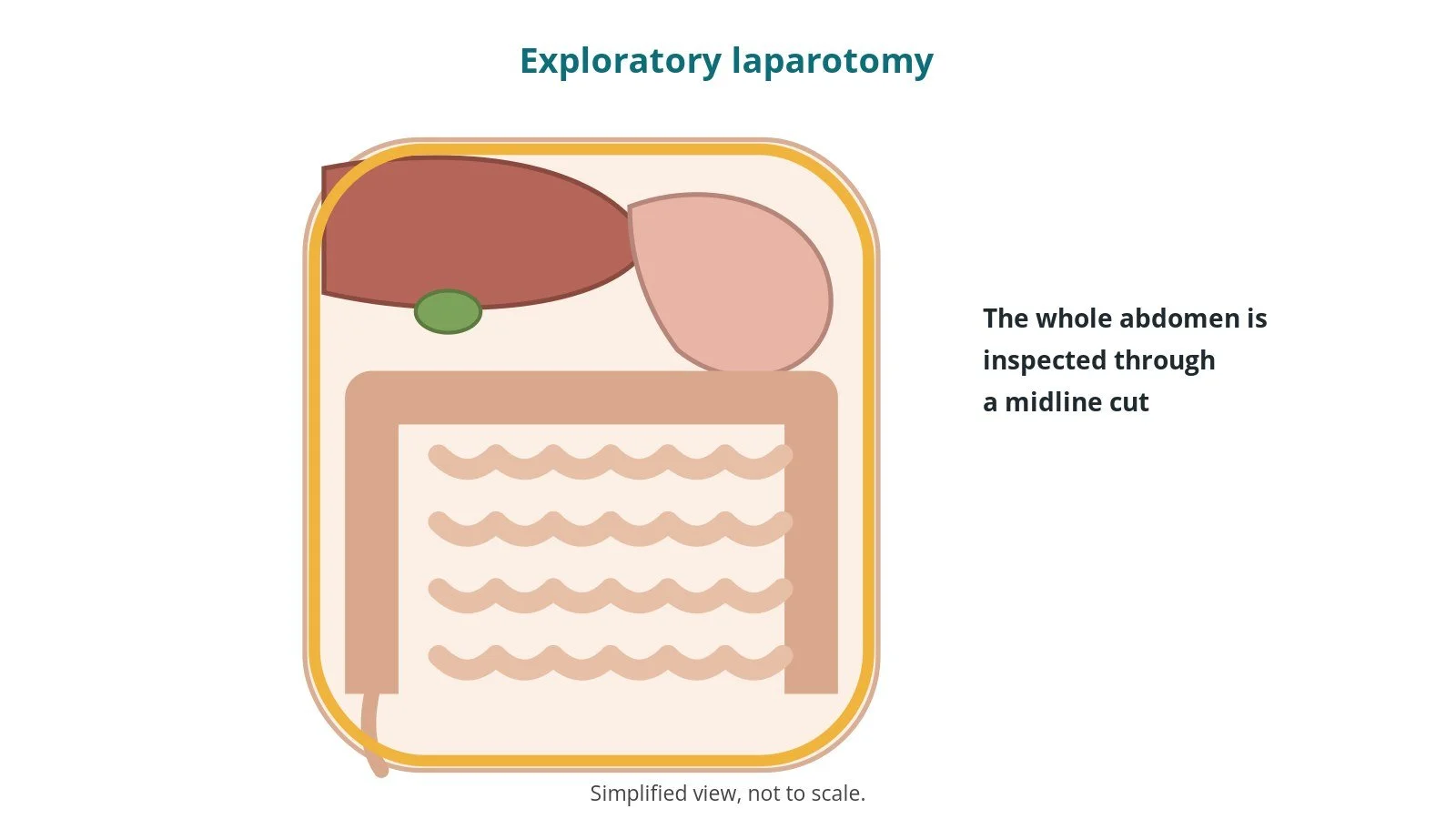
Flow is checked, the cuts closed and dressed. Final counts are confirmed, the shunt details recorded, and the patient goes to recovery with neuro checks.
Recovery and aftercare
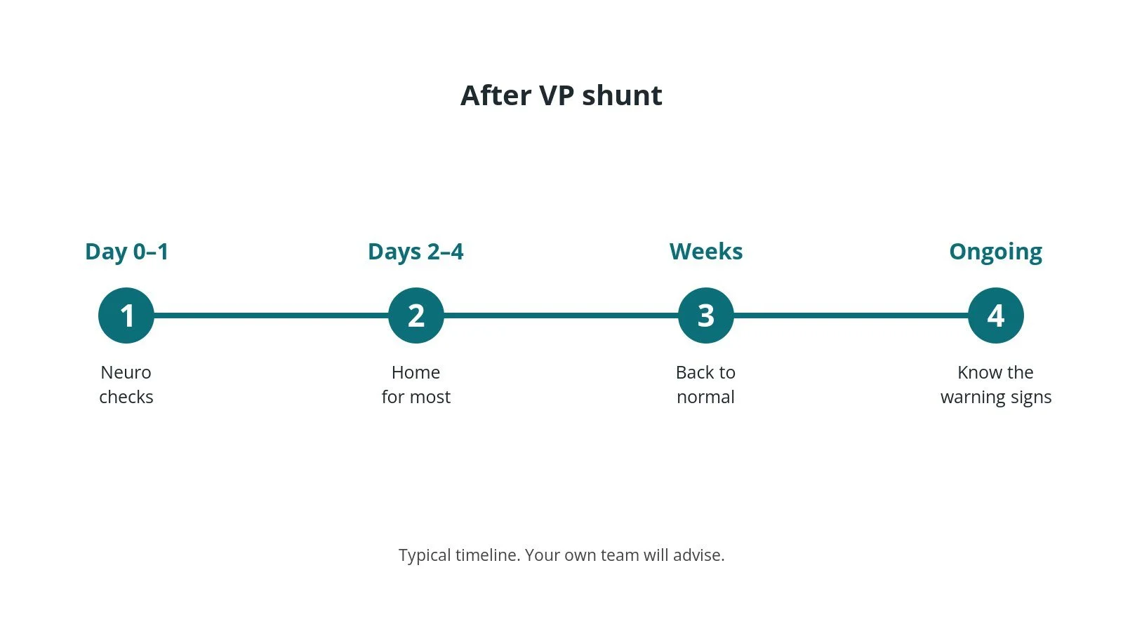
- First days: neuro checks and wound care.
- Going home: often 2–4 days.
- Warning signs: headache, vomiting, drowsiness or fever can mean a problem; seek help quickly.
- Adjustable valves: magnets and MRI can change some settings; tell staff you have a shunt.
- Activity: gradually back to normal.
- Follow-up: clinic review.
Call 999 if you have a seizure, sudden severe headache, confusion, drowsiness, weakness or collapse. Contact your team if you have a fever, leaking wound, worsening headache or vomiting.
Risks and complications
Your team will explain the risks that apply to you. Possible problems include:
- shunt blockage
- infection
- over- or under-drainage causing headaches
- bleeding in the brain
- abdominal problems (rare)
- needing a further operation
Theatre kit list (printable)
Example kit list
Every theatre has its own protocol, and sizes and doses vary.
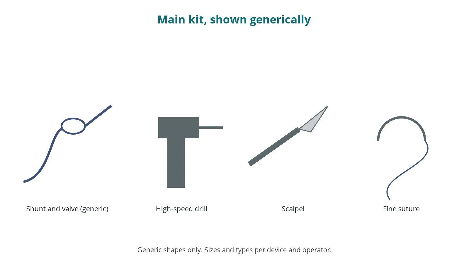
- shunt system with valve, type and setting per device and operator
- tunnelling device
- high-speed drill or perforator
- fine instruments
- antibiotic and infection precautions, as per centre protocol
- navigation or ultrasound, per centre
- closure sutures and dressings
Procedural steps
- Team brief, WHO sign in; shunt check; anaesthesia
- Head turned; prep head to abdomen; count
- Head cut and burr hole; abdominal cut
- Tunnel; valve connected
- Ventricle tube placed; flow checked; abdominal end placed
- Closure; counts; shunt log; sign out and handover
First time in theatre?
💡 First-time tip
- Know your role. Scrub, circulating, anaesthetic and ODP each have their own checks; ask at the team brief.
- Counts matter. Swabs, needles and instruments are counted before, during and at closure; speak up if one is missing.
- Infection control. Limit door opening and keep the shunt in its packet until needed.
- Shunt log. Record the shunt type and valve setting.
- Long prep. The prep runs from head to abdomen; check the drapes cover the whole route.
FAQ
Is the shunt permanent?
Usually, yes; some need revision over time.
Can I have an MRI?
Usually, but some valves need resetting afterwards.
Can I feel it?
You may feel the valve under the scalp.
Related guides
Sources
All checked on 14 October 2026. Each page's own date is shown where it gives one.
- NHS. Hydrocephalus. nhs.uk/conditions/hydrocephalus
- StatPearls (NCBI Bookshelf), search: ventriculoperitoneal shunt. ncbi.nlm.nih.gov/books
- Society of British Neurological Surgeons. www.sbns.org.uk
- World Health Organization. Surgical Safety Checklist. who.int
Disclaimer
Education only. This page is for general education for healthcare students, theatre staff and curious readers. It isn't medical advice and doesn't replace advice from your own doctor, surgical team or local unit protocols. Equipment and medicines are described generically. No product or manufacturer is recommended. If you think someone is having a medical emergency, call 999.
Disclosure: I work in the medical device industry. This page is independent education and isn't sponsored or reviewed by any company.
All diagrams on this page are original illustrations created for rodnieoro.com.