Mitral valve repair and replacement: an OR 101 guide
Reviewed by Rodnie Oro, cath lab charge nurse · Last reviewed: October 2026 · Education only, not medical advice
Mitral valve surgery in one minute
What it is: heart surgery to repair a leaking mitral valve (often with a supporting ring), or to replace it with a new valve.
Who it is for: people with severe mitral regurgitation or stenosis where the heart team agrees surgery is the best option.
Main risks: bleeding, irregular heartbeat, infection, stroke, a valve that still leaks; rarely death.
Usually: under general anaesthetic, 3–5 hours, about a week in hospital.
On this page: What is mitral valve surgery? · Who is it for? · When is it done? · Step by step · Recovery · Risks · Kit list · First time in theatre? · FAQ
What is mitral valve surgery?
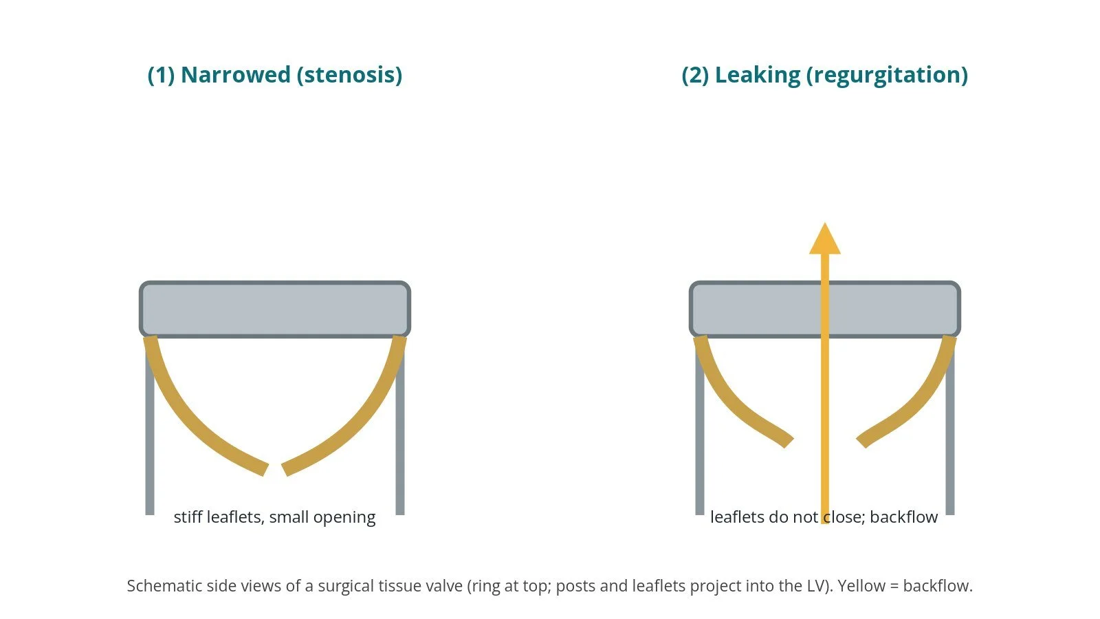
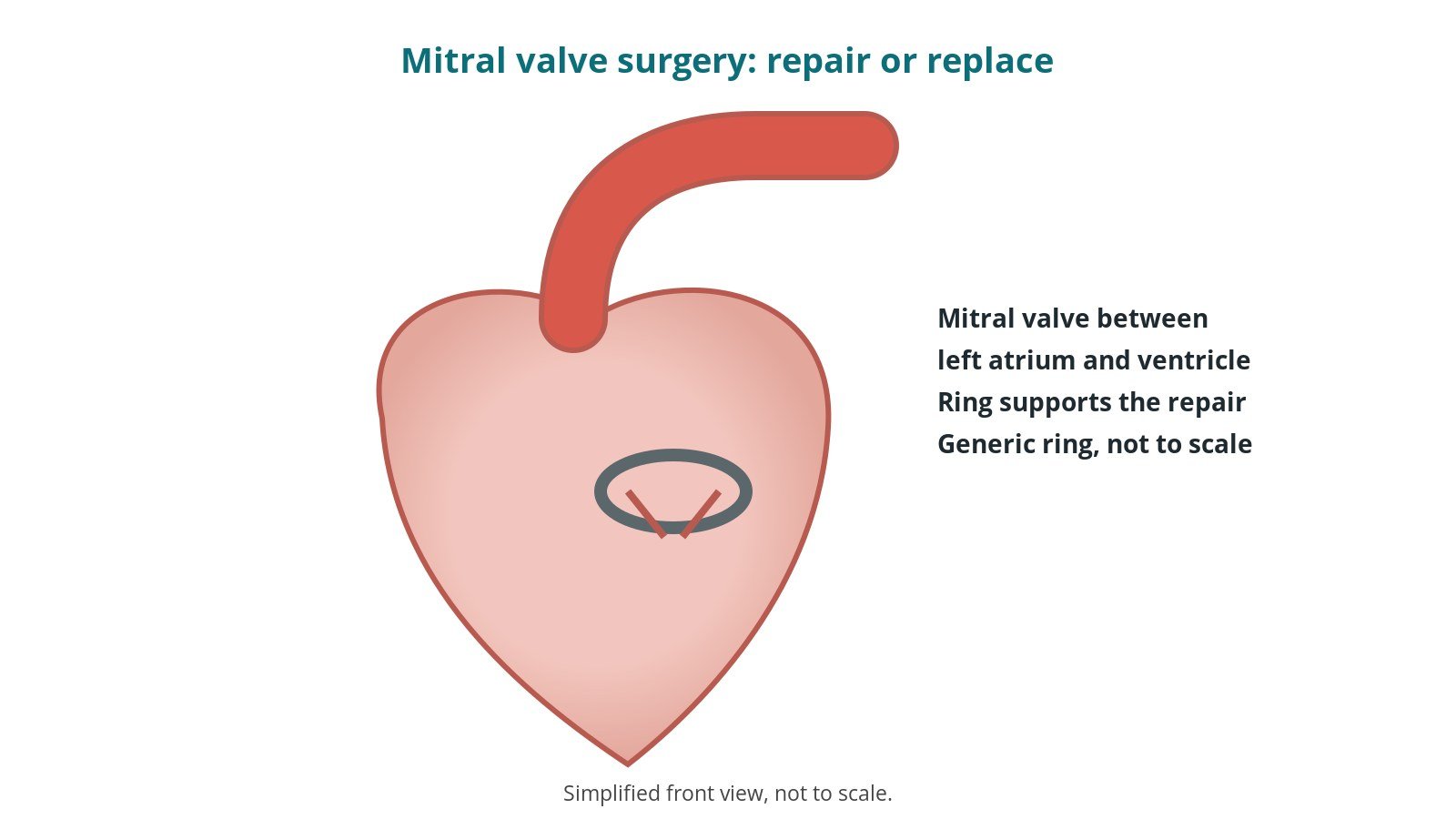
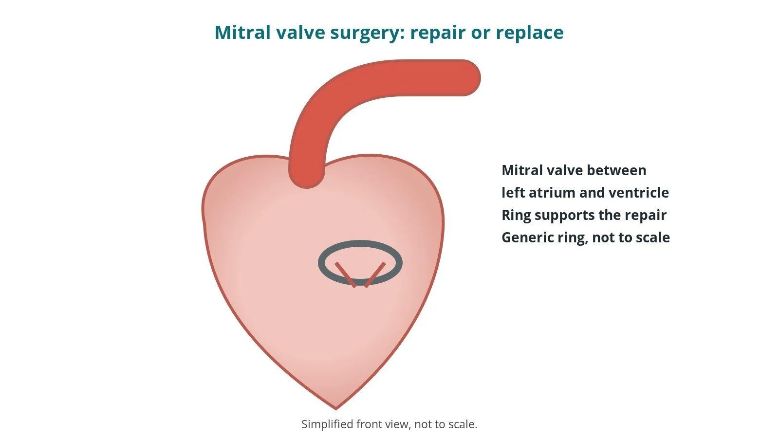
The mitral valve sits between the left atrium and the left ventricle. It can leak (regurgitation), for example when a leaflet is floppy or a supporting chord breaks, or become narrowed (stenosis).
There are two main approaches:
- Repair: the surgeon reshapes the leaflets, replaces broken chords with fine artificial chords and usually adds an annuloplasty ring to support the valve; repair is preferred where possible, per surgeon
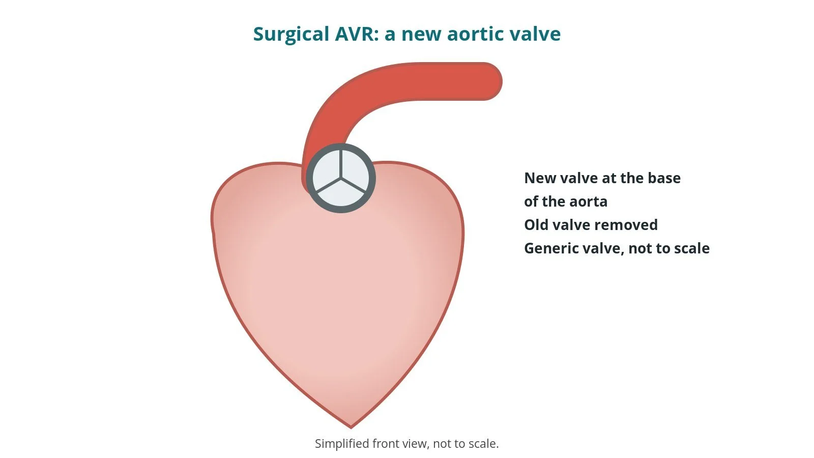
- Replacement: the valve is replaced with a mechanical or tissue valve, often keeping parts of the original supporting structures
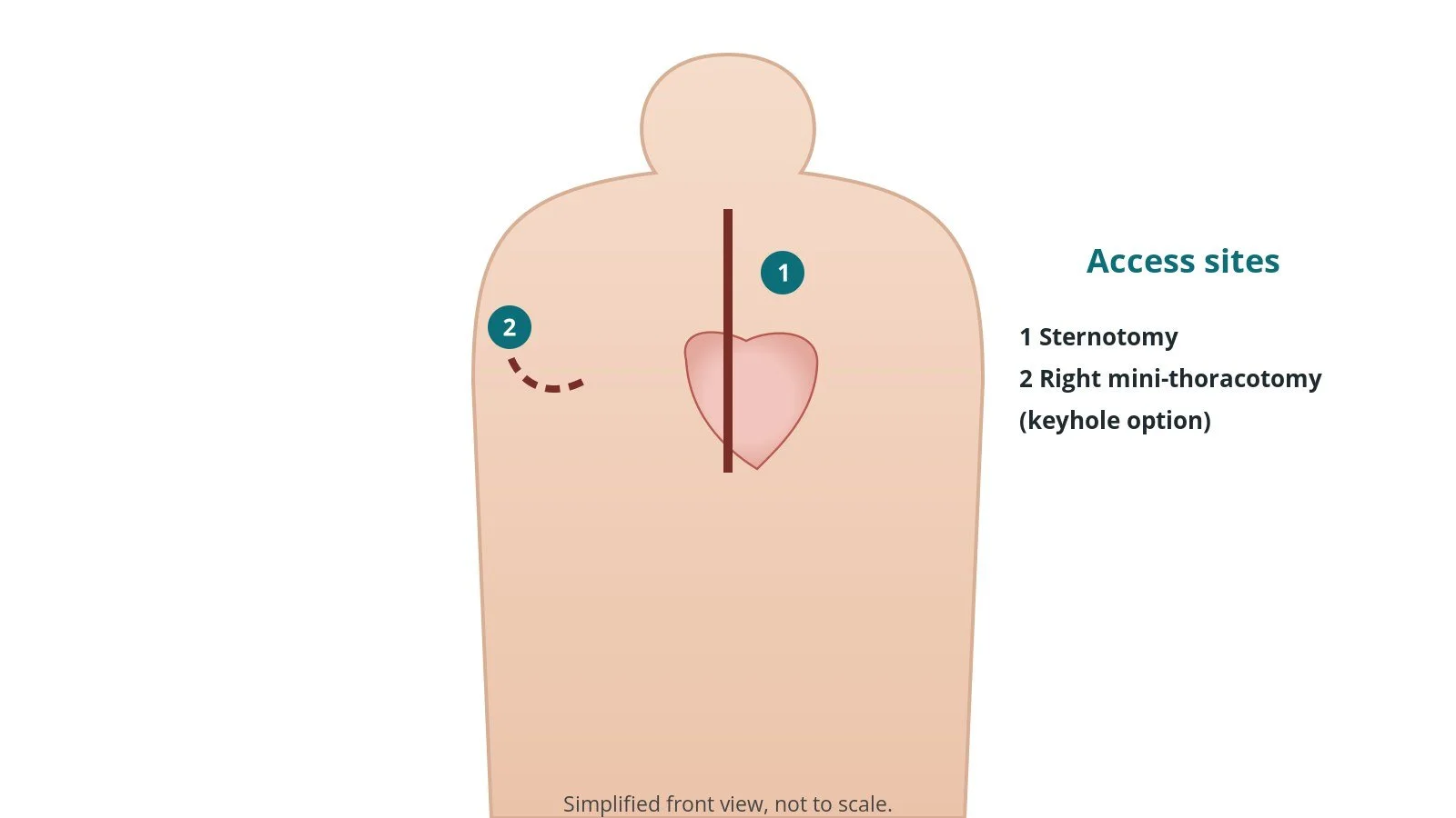
A heart-lung (bypass) machine supports the circulation while the heart is stopped. Surgery can be through the breastbone or through a small cut in the right side of the chest (minimally invasive), per surgeon and centre. Catheter-based options exist for some people.
New to the acronyms? See the Cath Lab Abbreviations A–Z.
Who is it for?
It may be considered for people with:
- severe mitral regurgitation with symptoms, or affecting heart size or function
- severe mitral stenosis not suitable for balloon treatment
- mitral disease plus other heart surgery needed
- infection of the valve (endocarditis), in some cases
The decision is made by your heart team, who will talk through the reasons, the alternatives (including catheter-based options) and the risks with you.
When is it done?
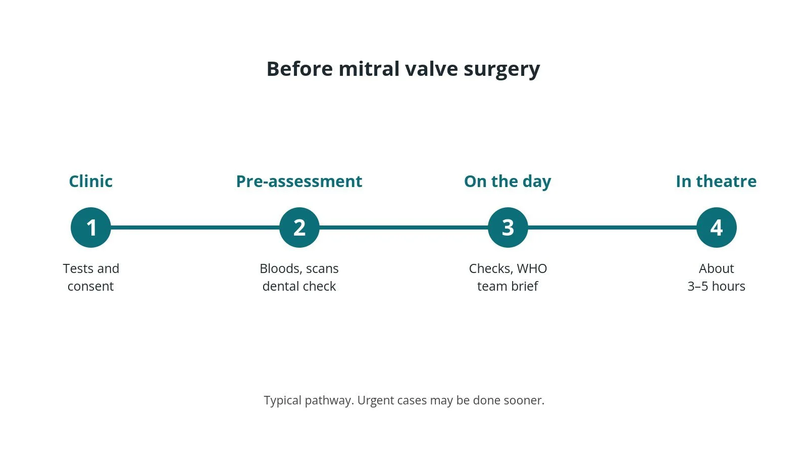
Most operations are planned; some are urgent. Before it, you usually have:
- an echo, often including a TOE, and a coronary angiogram or CT
- blood tests, including kidney function, clotting and group and save / crossmatch
- an ECG and chest X-ray
- a dental check to treat any infection before surgery
- a review of your medicines, especially blood thinners, as per centre protocol
- a chance to ask questions and give consent, including what happens if repair is not possible
Step by step
1. Getting ready
Theatre team brief and WHO checklist first. The anaesthetic team and ODP place large cannulas, an arterial line and a central line, give the general anaesthetic and insert a breathing tube and a heart ultrasound probe (TOE), drugs as per centre protocol. The scrub practitioner sets up the main trolley and valve instruments; the circulating practitioner checks positioning, pressure areas, diathermy plate, warming and that the implant sizes are available; the perfusionist primes and checks the bypass circuit.
2. Access
Either a median sternotomy, or a right mini-thoracotomy (small cut in the right side of the chest) with camera assistance and bypass connected through the groin, per surgeon and centre. Counts are recorded, as per centre protocol.
3. Repairing or replacing the valve
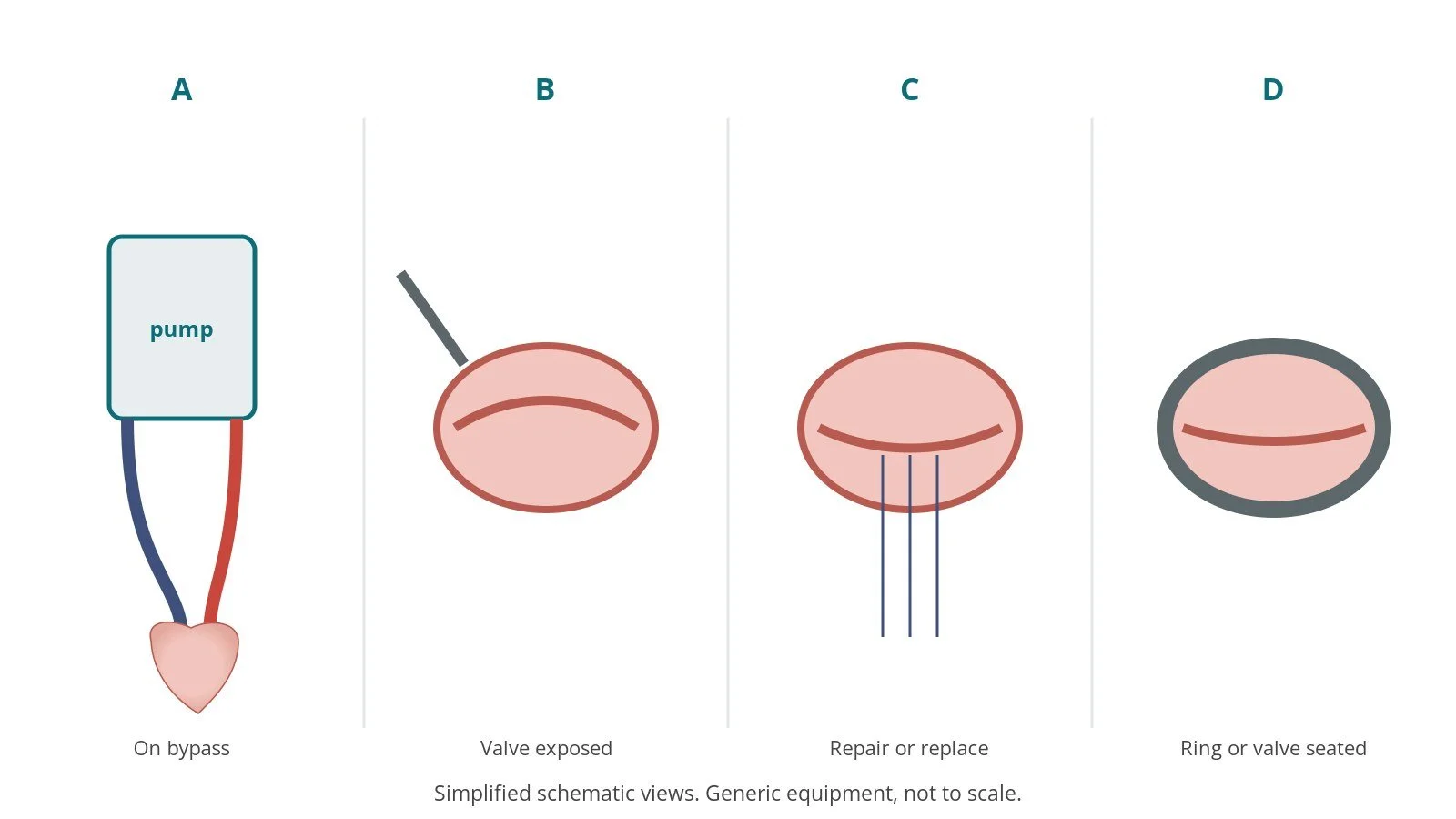
- A. On bypass. Heparin is given (as per centre protocol), cannulas placed and the heart-lung machine started; the aorta is clamped and cardioplegia stops the heart.
- B. Valve exposed. The left atrium is opened and a retractor lifts its wall to show the mitral valve; the surgeon inspects every leaflet.
- C. Repair or replace. Leaflets are reshaped and new chords placed, or the valve is cut out for replacement; sizers measure the opening, size per device and operator.
- D. Ring or valve seated. Stitches are placed around the valve ring and through the ring or new valve, which is lowered and tied; the valve is tested with saline.
After bypass the team checks the result with TOE. If a repair still leaks, the surgeon may go back on bypass to adjust it or replace the valve.
4. Finishing
The heart is restarted and the team checks the valve with TOE before coming off bypass. Heparin is reversed (as per centre protocol). Temporary pacing wires and chest drains are placed, the breastbone is closed with steel wires, and the skin is closed. Implant details are recorded, final counts confirmed, and the patient goes to cardiac intensive care with a full handover.
Recovery and aftercare
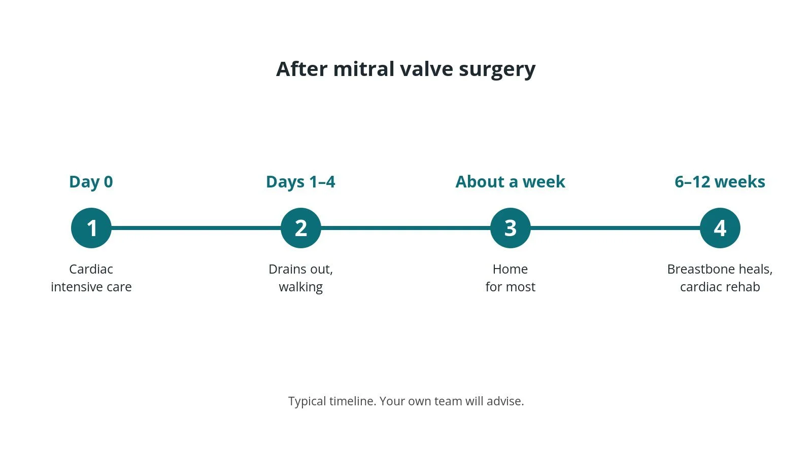
- First hours: in cardiac intensive care; the breathing tube usually comes out within hours, as per centre protocol.
- First days: drains, lines and pacing wires come out; physiotherapy and walking start early.
- Going home: often around 5–7 days after the operation.
- Breastbone: no heavy lifting, pushing or pulling for about 6–12 weeks; ask your team. Keyhole approaches may recover faster.
- Medicines: some people need blood thinners after valve surgery, short term or lifelong; take them exactly as prescribed.
- Dental care and infection: good dental hygiene matters; tell dentists and doctors you have a heart valve.
- Follow-up: clinic review, echo scans and cardiac rehabilitation where offered.
Call 999 if you have chest pain, breathlessness, fainting or signs of a stroke. Contact your team if you have a fever, or a wound becomes red, hot or leaky.
Risks and complications
Your team will explain the risks that apply to you. Possible problems include:
- bleeding needing blood transfusion or a return to theatre
- irregular heartbeat, especially atrial fibrillation; some people need a permanent pacemaker
- wound infection, or infection of the new valve (endocarditis)
- kidney problems, often temporary
- confusion after surgery, or stroke
- a valve that still leaks, needing further treatment
- death (uncommon)
Theatre kit list (printable)
Example kit list
Every theatre has its own protocol, and sizes and doses vary.
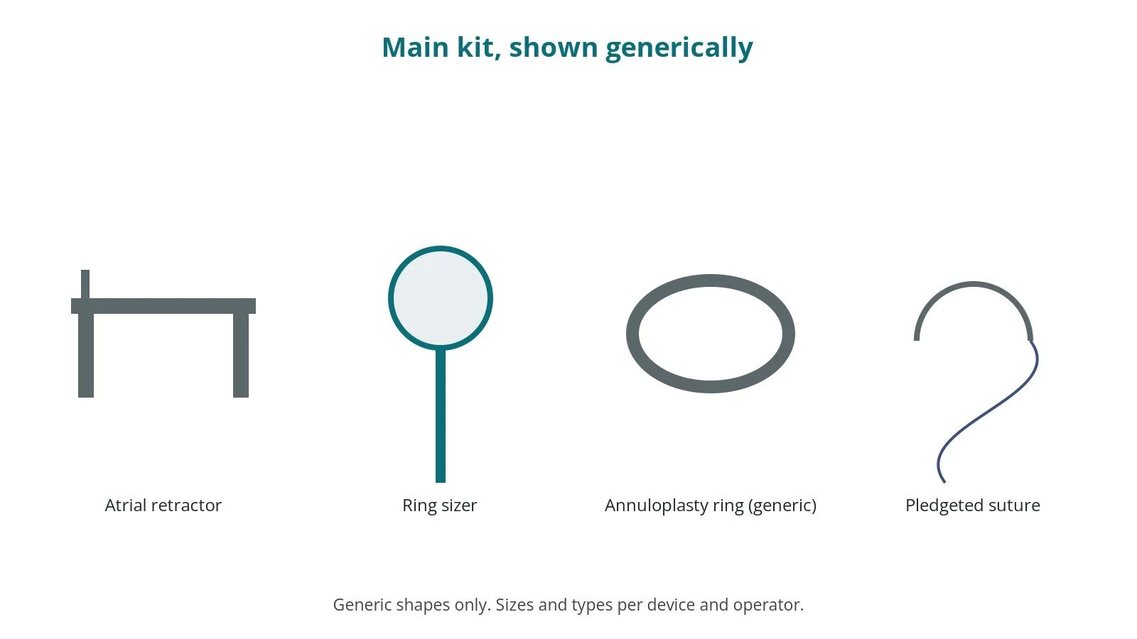
- cardiac major instrument set, sternal saw and sternal retractor (or minimally invasive set and soft-tissue retractor)
- left atrial retractor and long mitral instruments
- annuloplasty ring sizers and ring, or valve sizers and prosthetic valve, size per device and operator
- artificial chord sutures and pledgeted valve sutures, per surgeon
- saline test syringe or bulb
- bypass circuit and cannulas, sizes per device and operator; groin cannulas for keyhole approach
- cardioplegia set and solution, as per centre protocol
- heparin and its reversal, as per centre protocol
- temporary pacing wires and pacing box, chest drains and drainage system
- implant log and labels
Procedural steps
- Team brief, WHO sign in; anaesthesia, lines and TOE
- Prep and drape; counts; sternotomy or mini-thoracotomy
- Heparin; clotting check; cannulation; on bypass
- Cross-clamp, cardioplegia; left atrium opened; valve inspected
- Repair (leaflets, chords, ring) or replacement; implant check; saline test
- Atrium closed; de-air, clamp off, TOE check, wean; reversal; pacing wires, drains; counts; closure; sign out and handover
First time in theatre?
💡 First-time tip
- Know your role. Scrub, circulating, anaesthetic/ODP and perfusion each have their own checks; ask at the team brief.
- Implant check. The valve or ring type and size are read back out loud and checked before opening; record them in the implant log.
- Counts matter. Swabs, needles and instruments are counted before, during and at closure; speak up if one is missing.
- Keep valves wet. Some tissue valves must be rinsed and kept moist, as per the instructions for use; don't let them dry out.
- Pacing and drains. Have the pacing box and drainage system ready before the chest is closed.
FAQ
Is repair better than replacement?
Where a good repair is possible, surgeons usually prefer it. Your team will explain what is likely in your case.
What is the ring?
A flexible or firm ring sewn around the valve to support it and help the leaflets meet.
Can it be done through a small cut?
In some centres and for some people, yes, through the right side of the chest.
Related guides
- Mitral TEER (transcatheter edge-to-edge repair)
- TMVI (transcatheter mitral valve-in-valve)
- Balloon mitral valvuloplasty
- Cath Lab Abbreviations A–Z
Sources
All checked on 14 October 2026. Each page's own date is shown where it gives one.
- Praz F, et al. 2025 ESC/EACTS Guidelines for the management of valvular heart disease. Eur Heart J 2025;46(44):4635–4736. PubMed 40878295
- NHS. Mitral valve problems. nhs.uk/conditions/mitral-valve-problems
- StatPearls (NCBI Bookshelf), search: mitral valve repair. ncbi.nlm.nih.gov/books
- World Health Organization. Surgical Safety Checklist. who.int
Disclaimer
Education only. This page is for general education for healthcare students, theatre staff and curious readers. It isn't medical advice and doesn't replace advice from your own doctor, surgical team or local unit protocols. Equipment and medicines are described generically. No product or manufacturer is recommended. If you think someone is having a medical emergency, call 999.
Disclosure: I work for a company that makes a device used in this procedure. This page is independent education, isn't sponsored or reviewed by any company, and doesn't recommend any product.
All diagrams on this page are original illustrations created for rodnieoro.com.