Lobectomy and wedge resection: an OR 101 guide
Reviewed by Rodnie Oro, cath lab charge nurse · Last reviewed: October 2026 · Education only, not medical advice
Lobectomy and wedge resection in one minute
What it is: lung surgery to remove a whole lobe of the lung (lobectomy) or a small wedge-shaped piece (wedge resection).
Who it is for: people with a lung cancer, a suspicious nodule, or some infections or damaged areas of lung.
Main risks: air leak, bleeding, chest infection, irregular heartbeat; rarely death.
Usually: under general anaesthetic, 1–4 hours, often 2–5 days in hospital.
On this page: What are lobectomy and wedge resection? · Who is it for? · When is it done? · Step by step · Recovery · Risks · Kit list · First time in theatre? · FAQ

What are lobectomy and wedge resection?
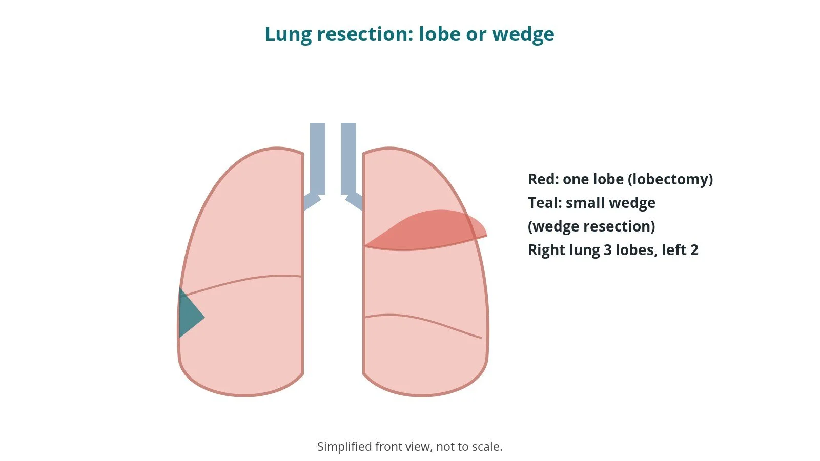
The right lung has three lobes and the left lung two. Each lobe has its own artery, vein and airway (bronchus).
- Lobectomy: the surgeon divides the lobe's artery, vein and airway one by one and removes the whole lobe, usually with nearby lymph nodes for testing
- Wedge resection: a small, wedge-shaped piece of lung around a nodule is removed with a stapler, without dividing the main vessels
- Segmentectomy: in between the two, removing one segment of a lobe (not covered in detail here)
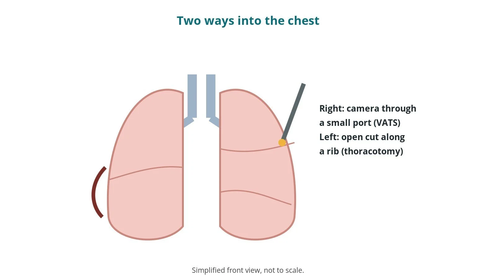
Most operations are done by keyhole surgery (VATS or robotic) through small cuts; some need an open thoracotomy. See the Thoracotomy and VATS guide.
New to the acronyms? See the Cath Lab Abbreviations A–Z.
Who is it for?
It may be considered for people with:
- an early lung cancer suitable for surgery (usually lobectomy)
- a lung nodule that needs removing to find out what it is (often wedge resection)
- some infections, abscesses or damaged areas of lung
- some cancers that have spread to the lung from elsewhere
The decision is made by your specialist (multidisciplinary) team, who will talk through the reasons, the alternatives (such as radiotherapy) and the risks with you.
When is it done?
Most operations are planned; some are urgent. Before it, you usually have:
- CT and often PET-CT scans, and sometimes a biopsy
- breathing (lung function) tests and sometimes an exercise test
- blood tests, an ECG and sometimes an echo
- a review of your medicines, especially blood thinners, as per centre protocol
- advice to stop smoking and keep active before surgery
- a chance to ask questions and give consent

Step by step
1. Getting ready
Theatre team brief and WHO checklist first. The anaesthetic team and ODP give a general anaesthetic and place a double-lumen tube (or blocker) so one lung can be deflated, plus an arterial line and pain relief such as a nerve block or epidural, as per centre protocol. The patient is turned onto their side (lateral position) with careful padding; the circulating practitioner checks pressure areas, the diathermy plate, warming and the correct side marking. The scrub practitioner sets up the thoracic and camera trolleys.
2. Access
Usually VATS: one to four small cuts between the ribs on the side of the chest for a camera and instruments. Some centres use a robot. Open surgery uses a thoracotomy, a longer cut between the ribs with a rib spreader. Counts are recorded, as per centre protocol.

3. Removing the lung tissue
- A. One lung deflated. The lung on the operating side is deflated through the double-lumen tube, giving room to work.
- B. Vessels and airway. For a lobectomy, the lobe's vein, artery and airway are divided one by one, usually with a stapler, per surgeon.
- C. Lobe or wedge removed. The lobe, or a stapled wedge for a wedge resection, is placed in a bag and removed through one of the cuts.
- D. Leak test and nodes. The lung is re-inflated under saline to check for air leaks; lymph nodes are taken for testing.
The scrub practitioner tracks stapler reloads; specimens are labelled with side and site and checked before they leave theatre.

4. Finishing
A chest drain is placed and connected to a drainage system. Local anaesthetic or a nerve catheter may be placed for pain relief. The cuts are closed, final counts confirmed, and the patient wakes and goes to recovery with a full handover.
Recovery and aftercare
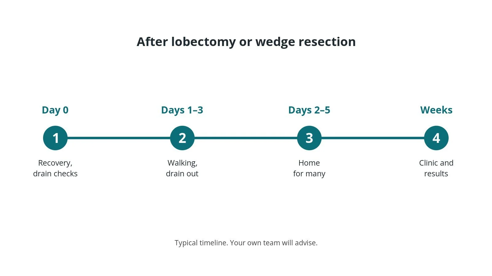
- First hours: in recovery, then a ward or high-dependency bed; oxygen, pain relief and chest drain checks, as per centre protocol.
- Breathing and walking: physiotherapy, deep breathing and walking start on day one.
- Chest drain: usually stays until air leak and fluid settle, often 1–3 days.
- Going home: often 2–5 days after keyhole surgery, longer after open surgery.
- Activity: avoid heavy lifting for several weeks; ask your team.
- Results: tissue results are discussed at clinic, usually within a few weeks.
Call 999 if you have severe breathlessness, chest pain or coughing up a lot of blood. Contact your team if you have a fever, or a wound becomes red, hot or leaky.
Risks and complications
Your team will explain the risks that apply to you. Possible problems include:
- air leak from the lung, keeping the chest drain in longer
- bleeding, rarely needing a return to theatre
- chest infection or pneumonia
- irregular heartbeat, usually atrial fibrillation
- wound pain or numbness that can last months
- blood clots in the legs or lungs
- conversion from keyhole to open surgery
- death (uncommon)
Theatre kit list (printable)
Example kit list
Every theatre has its own protocol, and sizes and doses vary.
- thoracic instrument set; VATS instruments (long graspers, scissors, dissectors)
- camera stack with scope (often 30 degree), light source and warmer
- wound protector or ports, per surgeon
- surgical staplers and reloads, sizes per device and operator
- specimen retrieval bag
- energy device for dissection, per surgeon
- thoracotomy tray and rib spreader available for conversion
- chest drain, drainage system and suction
- local anaesthetic for nerve block, as per centre protocol
- specimen pots, labels and request forms

Procedural steps
- Team brief, WHO sign in; side marked; anaesthesia, double-lumen tube, lines
- Lateral position, padding; prep and drape; counts
- Ports placed; one-lung ventilation; inspection
- Dissection; vessels and airway stapled (lobectomy) or wedge stapled
- Specimen bagged and removed; nodes sampled; leak test
- Chest drain; block; closure; counts; specimen check; sign out and handover
First time in theatre?
💡 First-time tip
- Correct side. The side is marked and checked at sign in and again before the cut; join in the check.
- One-lung ventilation. Anaesthetic and ODP confirm tube position with a camera after turning; stay out of the way at this moment.
- Camera first. Know how to white-balance the camera and warm the scope to stop fogging.
- Specimens. Label every specimen with side and site and check it with the scrub practitioner before it leaves theatre.
- Open set ready. A thoracotomy tray stays available in case keyhole surgery has to convert to open.
FAQ
Can I breathe normally with less lung?
Most people adapt well; tests before surgery check you will have enough lung function.
Will I have a scar?
Keyhole surgery leaves a few small scars on the side of the chest.
When will I know the results?
Tissue results usually take one to two weeks and are discussed at clinic.
Related guides
Sources
All checked on 14 October 2026. Each page's own date is shown where it gives one.
- NICE. Lung cancer: diagnosis and management (NG122). nice.org.uk/guidance/ng122
- NHS. Lung cancer: treatment. nhs.uk/conditions/lung-cancer/treatment
- StatPearls (NCBI Bookshelf), search: lobectomy. ncbi.nlm.nih.gov/books
- World Health Organization. Surgical Safety Checklist. who.int
Disclaimer
Education only. This page is for general education for healthcare students, theatre staff and curious readers. It isn't medical advice and doesn't replace advice from your own doctor, surgical team or local unit protocols. Equipment and medicines are described generically. No product or manufacturer is recommended. If you think someone is having a medical emergency, call 999.
Disclosure: I work in the medical device industry. This page is independent education and isn't sponsored or reviewed by any company.
All diagrams on this page are original illustrations created for rodnieoro.com.