Chronic total occlusion (CTO PCI): a Cath Lab 101 guide
Reviewed by Rodnie Oro, cath lab charge nurse · Last reviewed: October 2026 · Education only, not medical advice
CTO in one minute
What it is: a chronic total occlusion (CTO) is a coronary artery that has been completely blocked for about 3 months or more. Small natural bypass vessels (collaterals) often keep some blood flowing to the heart muscle beyond the blockage. CTO PCI is a specialist keyhole (catheter) procedure to reopen the artery with wires, balloons and stents.
Who it is for: people whose angina or breathlessness continues despite medicines, or who have a large area of reduced blood supply (ischaemia) in living heart muscle fed by the blocked artery. A specialist heart team weighs it against medicines alone or bypass surgery.
Main risks: a tear or leak in the artery (perforation), fluid around the heart (tamponade), injury to the artery supplying the collaterals, bleeding from the access site, and strain on the kidneys from contrast dye. The risks are higher than for a routine stent.
Usually: local anaesthetic with sedation, often 2 to 4 hours or more, then home the same or next day for many people.
On this page: What is a CTO? · Who is it for? · When is it done? · Step by step · Recovery · Risks · Kit list · First time in the lab? · FAQ
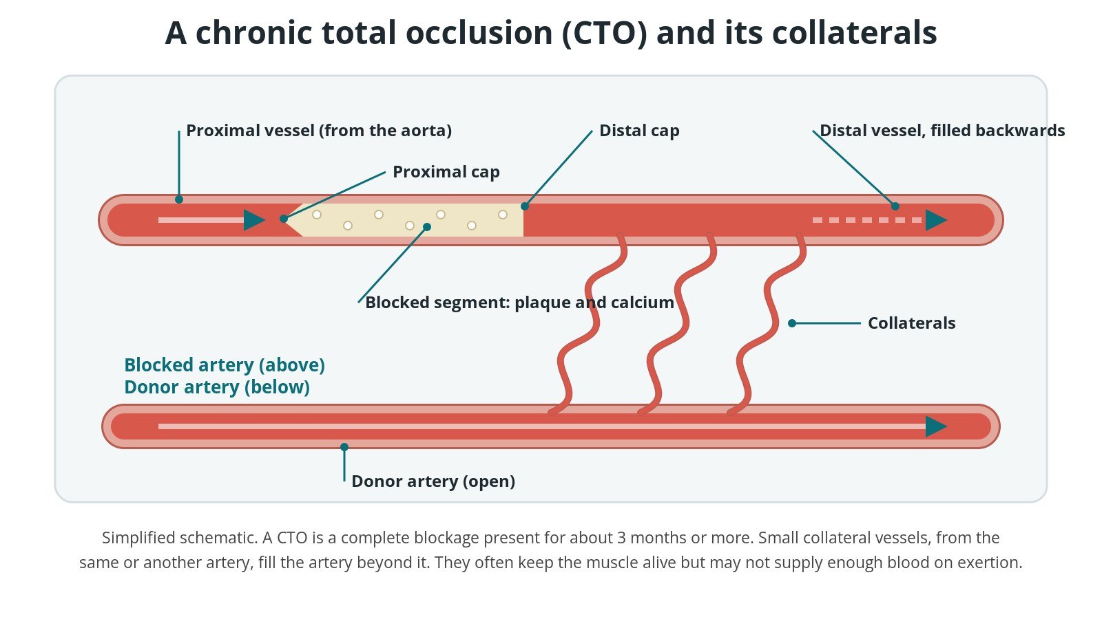
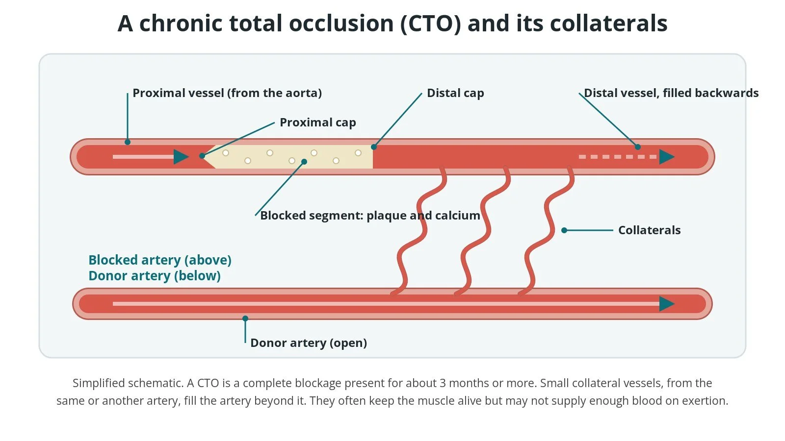
A CTO: (1) proximal cap; (2) blocked segment; (3) distal cap; (4) collaterals from a donor artery fill the artery beyond.
What is a CTO?
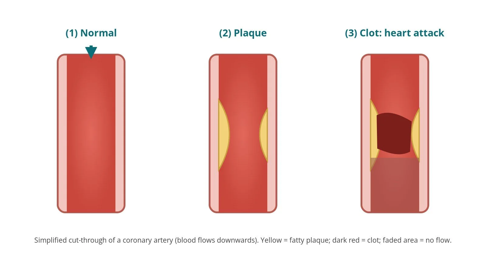
The coronary arteries run over the surface of the heart and supply the heart muscle with blood. In coronary artery disease, fatty, calcified plaque narrows them. Sometimes an artery closes completely, either suddenly during a heart attack or slowly over time. When a complete blockage has been there for about 3 months or more, it is called a chronic total occlusion (CTO).
A CTO has a few parts that matter for treatment:
Proximal cap: the hard start of the blockage, nearest the aorta. It may be tapered, blunt or hard to see (ambiguous), especially at a side branch.
Occluded segment: the blocked length, often with calcium and bends.
Distal cap and distal vessel: the end of the blockage, and the artery beyond it that needs to be reached.
Collaterals: small vessels, from the same artery or from another one, that grow around the blockage and fill the artery beyond it. They often keep the heart muscle alive but may not supply enough blood during exercise.
CTOs are common in people having coronary angiograms. Many cause no symptoms, but some cause angina or breathlessness that medicines do not control.
New to the acronyms? See the Cath Lab Abbreviations A–Z. For routine stenting, see the PCI guide.
Who is it for?
The decision is made by a heart team, usually an interventional cardiologist with CTO experience, a cardiac surgeon and the person's own cardiologist. European (ESC 2024 chronic coronary syndromes) guidance and the EuroCTO Club and global expert consensus documents broadly agree that:
everyone with a CTO should first be on the best medical treatment for angina and for preventing future heart problems
CTO PCI is considered when angina or ischaemia continues despite medicines, in the area of heart muscle supplied by the blocked artery
the heart muscle beyond the blockage should be alive (viable). Tests such as cardiac MRI, stress echo or nuclear scans can show this
the choice between CTO PCI, bypass surgery (CABG) or medicines alone depends on symptoms, the other arteries, the person's overall health and their wishes. Bypass may be better when several arteries are badly diseased
CTO PCI is done by experienced operators in centres with the right equipment and on-site support
The main aim of CTO PCI is to improve symptoms and quality of life. People should be told honestly about the chance of success, the risks, and that more than one attempt is sometimes needed.
When is it done?
CTO PCI is a planned procedure. It is not usually done during an emergency. Before it, the team usually:
reviews the previous angiogram in detail, ideally with injections of both the blocked artery and the artery supplying the collaterals
may do a CT coronary angiogram to show the course of the blocked segment, the calcium and the length of the blockage
assesses symptoms, ischaemia and viability with stress or MRI tests
checks kidney function and bloods, and reviews medicines including blood thinners and diabetes tablets, as per centre protocol
plans contrast and radiation limits for the case and agrees the strategy and back-up options
talks through the benefits, the chance of success and the risks before consent
Step by step
1. Set-up and access
Most CTO PCIs are done under local anaesthetic with light sedation. The procedure can be long, so pressure-area care, padding and comfort matter. Access is usually through two arteries, the radial at the wrist and/or the femoral in the groin, often both sides. Sheath and guide catheter sizes are per device and operator. Anticoagulation is given and checked regularly with blood tests (ACT), as per centre protocol.
2. Dual injection and planning
A guide catheter is placed in the blocked artery and a second in the artery that supplies the collaterals. Injecting contrast through both at once (dual injection) shows the blockage from both ends: the proximal cap, the length, the vessel beyond it and the collateral channels. Without this, the operator can see only one side of the problem.
Most experienced teams use a structured hybrid approach (a step-by-step decision method). It looks at four things:
whether the proximal cap is clear or ambiguous
the length of the blockage
the quality of the distal vessel, its size, branches and disease
whether there are collaterals suitable for a retrograde approach
The team chooses a first strategy from these features and switches early to another strategy if it is not working. This aims to improve success while keeping time, contrast and radiation down.
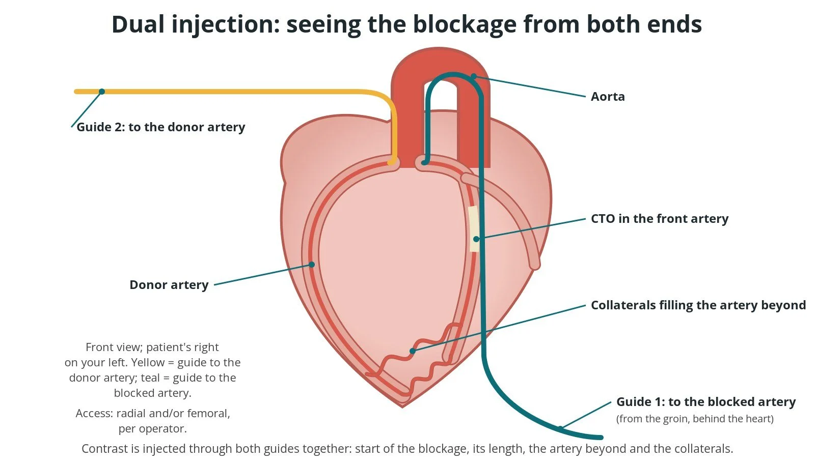
Dual injection: one guide in the blocked artery, one in the donor artery. Injecting both together shows the blockage from both ends.
3. Crossing strategies
A. Antegrade wire escalation. Working forwards from the start of the blockage, the operator steers a guidewire through it, supported by a microcatheter (a thin catheter that backs up the wire and allows wires to be swapped). If a soft wire does not cross, stiffer or more penetrating wires are tried step by step. This is often the first choice for shorter blockages with a clear entry point.
B. Antegrade dissection and re-entry (ADR). The wire or a dedicated blunt crossing catheter is passed deliberately within the artery wall (the subintimal space) alongside the blockage. Then, beyond it, a re-entry device (for example a flat balloon with side exit ports) or a specialised wire is used to puncture back into the true channel of the artery. This is often used for longer blockages with a good distal vessel.
C. Retrograde approach. The operator reaches the blockage from the far end, by passing a wire and microcatheter through a suitable collateral from another artery or a bypass graft. The blockage is then crossed backwards, either by wiring or by joining the two channels with balloon inflations from the front (reverse controlled dissection and re-entry). Finally, a long wire is passed through to the front guide catheter (externalisation) so equipment can be delivered from the front. This is often used when the proximal cap is ambiguous or the distal vessel is poor.
Operators often use more than one strategy in the same case. Extra support may come from guide extension catheters or from anchoring balloons in side branches. Wire and device choices are per operator.
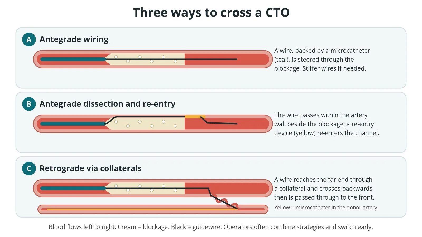
A: antegrade wiring. B: antegrade dissection and re-entry (ADR). C: retrograde approach via collaterals. Operators often combine them.
4. Imaging, balloon and stent
Confirm the wire position. Once across, the operator confirms the wire is in the true channel of the distal artery, using contrast injections and often intravascular imaging: either IVUS (ultrasound inside the artery) or OCT (light-based imaging). IVUS is often used in CTOs because it can guide wiring from inside the artery and does not need extra contrast flushing.
Prepare the vessel. The blockage is opened with balloons, sometimes with extra tools for heavy calcium, as per operator.
Stent. Drug-eluting stents are placed to hold the artery open, often over a long length. Imaging helps choose the size and length, and checks that the stents are fully expanded and the edges are healthy.
Check the result. Final dual injections check the flow, the side branches, and any tear or leak. In a retrograde case, the collateral channel and donor artery are checked carefully for injury before equipment is removed.
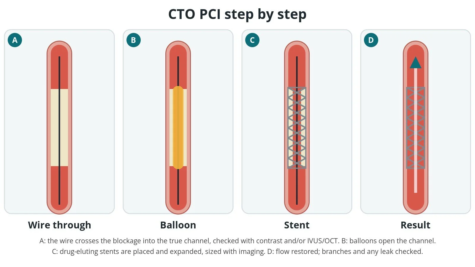
A: wire through. B: balloon. C: drug-eluting stent, sized with imaging. D: flow restored.
5. Finishing
The wires and catheters are removed. The wrist is closed with a compression band, and the groin with pressure or a closure device, per operator. Total contrast volume, radiation dose and procedure time are recorded. If the radiation skin dose is high, the person is told to look out for skin changes on the back and is followed up as per centre protocol.
If the artery cannot be opened safely, the team stops. A second attempt, sometimes with a different strategy, can be planned later.
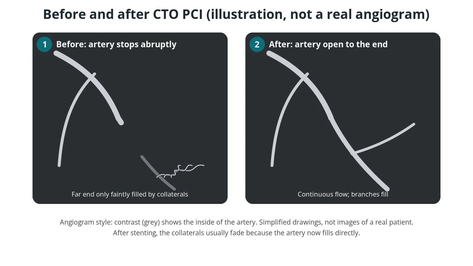
Illustration, not a real angiogram. Before: the artery stops abruptly. After: open to the end; the collaterals fade.
Recovery and aftercare
First hours. Rest, with checks of the wrist or groin, blood pressure, heart rhythm, ECG and any chest pain, as per centre protocol. Some centres do an echo if there is any concern about fluid around the heart.
Kidneys. Fluids may be given and kidney blood tests repeated, especially if kidney function was reduced before.
Going home. Many people go home the same day or the next day, and some stay longer.
Access site. Avoid heavy lifting and strenuous exercise for at least a week, or as your team advises. Keep the site clean and dry.
Medicines. Antiplatelet medicines keep the stents open. Do not stop them unless your cardiologist tells you to. Your team will tell you which to take and for how long.
Skin. If you were told your radiation dose was high, watch for redness or soreness on your back over the following weeks and tell your team.
Follow-up. A clinic review, and cardiac rehabilitation if offered. Symptoms are reassessed over the following weeks and months.
Driving (UK). DVLA guidance after a planned (elective) PCI: car and motorcycle drivers must not drive for at least 1 week, and need not tell the DVLA. Bus and lorry drivers must not drive, must tell the DVLA, and may be relicensed after at least 6 weeks if they meet the tests. Check the current GOV.UK guidance.
Contact your team or NHS 111 if the wrist or groin swells, bleeds or becomes painful, or if you feel unwell. Call 999 for chest pain that does not settle, fainting or severe breathlessness.
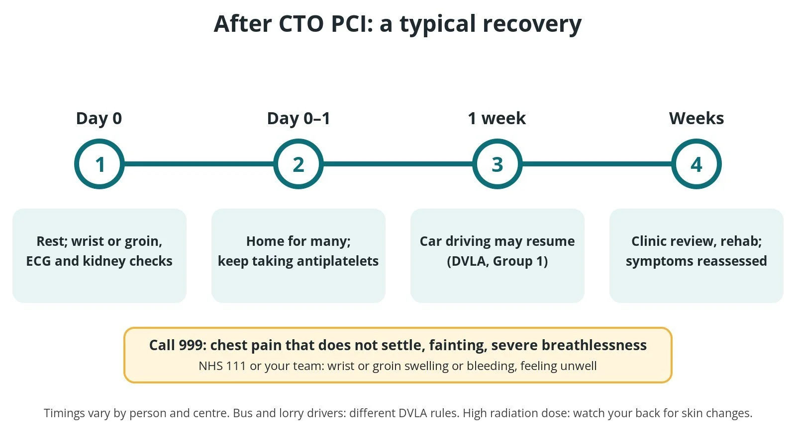
After CTO PCI: (1) rest and checks; (2) home for many, keep taking antiplatelets; (3) car driving after 1 week (DVLA Group 1); (4) review and rehab.
Risks and complications
CTO PCI is more complex than routine PCI, and the risks are higher. Possible problems include:
Perforation: a tear or leak in the coronary artery or a collateral channel, which can let blood leak around the heart
Tamponade: fluid or blood around the heart pressing on it. It may need a drain (pericardiocentesis), covered stents, coils, or rarely surgery
Injury to the donor artery, the artery supplying the collaterals, during a retrograde approach, which can reduce blood supply to a large area of heart
Heart attack during or after the procedure, including from blocked side branches
Bleeding or damage at the wrist or groin, especially with large or multiple sheaths
Kidney injury from the contrast dye, more likely if kidney function is already reduced
Radiation skin injury after long procedures (uncommon)
Stroke, emergency bypass surgery or death (rare)
Not being able to open the artery, which may mean another attempt or staying on medicines
Your team will explain how these apply to you.
Cath lab kit list (printable)
Drugs, doses and dilutions: always as per centre protocol. Equipment, sizes and choices also vary by unit and operator. Device and product names below are generic. Always check the sheath, wire and guide catheter sizes required in each product's instructions for use.
Example: Barts Heart Centre kit list — always follow your own centre protocol
Typical setup for CTO PCI, shared as one real-world example. It is not a protocol for your unit.
Scrub trolley
angiography pack, with an extra drape for bi-radial access
skin antiseptic applicators and clear dressings
saline bag and spike, as per centre protocol
two manifolds and two 3-way taps, colour-tagged (for example red for the retrograde side and blue for the antegrade side)
two PCI kits (inflation devices and short extensions)
contrast, as per centre protocol, and a Luer-lock syringe
two haemostasis valves
two radial or femoral sheaths, size per device and operator
small syringes for ACT checks
radiation shielding drape or pad, per operator
small and large gauzes
Additional equipment, as agreed at the team brief
guide catheters for both arteries, size and shape per operator
workhorse and specialist CTO guidewires (soft polymer-jacketed, tapered-tip and stiffer penetrating wires), per operator
microcatheters, including dual-lumen microcatheters, per operator
long exchange and externalisation wires
blunt dissection crossing catheter and re-entry system, per operator
guide extension catheters
snares
intravascular imaging catheter (IVUS or OCT), per operator
compliant, non-compliant and small balloons; calcium modification tools if needed, per operator
drug-eluting stents
bail-out kit ready: covered stents, coils or other embolisation material, pericardiocentesis kit, per centre protocol
Drugs: local anaesthetic, sedation, anticoagulation and its reversal, intracoronary vasodilators and emergency drugs, all as per centre protocol.
Procedural steps (Barts example)
Team brief: strategy, back-up plan, contrast and radiation limits agreed
Dual arterial access (radial and/or femoral), per operator
Anticoagulation as per centre protocol; ACT checked regularly
Guide catheters engaged in the blocked artery and the collateral donor artery
Dual injection to review the proximal cap, length, distal vessel and collaterals
First strategy chosen (antegrade wiring, ADR or retrograde); early switch if not progressing
Wire position in the true channel confirmed with contrast and/or intravascular imaging
Balloon preparation, then drug-eluting stents, sized with imaging
Final dual injection: flow, side branches, perforation check; donor artery and collateral checked after retrograde
Equipment removed; access sites closed; contrast, radiation dose and time recorded
Handover: access sites, kidney plan, skin-dose follow-up if needed
First time in the cath lab?
💡 First-time tip
Label everything. Two guides, two manifolds and two sets of wires: tag the antegrade and retrograde sides so nobody injects the wrong one. Count the contrast and the dose. Keep a running total and tell the operator when you reach the limits agreed at the brief. Keep the ACT on time. Long cases need regular checks, as per centre protocol. Have the bail-out kit ready. Know where the pericardiocentesis kit, covered stents and coils are, and check the echo machine is close. Watch the patient. New chest pain, a falling blood pressure or a change in the ECG can be an early sign of perforation or tamponade. Say so straight away.
FAQ
How is a CTO different from a normal narrowing?
A normal narrowing still lets some blood through, so a wire usually passes easily. A CTO is completely blocked, often hard and calcified, so special wires and techniques are needed.
Will I be awake?
Usually yes, with sedation and local anaesthetic. The procedure can be long, so tell the team if you are uncomfortable.
What if it can't be opened?
The team stops if it is not safe to continue. A second attempt, sometimes with a different technique, can be planned, or you may continue with medicines or be considered for surgery.
Why are two arteries used for access?
So the team can inject dye into both the blocked artery and the artery that feeds the collaterals, and sometimes approach the blockage from both ends.
How long will the stents last?
Stents stay in for life. Taking your antiplatelet medicines, not smoking and controlling your blood pressure, cholesterol and diabetes all help keep them open.
Related Cath Lab 101 guides
PCI (coronary angioplasty and stents): start here for routine stenting
Sources
All checked on 10 October 2026. Each page's own date is shown where it gives one.
Vrints C, et al. 2024 ESC Guidelines for the management of chronic coronary syndromes. Eur Heart J 2024;45(36):3415–3537. PubMed 39210710
Galassi AR, et al. Percutaneous recanalisation of chronic total occlusions: 2019 consensus document from the EuroCTO Club. EuroIntervention 2019;15(2):198–208. PubMed 30636678
Brilakis ES, et al. Guiding principles for chronic total occlusion percutaneous coronary intervention: a global expert consensus document. Circulation 2019;140(5):420–433. PubMed 31356129
British Cardiovascular Intervention Society (BCIS). Chronic total occlusion: public information. bcis.org.uk
NHS. Coronary angioplasty. nhs.uk
British Heart Foundation. Coronary angioplasty and stents. bhf.org.uk
DVLA / GOV.UK. Cardiovascular disorders: assessing fitness to drive. gov.uk (last updated 7 Nov 2025)
Disclaimer
Education only. This page is for general education for healthcare students, cath lab staff and curious readers. It isn't medical advice and doesn't replace advice from your own doctor, heart team or local unit protocols. Equipment and medicines are described generically. No product or manufacturer is recommended. If you think someone is having a heart emergency, call 999.
Disclosure: I work in the medical device industry. This page is independent education and isn't sponsored or reviewed by any company.
All diagrams on this page are original illustrations created for rodnieoro.com.