Left main and bifurcation PCI: a Cath Lab 101 guide
Reviewed by Rodnie Oro, cath lab charge nurse · Last reviewed: November 2026 · Education only, not medical advice
Left main and bifurcation PCI in one minute
What it is: PCI to a narrowing where a coronary artery divides (a bifurcation), including the left main stem, which supplies most of the left side of the heart.
Who it is for: People with a bifurcation or left main narrowing that the heart team has decided is best treated with PCI rather than bypass surgery or medicines alone.
Main risks: Closure of the side branch, low blood pressure or a dangerous rhythm while the left main is blocked by a balloon, bleeding, artery tear, and stent problems later. Your team will explain your own risk.
Usually: Awake, under local anaesthetic, about 1.5 to 2.5 hours; often one night in hospital.
On this page: What is left main and bifurcation PCI? · Who is it for? · When is it done? · Step by step · Recovery · Risks · Kit list · First time in the lab? · FAQ
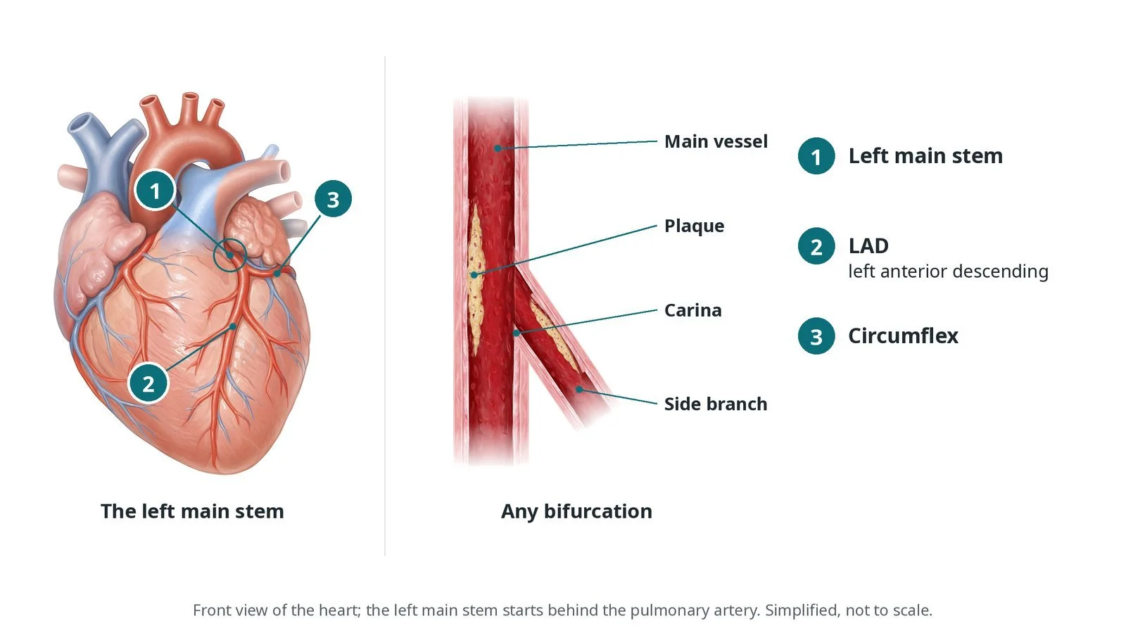
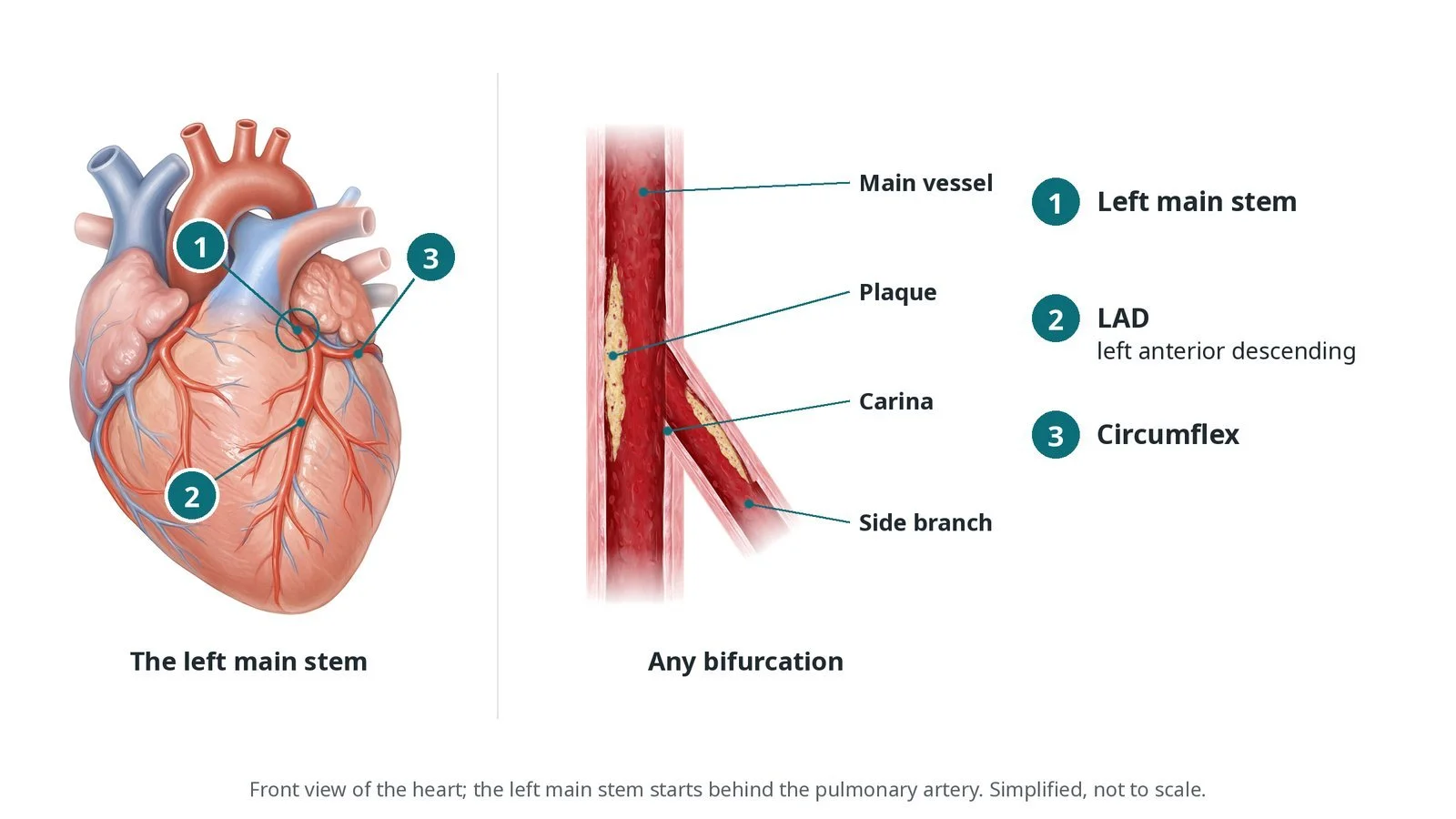
(1) The left main stem divides into (2) the LAD and (3) the circumflex. Any bifurcation has a main vessel, a side branch and the carina where they divide; plaque often sits on the walls opposite the carina.
What is left main and bifurcation PCI?
A bifurcation is where a coronary artery divides into a main vessel and a side branch. The point where they split is the carina. Plaque usually sits on the walls opposite the carina and can involve one or both branches.
The left main stem is short and wide. It divides into the left anterior descending (LAD) and the circumflex, and together they supply most of the left ventricle. A problem here affects a large area of heart muscle, so planning matters.
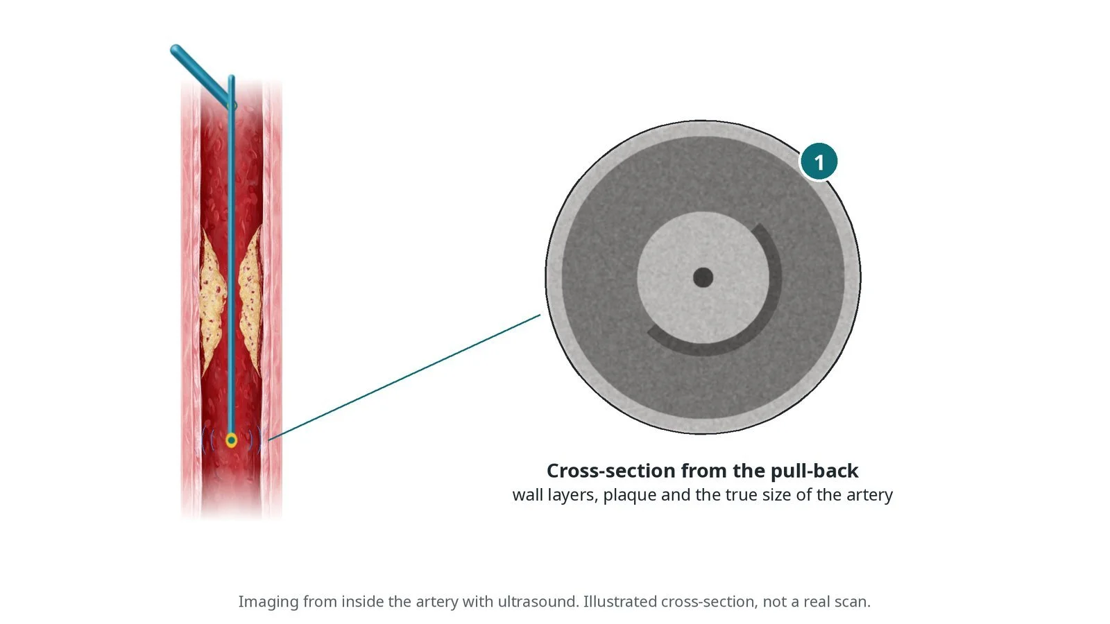
Most bifurcations are treated with a provisional strategy: one stent in the main vessel, with a second stent in the side branch only if it is needed. Complex side-branch disease may need a planned two-stent technique such as DK-crush, culotte, or T and protrusion (TAP). Intravascular imaging (IVUS or OCT) is strongly favoured in current ESC guidance for left main and complex bifurcation PCI, to size and check the stents.
New to the acronyms? See the Cath Lab Abbreviations A–Z.
Who is it for?
For left main disease, the heart team (cardiologists and surgeons) discusses PCI and bypass surgery with you. The choice depends on the anatomy, other illnesses and your preference.
For other bifurcations, the operator plans the strategy from the angiogram and imaging.
Who it may not suit: Some people with complex left main and multivessel disease do better with bypass surgery. The heart team advises.
The decision is made by your heart team, who will talk through the reasons, the alternatives and the risks with you.
When is it done?
Planned, after heart team discussion, or urgently in a heart attack. Before it the team checks kidney function, blood thinners and heart pumping function, and decides whether heart support (such as a heart pump or balloon pump) should be on standby or placed first.
gives you a chance to ask questions and give consent
arranges blood tests, including kidney function and a blood count, and an ECG
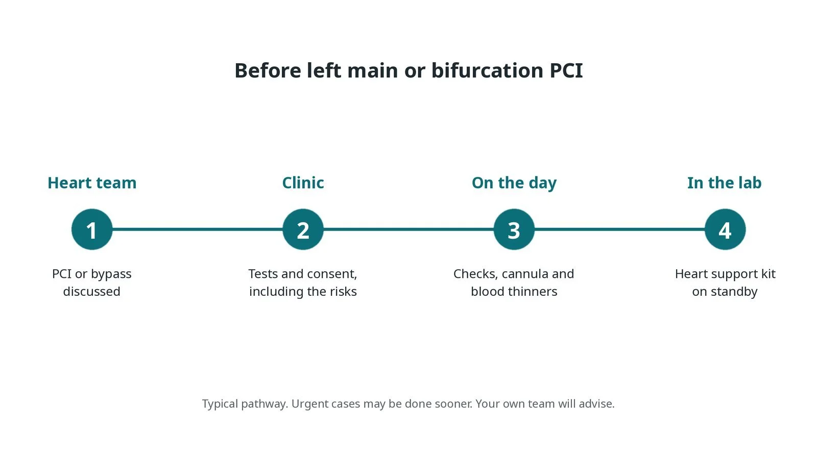
Before left main or bifurcation PCI: the heart team discusses PCI or bypass surgery; tests and consent; checks, a cannula and blood thinners on the day; heart support kit on standby in the lab.
Step by step
1. Getting ready
Monitoring, defibrillator pads on and a check that heart support equipment is ready if planned. Large-lumen guides may be used so that two balloons or a stent and balloon fit together.
Watch: Blocking the left main with a balloon can cause sudden low blood pressure or a dangerous rhythm. Call out the pressure and rhythm during each inflation.
2. Access and imaging
Radial or femoral access, per operator; femoral is often used for larger guides or if heart support may be needed. Heparin as per centre protocol. Two guidewires are placed, one in each branch. IVUS or OCT may be used to measure the vessels and plaque.
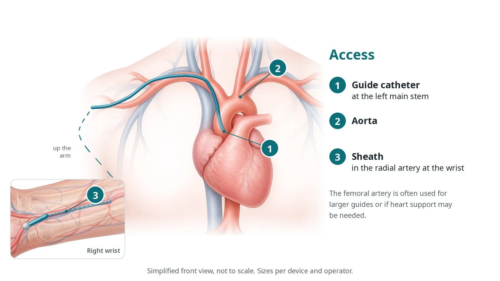
Access: (1) guide catheter at the left main stem; (2) aorta; (3) sheath in the radial artery at the wrist. The femoral artery is often used for larger guides or if heart support may be needed.
3. Provisional stenting
A. Two wires. One wire goes into the main vessel and one into the side branch.
B. Main vessel stent. A stent is placed across the side branch opening. The side branch wire is left 'jailed' behind the stent to keep access and mark the branch.
C. Proximal optimisation. A short balloon, sized to the larger proximal vessel, is inflated from the carina upwards so the stent fits the wider part of the artery.
D. Kissing balloons. If the side branch needs it, the jailed wire is exchanged for a wire through the stent struts, and balloons in both branches are inflated together, often followed by another proximal optimisation.
Watch: Loss of flow down the side branch with chest pain or ECG changes needs urgent action. Tell the operator at once.
4. If a second stent is needed
If the side branch remains significantly narrowed or has a tear after provisional stenting, a second stent is placed using a technique such as T, TAP or culotte. In a planned two-stent strategy, DK-crush or culotte may be used from the start, per operator.
5. Final checks
Imaging checks that the stents are fully expanded, well opposed and that the edges are healthy. A final angiogram checks both branches. The sheath is removed and haemostasis achieved, as per centre protocol.
Recovery and aftercare
First hours: heart rhythm, blood pressure and access site checks, as per centre protocol.
Going home: often after one night in hospital.
Medicines: take antiplatelet medicines exactly as advised; stopping them early can cause the stent to clot.
Activity: avoid heavy lifting with the wrist or leg used for a few days.
Driving: DVLA Group 1 rules usually allow car driving about one week after a planned PCI if you are well; ask your team.
Follow-up: clinic review and cardiac rehabilitation where offered.
Call 999 if you have chest pain that does not settle, breathlessness, fainting or heavy bleeding.
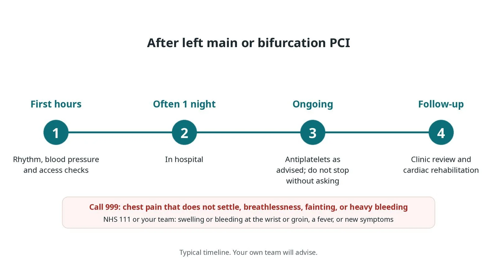
After left main or bifurcation PCI: (1) rhythm, blood pressure and access checks; (2) often one night in hospital; (3) antiplatelets as advised, not stopped without asking; (4) clinic review and cardiac rehabilitation.
Risks and complications
Your team will explain the risks that apply to you. Possible problems include:
Bleeding at the access site
Side branch narrowing or closure (uncommon)
Low blood pressure or a dangerous rhythm during inflations (uncommon)
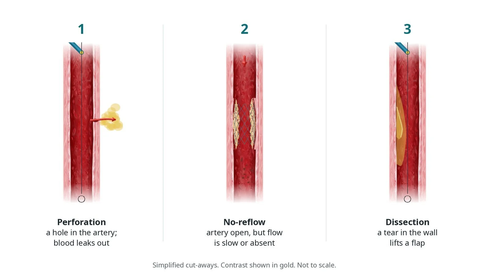
Tear or perforation of the artery (rare)
Heart attack around the procedure (uncommon)
Stent clot or narrowing later (uncommon): Antiplatelet medicines lower this risk.
Kidney strain from contrast
Cath lab kit list (printable)
Barts example kit list
An example from one UK centre, written generically. Every lab has its own protocol, and sizes and doses vary.
radial or femoral sheaths, size per operator
large-lumen guide catheter, per operator
two or more coronary guidewires, including one suitable for rewiring through stent struts
semi-compliant and non-compliant balloons, including a short proximal optimisation balloon
drug-eluting stents, sizes per operator
two inflation devices
IVUS or OCT catheter and console
heart support device (balloon pump or microaxial pump) on standby, as planned
defibrillator pads and emergency drugs
heparin, GTN and contrast, as per centre protocol
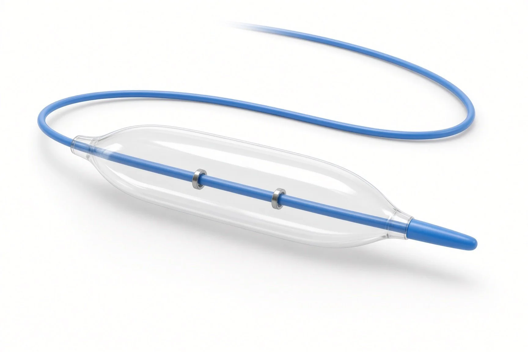
Non-compliant balloons.
IVUS or OCT catheter.
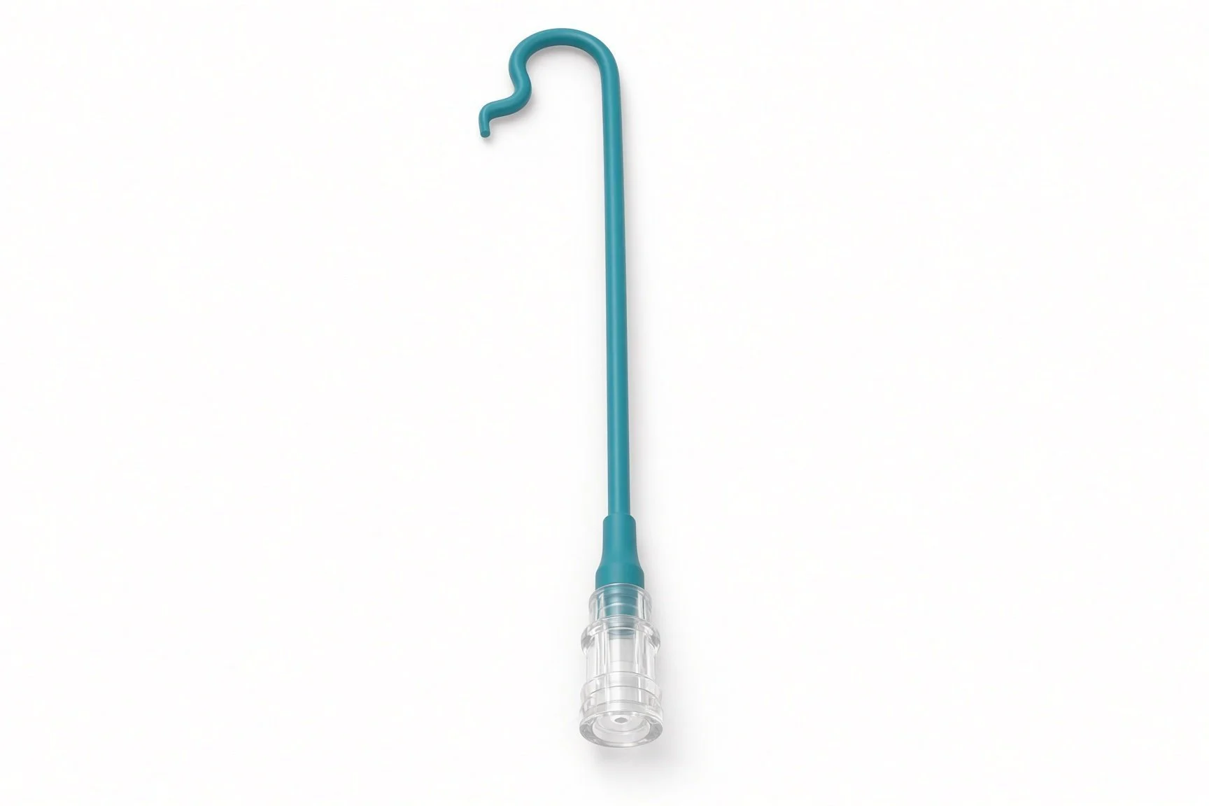
Large-lumen guide catheter.
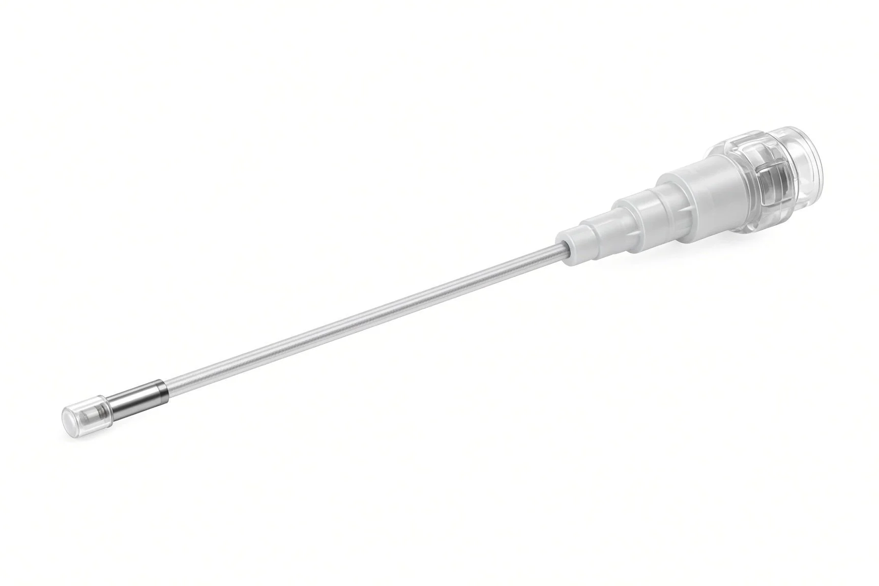
IVUS or OCT catheter.
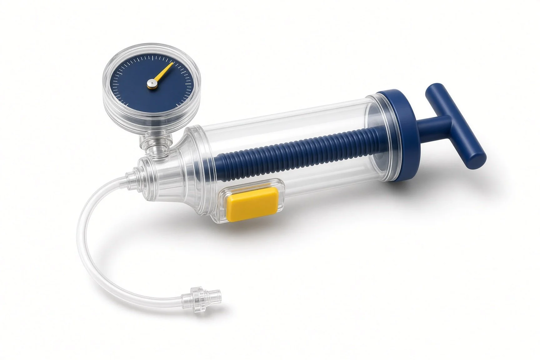
Inflation devices.
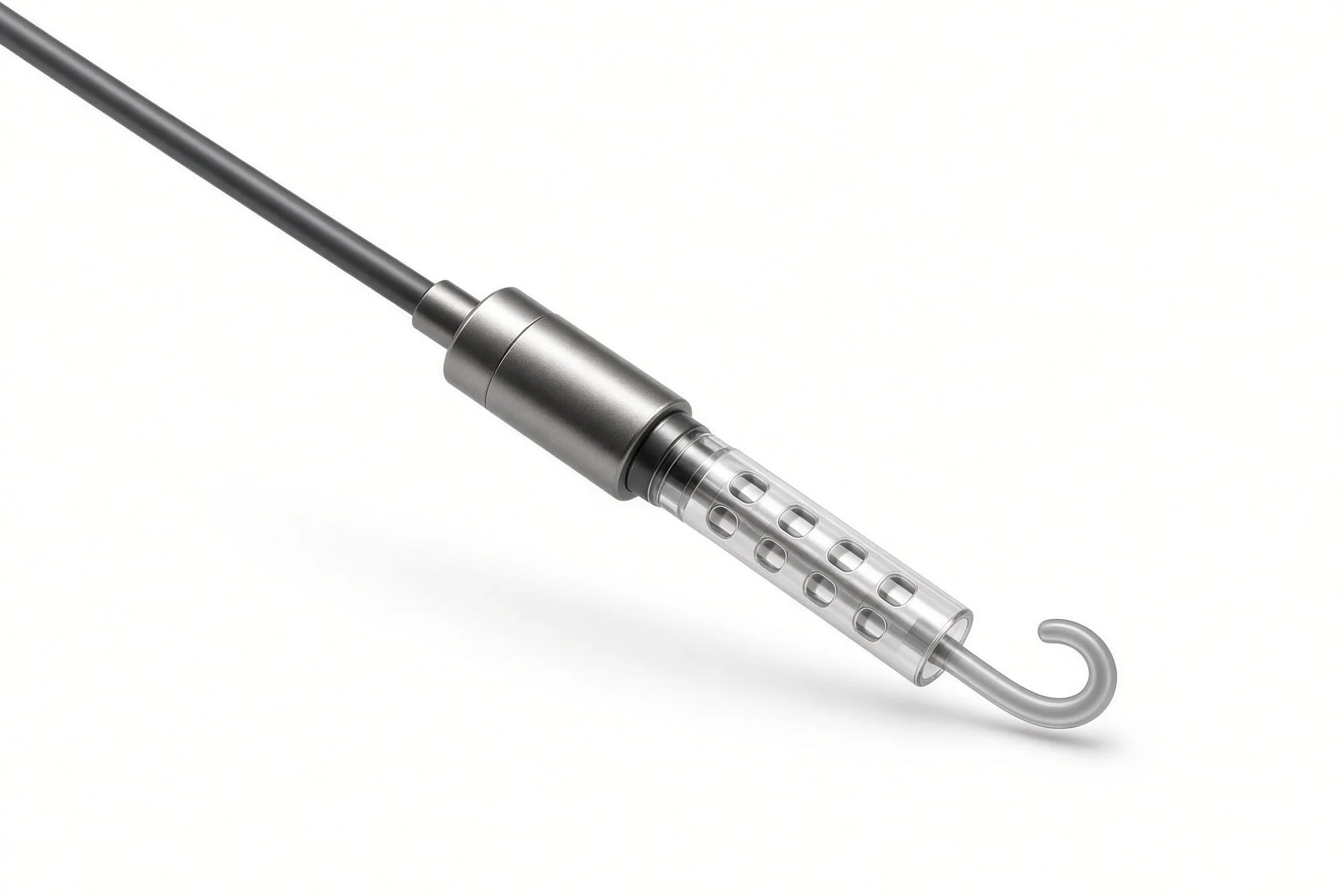
Heart support device on standby.
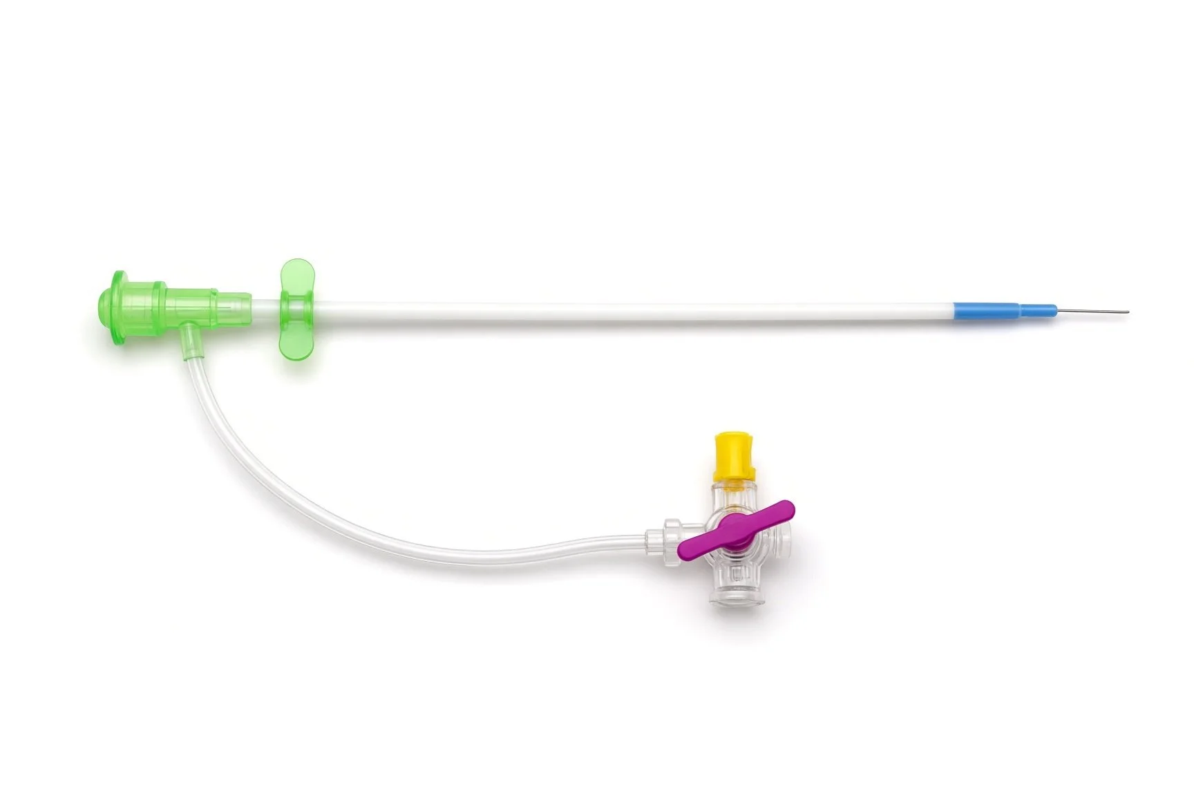
Arterial sheath.
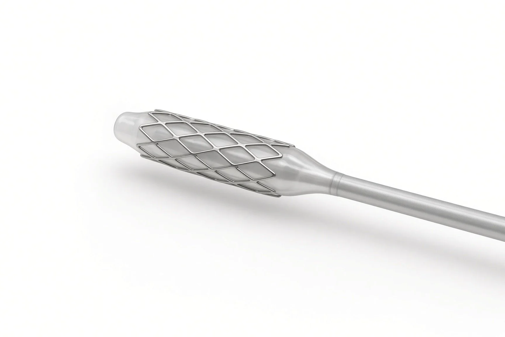
Drug-eluting stents.
Heart support device on standby.
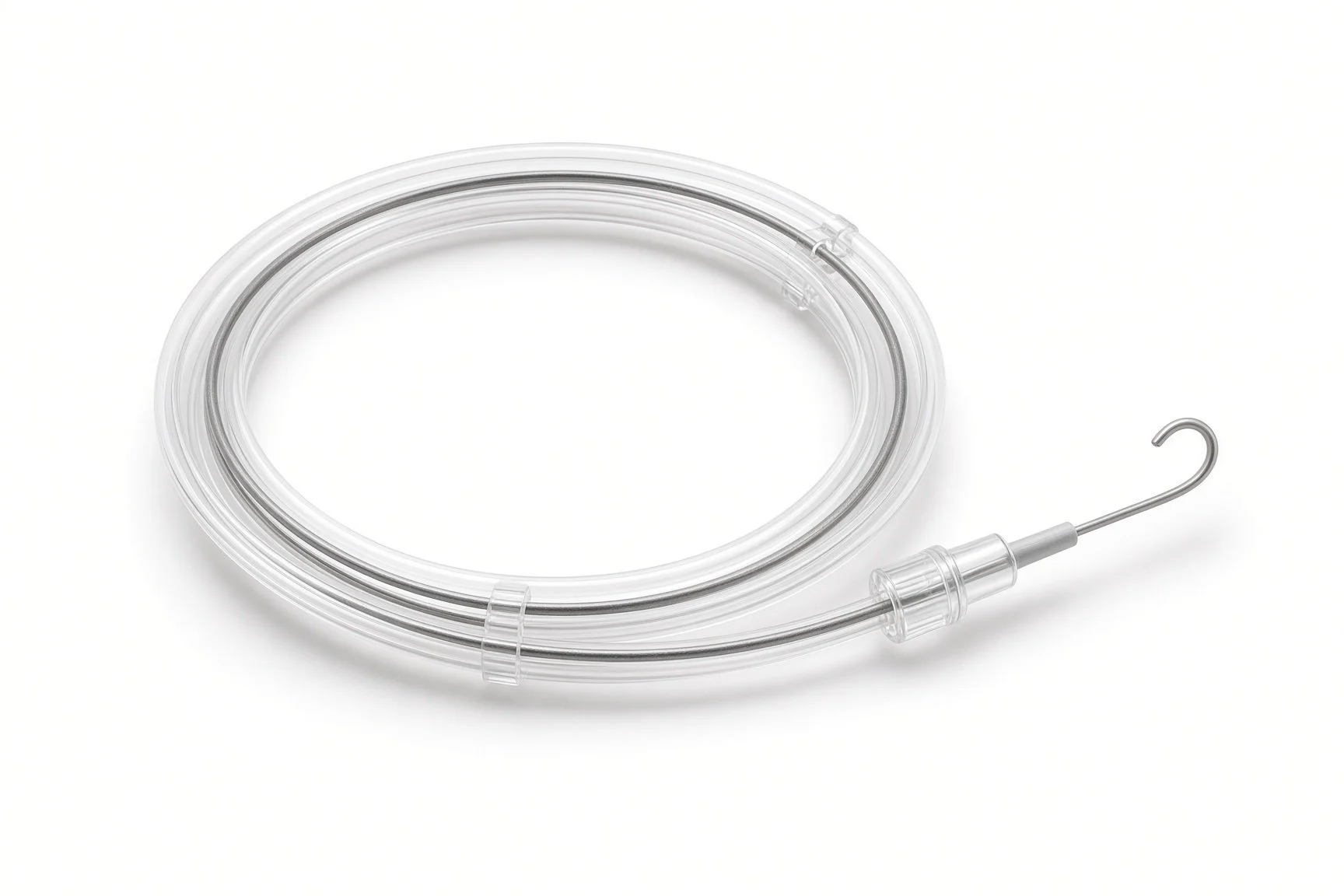
Two coronary guidewires.
Procedural steps (Barts example)
Getting ready
Access and imaging
Provisional stenting
If a second stent is needed
Final checks
First time in the cath lab?
Know which wire is which. Two wires look the same on the table. Label them and keep them apart.
Prepare two inflation devices. Kissing balloons need two balloons inflated and deflated together on the operator's call.
Watch the pressure during left main inflations. Blood pressure can fall quickly. Say the numbers out loud.
Know where heart support is. Even if not planned, know where the balloon pump or pump catheter is and who sets it up.
Label and fix both wires on the table so they cannot be mixed up or pulled back.
Have the proximal optimisation balloon open and ready. It is sized to the proximal vessel, not to the stent.
When two balloons are used together, call out each inflation pressure and deflation clearly.
FAQ
Why not bypass surgery?
For some people PCI and surgery give similar results; for others surgery is better. The heart team discusses this with you.
Will I have one stent or two?
Usually one, with a second only if the side branch needs it. Some complex cases plan two from the start.
What is a 'kissing balloon'?
Two balloons, one in each branch, inflated together to open both and shape the stent at the split.
Why is imaging used inside the artery?
It measures the artery and checks that the stent is fully expanded, which matters most in the left main.
Related Cath Lab 101 guides
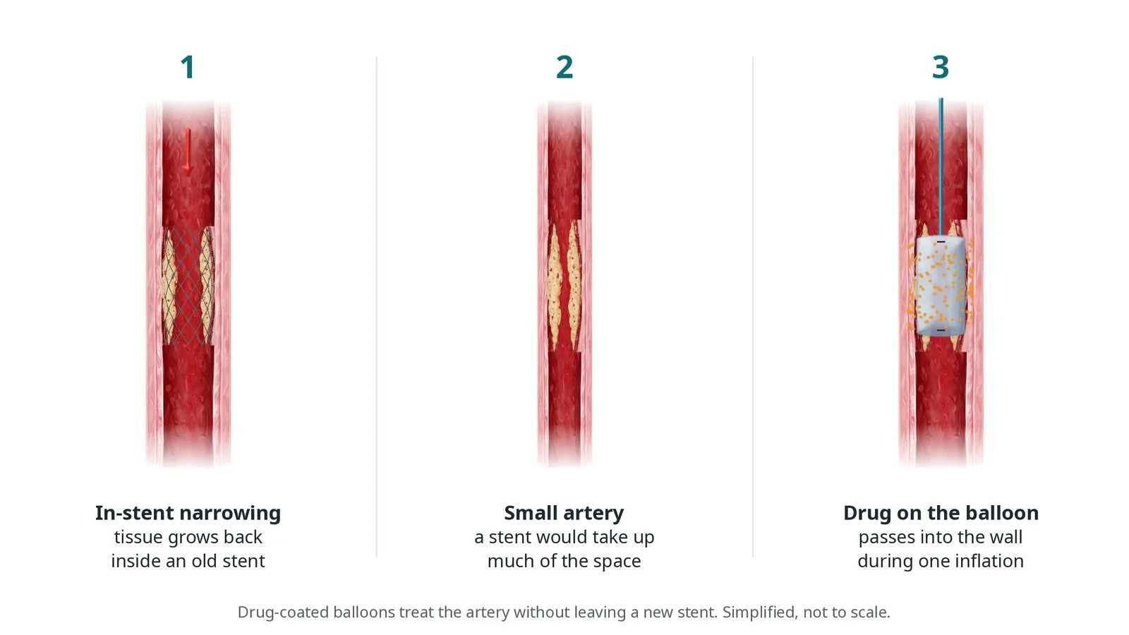
Drug-coated balloon PCI (coming soon)
Sources
All checked on 12 November 2026. Each page's own date is shown where it gives one.
Neumann FJ, et al. 2018 ESC/EACTS Guidelines on myocardial revascularization. Eur Heart J 2019
Vrints C, et al. 2024 ESC Guidelines for the management of chronic coronary syndromes. Eur Heart J 2024;45(36):3415–3537. PubMed 39210710
Banning AP, et al. Percutaneous coronary intervention for obstructive bifurcation lesions: the 14th consensus document from the European Bifurcation Club. EuroIntervention 2019
StatPearls (NCBI Bookshelf), search: left main coronary artery disease. ncbi.nlm.nih.gov/books
Disclaimer
Education only. This page is for general education for healthcare students, cath lab staff and curious readers. It isn't medical advice and doesn't replace advice from your own doctor, heart team or local unit protocols. Equipment and medicines are described generically. No product or manufacturer is recommended. If you think someone is having a heart emergency, call 999.
Disclosure: I work for a company that makes a device used in this procedure. This page is independent education, isn't sponsored or reviewed by any company, and doesn't recommend any product.
All diagrams on this page are original illustrations created for rodnieoro.com.