Coronary perforation, no-reflow and dissection: a Cath Lab 101 guide
Reviewed by Rodnie Oro, cath lab charge nurse · Last reviewed: November 2026 · Education only, not medical advice
Coronary perforation, no-reflow and dissection in one minute
What it is: Three complications that can happen during PCI: a hole in the artery (perforation), poor flow despite an open artery (no-reflow), and a tear in the artery wall (dissection).
Who it is for: Staff working in PCI lists. Patients may read it if a complication happened during their procedure.
Main risks: Bleeding around the heart (tamponade), heart attack, dangerous rhythms, emergency surgery and, rarely, death. Quick, calm teamwork lowers the risk.
Usually: Rare. Managed in the lab by the operator and team, sometimes with emergency echo, a pericardial drain or surgery.
On this page: What is coronary perforation, no-reflow and dissection? · Who is it for? · When is it done? · Step by step · Recovery · Risks · Kit list · First time in the lab? · FAQ
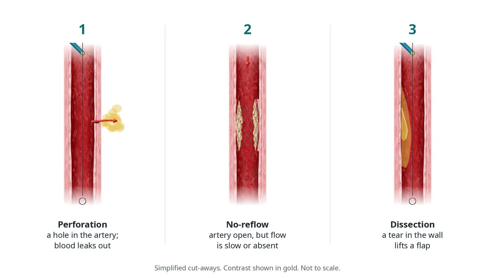
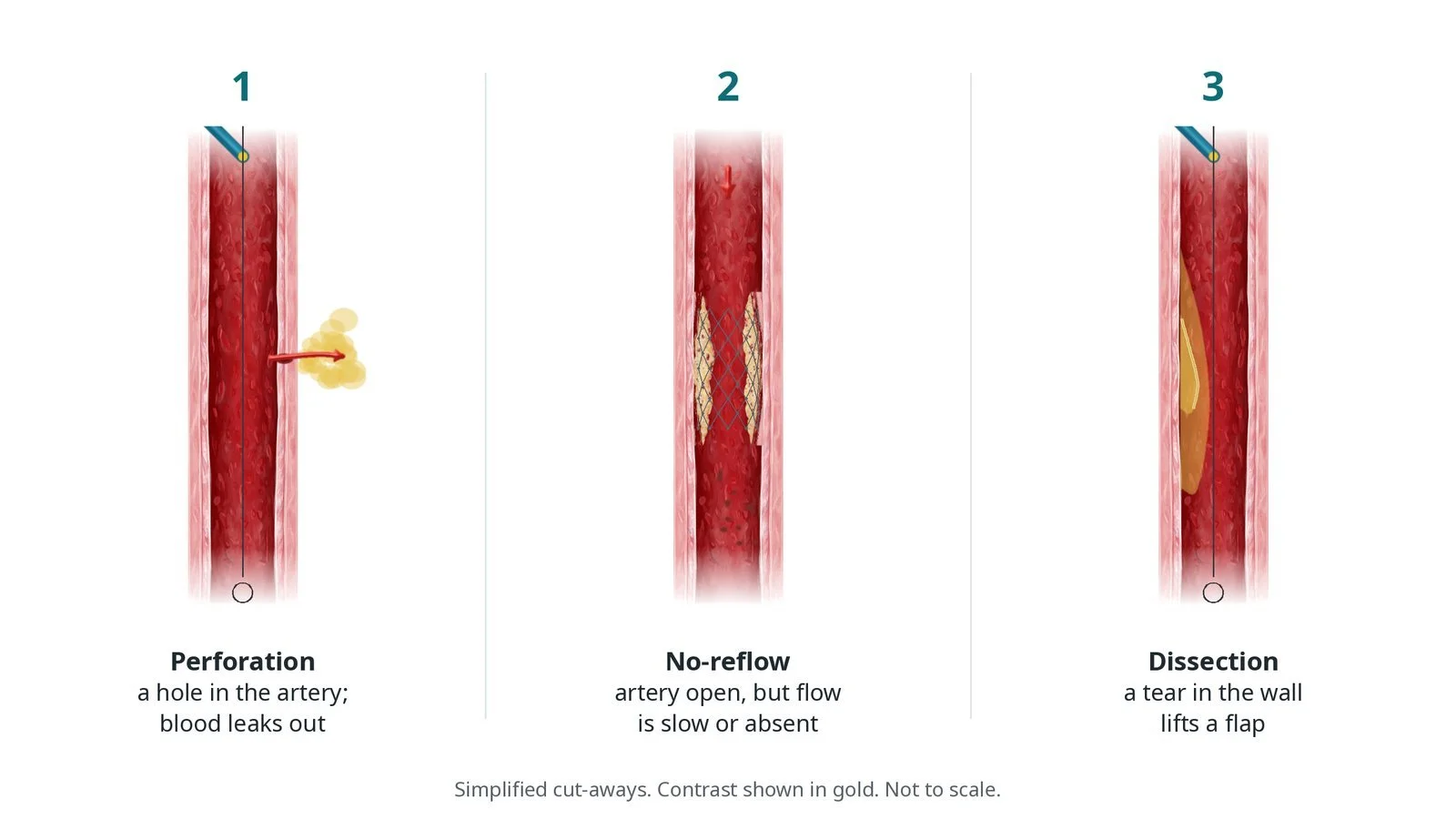
Three complications of PCI, with contrast shown in gold: (1) perforation, a hole that lets blood leak out; (2) no-reflow, where the artery is open but flow is slow or absent; (3) dissection, a tear that lifts a flap in the wall.
What is coronary perforation, no-reflow and dissection?
Perforation is a hole through the artery wall. It is more likely with complex PCI, such as chronic total occlusions, calcium treatment and oversized balloons. The Ellis classification describes it in steps: type I is a small crater outside the lumen with no leak; type II is a blush of contrast in the heart muscle or around the heart without a jet; type III is a clear jet of contrast through a hole of 1 mm or more; a separate type spills into a heart chamber or vein. Type III carries the highest risk of tamponade.
No-reflow is when the artery is open but blood barely flows beyond the treated segment. Debris, clot and spasm block the tiny vessels downstream. It is more common in heart attacks, vein grafts and atherectomy.
Dissection is a tear in the inner wall. A flap lifts and contrast may track into the wall. Small dissections are common after balloon inflation and may be left. Larger ones can narrow or close the artery (abrupt closure) and usually need a stent.
New to the acronyms? See the Cath Lab Abbreviations A–Z.
Who is it for?
Staff working in PCI lists. Patients may read it if a complication happened during their procedure.
The decision is made by your heart team, who will talk through the reasons, the alternatives and the risks with you.
When is it done?
gives you a chance to ask questions and give consent
arranges blood tests, including kidney function and a blood count, and an ECG
Step by step
1. Recognise and call it
Whoever sees a problem says it clearly: 'contrast leak', 'pressure falling', 'slow flow'. The operator confirms and the team leader allocates roles: one person calls for senior help and, if needed, the cardiac surgeons; one brings the echo; one prepares the pericardiocentesis tray; one records times and drugs.
2. Managing a perforation
A. Balloon tamponade. A balloon is inflated at low pressure at or just before the hole to stop the leak while the team prepares.
B. Covered stent. For a large hole in a main artery, a covered stent may be placed, sometimes through a second guide catheter so the balloon can stay inflated meanwhile.
C. Coils or fat. For a hole at the end of a small branch, coils, the patient's own fat or other material is delivered through a microcatheter to seal the branch.
D. Drain if needed. Echo checks for blood around the heart. If tamponade develops, a pericardial drain is placed. The operator decides whether to reverse heparin with protamine.
Watch: Falling blood pressure, a rising heart rate and a damped trace suggest tamponade. Tell the operator and get the echo and the pericardiocentesis tray immediately.
3. Managing no-reflow
The operator may give drugs into the small vessels through the guide or a microcatheter, such as adenosine, verapamil, nitroprusside or adrenaline, per operator. Blood pressure is supported. Clot may be removed. Each drug and time is recorded.
Watch: Avoid GTN or vasodilators that drop blood pressure further when the patient is already hypotensive; check with the operator.
4. Managing a dissection
Small, non-flow-limiting dissections may be left and checked again. Larger tears are covered with a stent after making sure the wire is in the true channel. If the artery closes suddenly, the operator rewires and stents it. A dissection caused by the guide catheter at the coronary opening can spread back into the aorta and needs urgent treatment.
Watch: Sudden chest pain, ST elevation and loss of flow after balloon inflation suggest abrupt closure. Keep the wire in place and tell the operator at once.
5. After the event
The patient is monitored closely, often in CCU, with repeat echo. Any drain stays until the fluid stops. The event is documented, reported through the local incident system, and explained to the patient (duty of candour). A team debrief helps everyone learn.
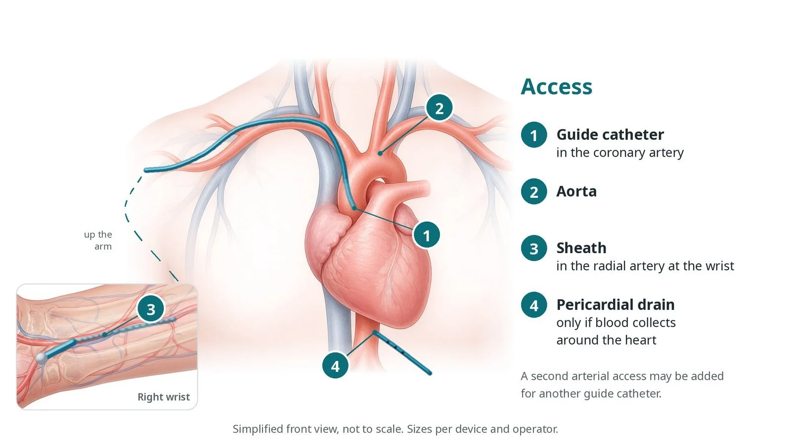
Access: (1) guide catheter in the coronary artery; (2) aorta; (3) sheath in the radial artery at the wrist; (4) a pericardial drain only if blood collects around the heart. A second arterial access may be added.
Recovery and aftercare
First hours: close monitoring of rhythm and blood pressure, often in CCU.
Echo: repeated to check for fluid around the heart; any drain is removed when it stops draining.
Going home: usually later than after a routine PCI; your team will advise.
Medicines: antiplatelets and other medicines are reviewed; take them exactly as advised.
Follow-up: clinic review, and an open explanation of what happened.
Driving: ask your team; DVLA rules depend on what was done and how your heart recovers.
Call 999 if you have chest pain that does not settle, breathlessness, fainting or heavy bleeding.
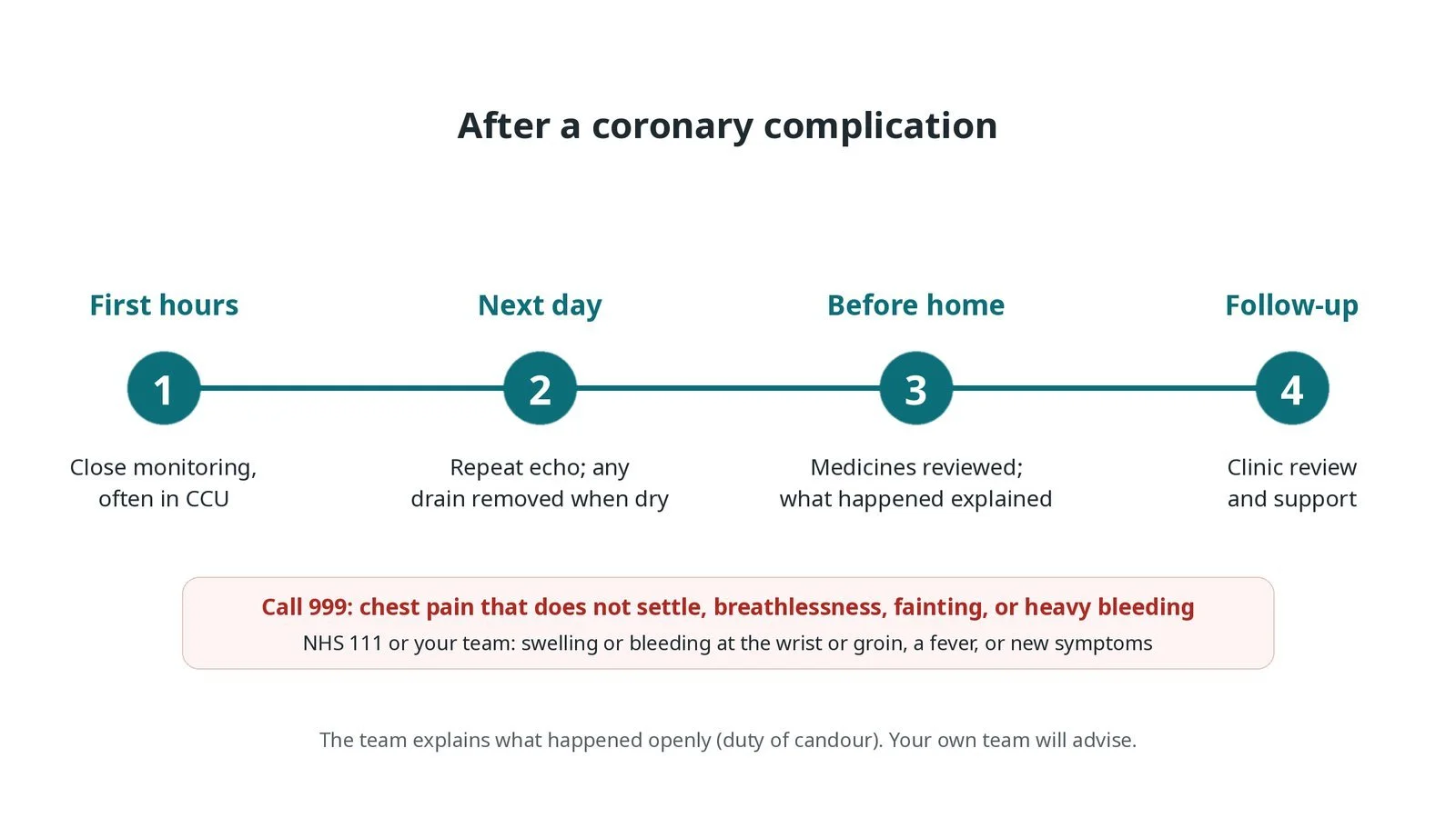
After a coronary complication: (1) close monitoring, often in CCU; (2) repeat echo and any drain removed when dry; (3) medicines reviewed and what happened explained; (4) clinic review and support.
Risks and complications
Your team will explain the risks that apply to you. Possible problems include:
Bleeding around the heart (tamponade) (uncommon): Treated with a pericardial drain.
Heart attack from loss of flow (uncommon)
Dangerous heart rhythms (uncommon)
Need for emergency heart surgery (rare)
Loss of a small branch after coil sealing (uncommon)
Death (rare)
Cath lab kit list (printable)
Barts example kit list
An example from one UK centre, written generically. Every lab has its own protocol, and sizes and doses vary.
covered coronary stents, sizes per operator
balloons for tamponade
second guide catheter and access set for a second guide
microcatheters
embolisation coils, and a set to harvest fat if used
pericardiocentesis set and pericardial drain
echo machine, ready in the lab
protamine, given only on the operator's request
no-reflow drugs such as adenosine, verapamil and adrenaline, as per centre protocol
aspiration catheter for clot, per operator
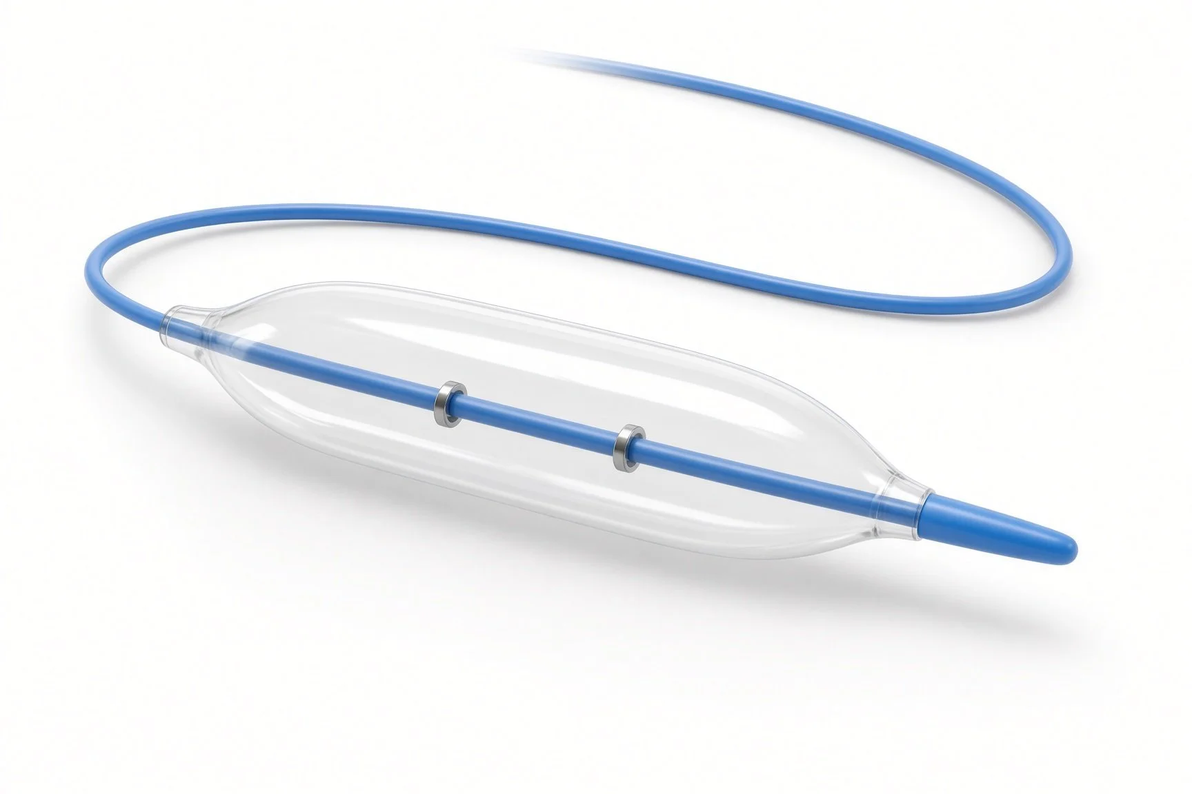
Balloons for tamponade.
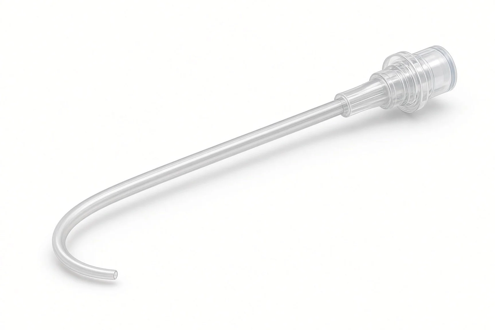
Microcatheter.
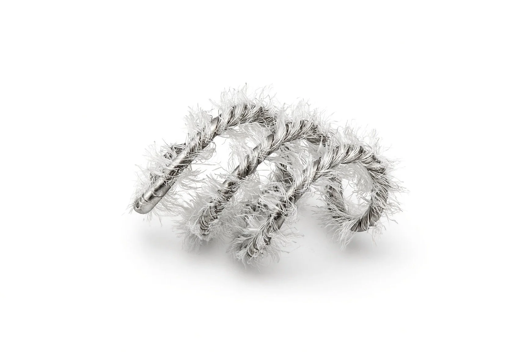
Embolisation coils.
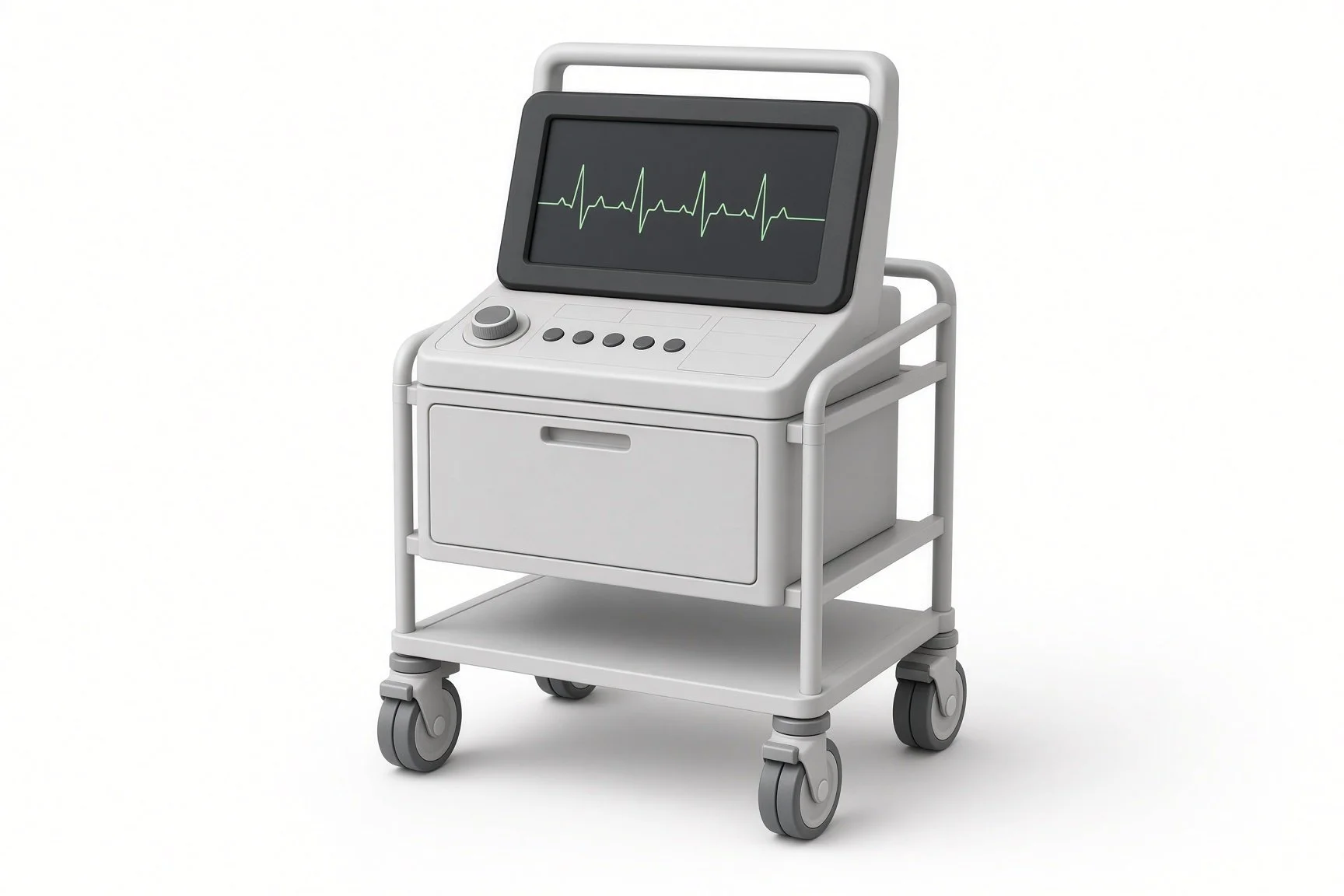
Echo machine.
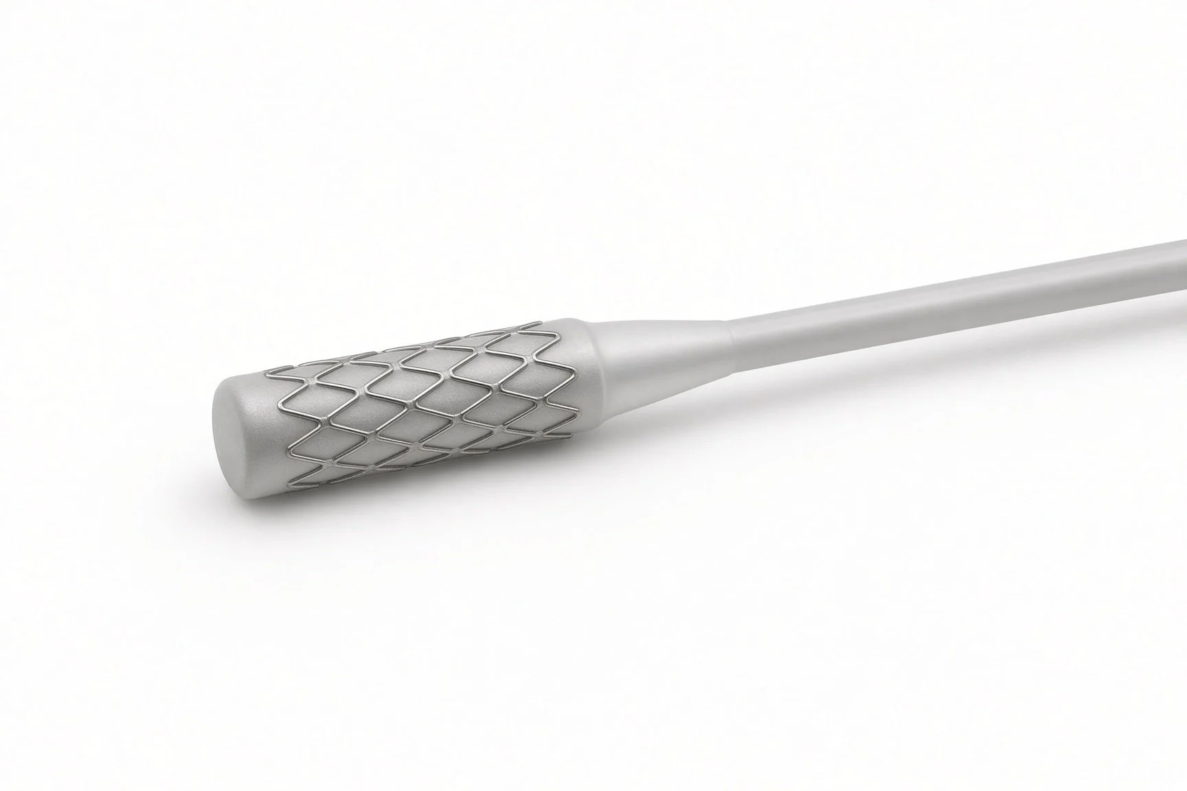
Covered stent.
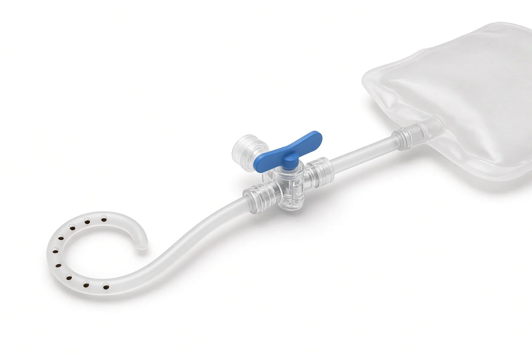
Pericardiocentesis set and drain.
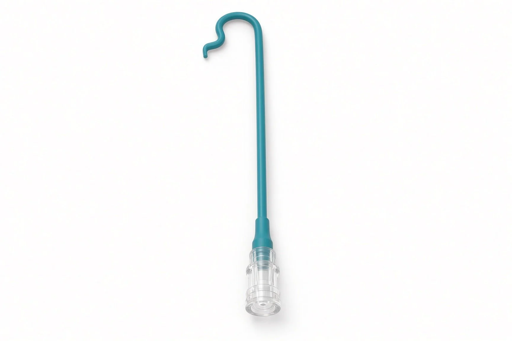
Second guide catheter.
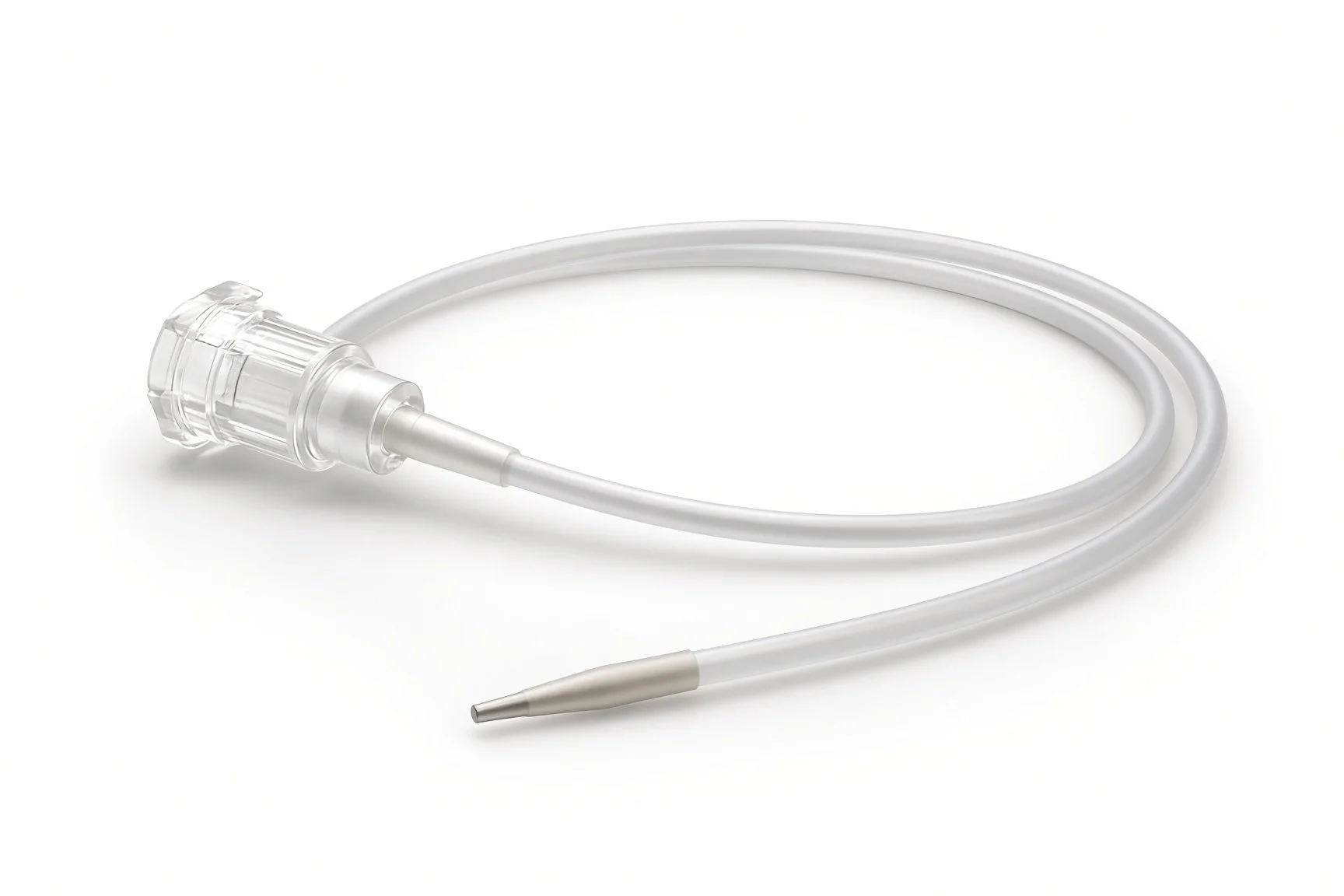
Microcatheter.
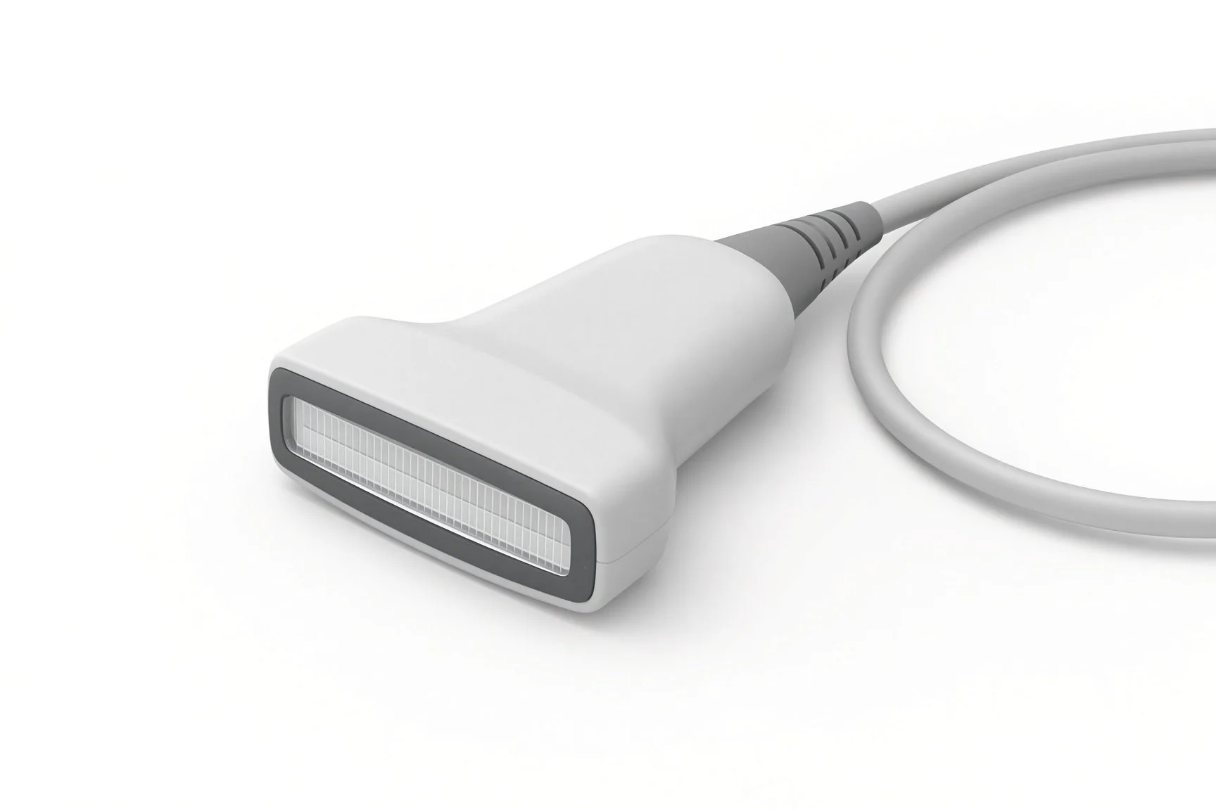
Echo machine.

Protamine, given by the operator.
Procedural steps (Barts example)
Recognise and call it
Managing a perforation
Managing no-reflow
Managing a dissection
After the event
First time in the cath lab?
Know where everything is. Find the covered stents, coils, microcatheters and pericardiocentesis tray before your first PCI list.
Say what you see. A clear 'pressure falling' or 'contrast leak' helps the operator act faster.
Keep a timeline. Write down times, drugs and devices as they happen; it guides care and the debrief.
Stay in your role. Calm, clear roles work better than everyone helping at once.
Agree in the team brief who does each role before the case, so no one has to decide under pressure.
Have the echo machine in the room and switched on at the first sign of a leak.
Do not give protamine unless the operator asks for it. The timing matters.
Prepare the drugs the operator names, label each syringe and read the label back before handing it over.
FAQ
How common are these complications?
Perforation and major dissection are rare. No-reflow is more common in heart attacks and vein grafts.
Will I need surgery?
Most are managed in the lab. Surgery is needed only rarely.
Why was a drain put in?
To remove blood that collected around the heart and stop it squeezing the heart.
Will I be told what happened?
Yes. The team will explain what happened and what was done (duty of candour).
Related Cath Lab 101 guides
Sources
All checked on 12 November 2026. Each page's own date is shown where it gives one.
Ellis SG, et al. Increased coronary perforation in the new device era. Circulation 1994
Lawton JS, et al. 2021 ACC/AHA/SCAI Guideline for Coronary Artery Revascularization. Circulation 2022
StatPearls (NCBI Bookshelf), search: coronary artery perforation. ncbi.nlm.nih.gov/books
Disclaimer
Education only. This page is for general education for healthcare students, cath lab staff and curious readers. It isn't medical advice and doesn't replace advice from your own doctor, heart team or local unit protocols. Equipment and medicines are described generically. No product or manufacturer is recommended. If you think someone is having a heart emergency, call 999.
Disclosure: I work for a company that makes a device used in this procedure. This page is independent education, isn't sponsored or reviewed by any company, and doesn't recommend any product.
All diagrams on this page are original illustrations created for rodnieoro.com.