Coronary function testing (CFR, IMR and acetylcholine): a Cath Lab 101 guide
Reviewed by Rodnie Oro, cath lab charge nurse · Last reviewed: November 2026 · Education only, not medical advice
Coronary function testing in one minute
What it is: A test done during a coronary angiogram for people with angina whose main arteries are not blocked. A special wire measures how well the small vessels work (CFR and IMR), and acetylcholine is given into the artery to look for spasm.
Who it is for: People with angina, or signs of reduced blood flow, and no obstructive coronary disease (ANOCA or INOCA), whose symptoms continue despite treatment, as selected by their cardiologist.
Main risks: Bleeding at the wrist or groin, chest pain and ECG changes during the acetylcholine test, a slow heart rate or heart block, and rarely damage to the artery. Your team will explain your own risk.
Usually: Awake, under local anaesthetic, from the wrist; about 45 to 60 minutes; home the same day for most.
On this page: What is coronary function testing? · Who is it for? · When is it done? · Step by step · Recovery · Risks · Kit list · First time in the lab? · FAQ
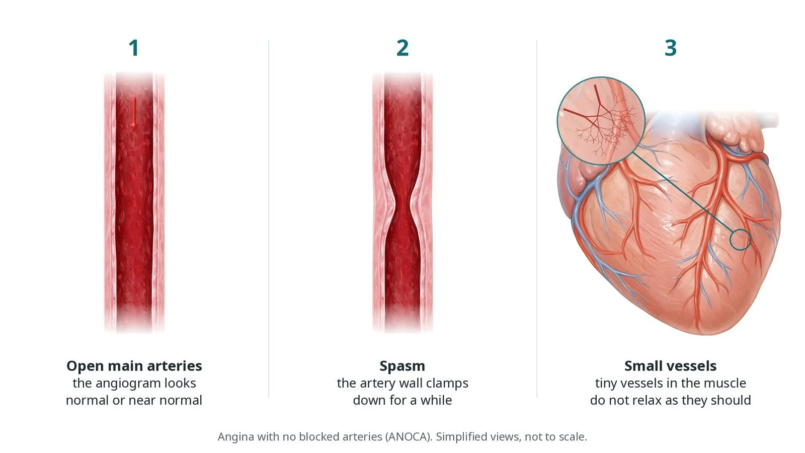
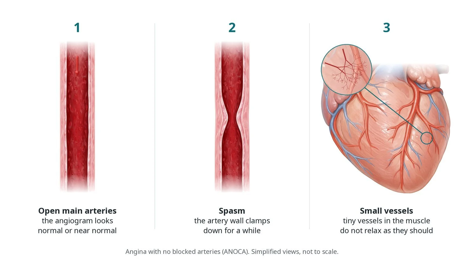
Angina with no blocked arteries: (1) the main arteries look open on the angiogram; (2) spasm, where the artery wall clamps down; (3) small vessels in the heart muscle that do not relax normally.
What is coronary function testing?
Many people with angina have an angiogram that shows no blocked arteries. This is called ANOCA (angina with no obstructive coronary arteries) or INOCA when there is evidence of reduced blood flow (ischaemia). The symptoms are real, and two common causes can be found with testing in the lab.
Coronary microvascular dysfunction: the tiny vessels inside the heart muscle do not relax properly when the heart needs more blood. The large arteries look normal on the angiogram because these vessels are too small to see.
Vasospasm: the muscle in the artery wall clamps down for a while, narrowing a large artery (epicardial spasm) or the small vessels (microvascular spasm).
A pressure-temperature wire in the artery measures flow at rest and with adenosine, which opens the small vessels. Coronary flow reserve (CFR) shows how much flow can rise. The index of microcirculatory resistance (IMR) shows how much the small vessels resist flow. Acetylcholine, given into the artery, makes a healthy artery relax but makes a spasm-prone artery clamp down.
New to the acronyms? See the Cath Lab Abbreviations A–Z.
Who is it for?
People with angina and no blocked arteries whose symptoms continue despite treatment.
It can be done straight after a normal-looking angiogram or as a planned test.
Who it may not suit: People with severe asthma may not have adenosine. People with significant narrowings need a different plan. The team decides case by case.
The decision is made by your heart team, who will talk through the reasons, the alternatives and the risks with you.
When is it done?
As a planned day case.
Before it, the team usually:
asks you to pause some heart medicines, such as calcium channel blockers and long-acting nitrates, for a set time, as advised
asks you to avoid caffeine before the test, because it blocks adenosine
checks for asthma or lung disease, which affect the choice of adenosine
gives you a chance to ask questions and give consent
arranges blood tests, including kidney function and a blood count, and an ECG
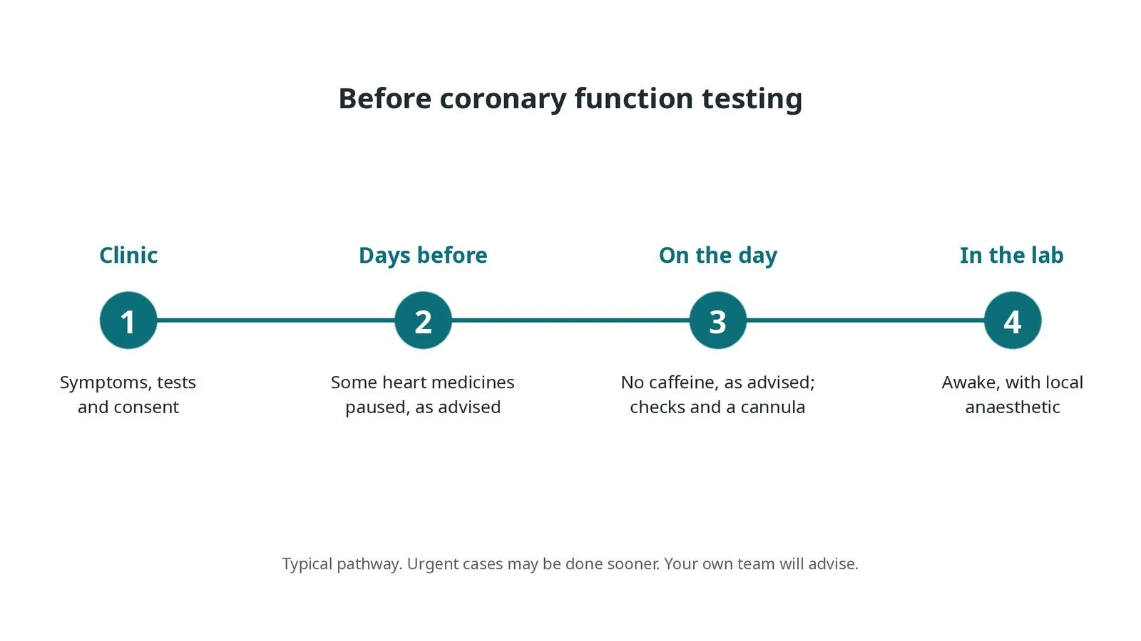
Before coronary function testing: symptoms, tests and consent; some heart medicines paused as advised; no caffeine as advised, checks and a cannula; awake with local anaesthetic.
Step by step
1. Getting ready
You lie on the X-ray table with ECG, blood pressure and oxygen monitoring. A 12-lead ECG is often used during the acetylcholine test, because changes on it are part of the result. Defibrillator pads, GTN and atropine are ready, and a temporary pacing wire is available, as per centre protocol.
2. Access and angiogram
A sheath goes into the radial artery at the wrist. Heparin is given as per centre protocol. An angiogram confirms there are no significant narrowings, and a guide catheter is placed in the left coronary artery, usually to test the LAD.
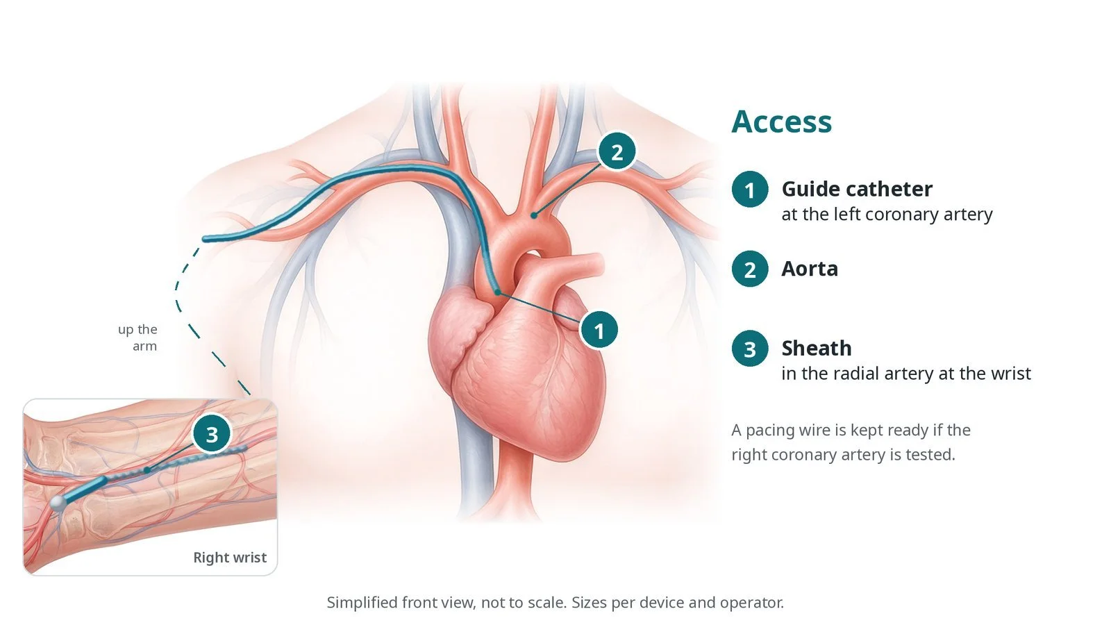
Access: (1) guide catheter at the left coronary artery; (2) aorta; (3) sheath in the radial artery at the wrist. A pacing wire is kept ready if the right coronary artery is tested.
3. Testing
A. Sensor wire. A pressure-temperature wire is passed down the artery and equalised at the guide tip first.
B. Adenosine. Small room-temperature saline flushes are given at rest and again during adenosine. The wire measures how long the saline takes to pass (transit time) and the pressure, giving CFR and IMR. Fractional flow reserve is often measured at the same time.
C. Acetylcholine. Acetylcholine is given into the artery in rising doses, as per centre protocol. After each dose the team records symptoms, the ECG and an angiogram.
D. Spasm and GTN. If the artery narrows with symptoms and ECG changes, GTN is given into the artery to relax it. GTN is also usually given at the end.
Watch: A slow heart rate or heart block can happen, especially if the right coronary artery is tested. Tell the operator at once; atropine and pacing must be ready.
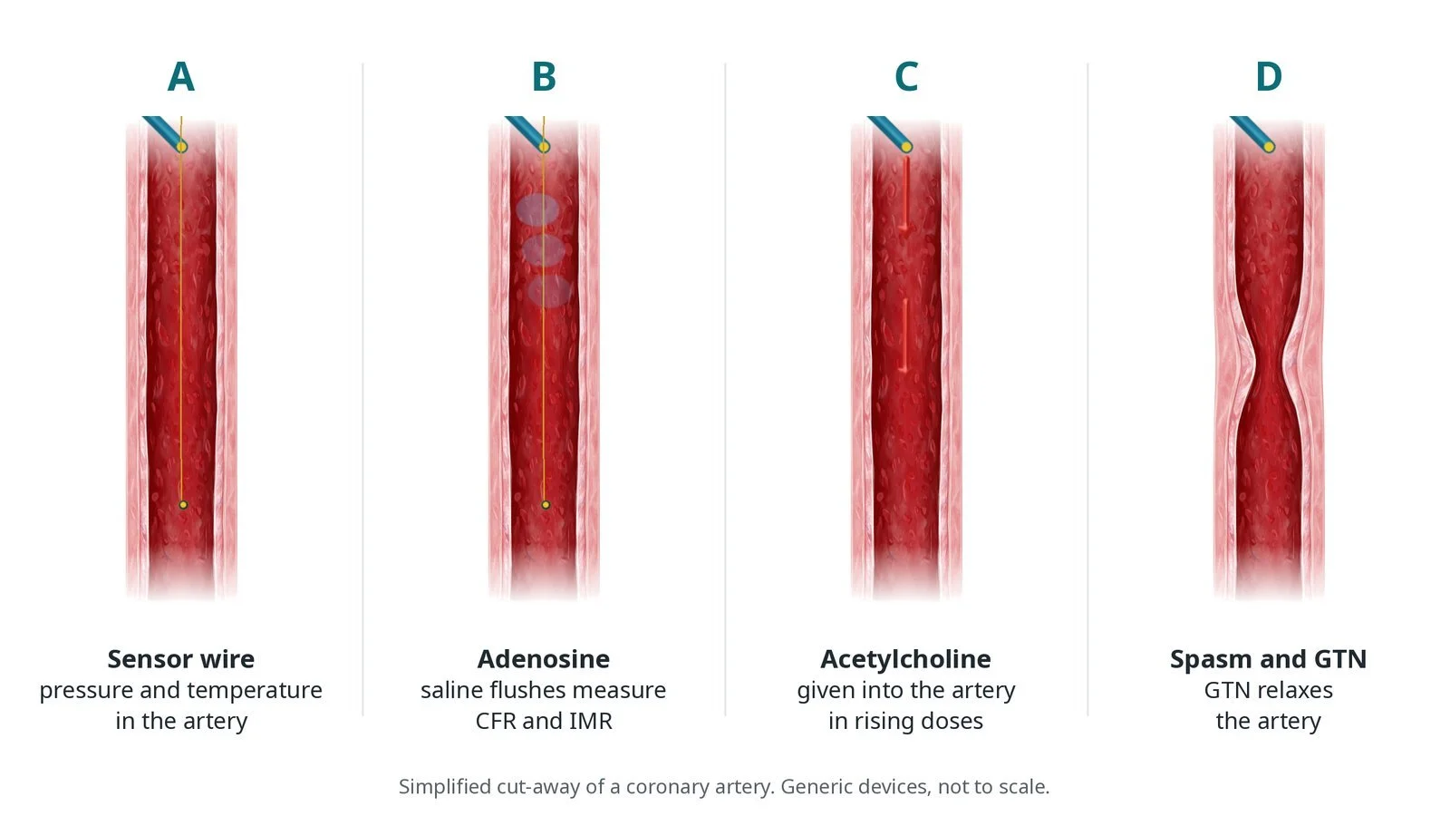
A: a pressure and temperature sensor wire in the artery. B: adenosine, with small saline flushes to measure CFR and IMR. C: acetylcholine into the artery in rising doses. D: if spasm occurs, GTN relaxes the artery.
4. Reading the result
The operator combines the measurements. Low CFR or high IMR suggests microvascular dysfunction. Narrowing of the large artery with the usual pain and ECG changes suggests epicardial spasm. Pain and ECG changes without large-artery narrowing suggest microvascular spasm. Cut-off values are set by your centre's protocol.
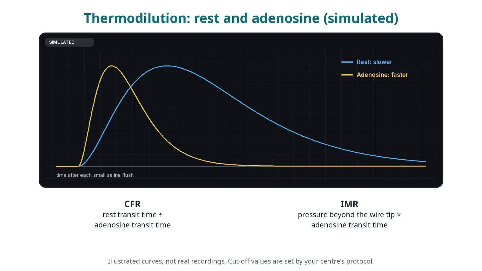
Illustrated thermodilution curves, not real recordings: saline passes faster with adenosine. CFR is the rest transit time divided by the adenosine transit time; IMR is the pressure beyond the wire tip multiplied by the adenosine transit time.
5. Finishing
The wire and catheter are removed. The radial sheath comes out and a wrist band is applied, as per centre protocol.
Watch: Ongoing chest pain or ECG changes after the test need GTN and the operator's review before the patient leaves the lab.
Recovery and aftercare
First hours: wrist, heart rhythm and blood pressure checks, as per centre protocol.
Going home: the same day for most.
Medicines: restart your usual medicines as advised; new medicines may be started to match the result.
Results: the team explains whether you have microvascular angina, vasospastic angina, both, or neither.
Activity: avoid heavy lifting with the wrist for a few days.
Driving: ask your team; DVLA rules depend on your diagnosis and how you feel.
Call 999 if you have chest pain that does not settle with your GTN, breathlessness, fainting or heavy bleeding from the wrist.
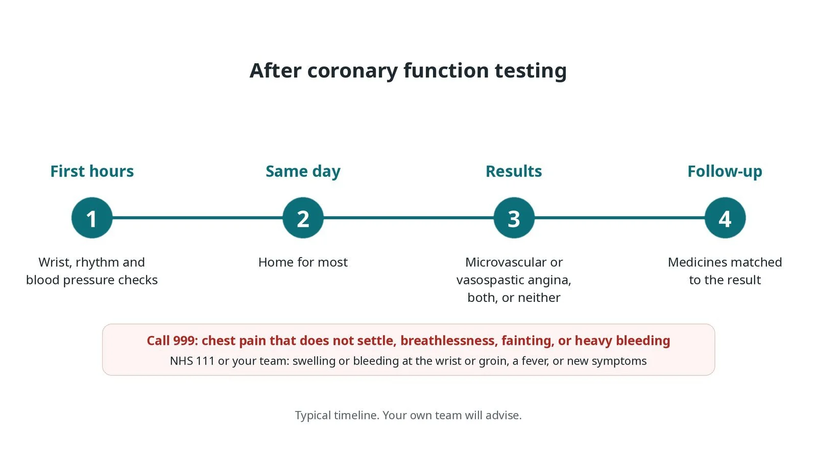
After coronary function testing: (1) wrist, rhythm and blood pressure checks; (2) home the same day for most; (3) the result: microvascular angina, vasospastic angina, both or neither; (4) medicines matched to the result.
Risks and complications
Your team will explain the risks that apply to you. Possible problems include:
Bleeding or bruising at the wrist
Chest pain during the acetylcholine test: Expected in some patients and usually relieved by GTN.
Slow heart rate or heart block (uncommon): Usually brief; atropine or pacing may be needed.
Flushing, breathlessness or tightness with adenosine: Short-lived and settles when it stops.
Spasm that is slow to settle (uncommon)
Damage to the artery from the wire (rare)
Cath lab kit list (printable)
Barts example kit list
An example from one UK centre, written generically. Every lab has its own protocol, and sizes and doses vary.
radial sheath and arterial access pack, size per operator
diagnostic catheters and a guide catheter for the left coronary artery
pressure-temperature sensor wire and its console, per device
room-temperature saline syringes for the thermodilution flushes
adenosine, as per centre protocol
acetylcholine, prepared in labelled steps as per centre protocol
GTN for the artery, and atropine
temporary pacing wire and pacing box, ready
12-lead ECG and defibrillator pads
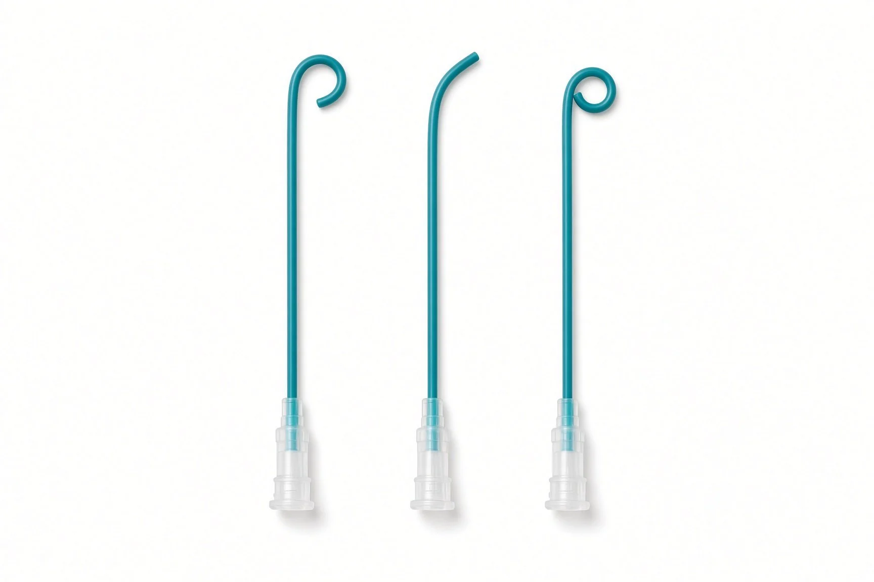
Diagnostic catheters and a guide catheter for the left coronary artery.
Procedural steps (Barts example)
Getting ready
Access and angiogram
Testing
Reading the result
Finishing
First time in the cath lab?
Know the protocol sheet. Coronary function testing follows a set order of drugs and measurements. Have the sheet on the trolley and tick off each step.
Watch the ECG and the patient together. During acetylcholine the result depends on symptoms, ECG changes and the angiogram at the same moment, so call out any change.
Be ready for a slow heart. Atropine and a pacing wire should be ready before the first acetylcholine dose.
Explain adenosine before it starts. It often causes flushing, breathlessness and chest tightness for a short while; warning the patient keeps them calm.
Write down the time of each drug and each measurement as it happens. The operator will ask for them when interpreting the result.
Prepare each acetylcholine dose exactly as the protocol says and label it clearly. The doses rise in steps and must not be mixed up.
Ask the patient to describe the pain each time: whether it feels like their usual angina is part of the result.
FAQ
Why test when the arteries look normal?
Because angina can come from the small vessels or from spasm, which the angiogram cannot show. Knowing the cause helps choose the right medicines.
Will the test bring on my pain?
It may. That is part of the test, and GTN is given to relieve it.
Why must I avoid caffeine?
Caffeine blocks adenosine, so the measurements would not be accurate.
What is the difference between CFR and IMR?
CFR shows how much flow can increase; IMR shows how much the small vessels resist flow. Together they show how the small vessels work.
Related Cath Lab 101 guides
Coronary sinus reducer (coming soon)
Sources
All checked on 12 November 2026. Each page's own date is shown where it gives one.
Vrints C, et al. 2024 ESC Guidelines for the management of chronic coronary syndromes. Eur Heart J 2024;45(36):3415–3537. PubMed 39210710
Kunadian V, et al. An EAPCI Expert Consensus Document on Ischaemia with Non-Obstructive Coronary Arteries. Eur Heart J 2020;41(37):3504–3520. PubMed 32626906
StatPearls (NCBI Bookshelf), search: coronary microvascular dysfunction. ncbi.nlm.nih.gov/books
Disclaimer
Education only. This page is for general education for healthcare students, cath lab staff and curious readers. It isn't medical advice and doesn't replace advice from your own doctor, heart team or local unit protocols. Equipment and medicines are described generically. No product or manufacturer is recommended. If you think someone is having a heart emergency, call 999.
Disclosure: I work for a company that makes a device used in this procedure. This page is independent education, isn't sponsored or reviewed by any company, and doesn't recommend any product.
All diagrams on this page are original illustrations created for rodnieoro.com.