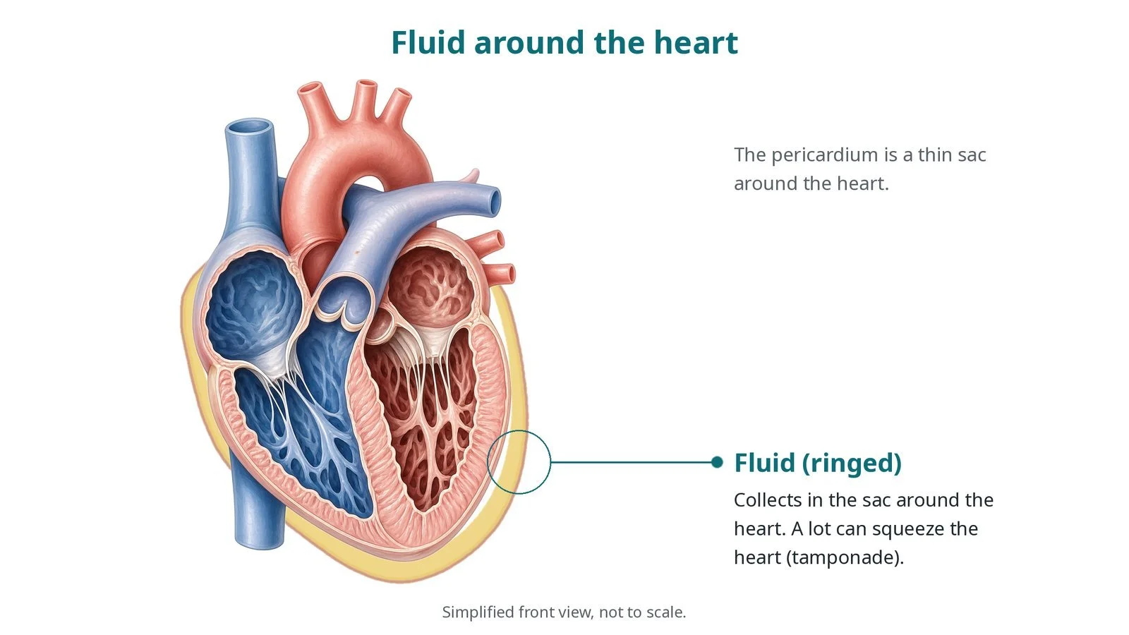
How fluid around the heart is drained with a needle and a small drain, guided by echo and X-ray. Original diagrams; steps, kit, risks and recovery.

Welcome to a hub where you can watch and learn anything about Cath Lab procedures.
How fluid around the heart is drained with a needle and a small drain, guided by echo and X-ray. Original diagrams; steps, kit, risks and recovery.
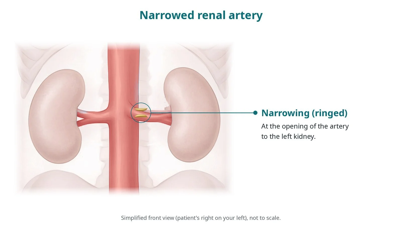
How a narrowed artery to the kidney is opened with a balloon-expandable stent, in selected patients. Original diagrams; steps, kit, risks and recovery.
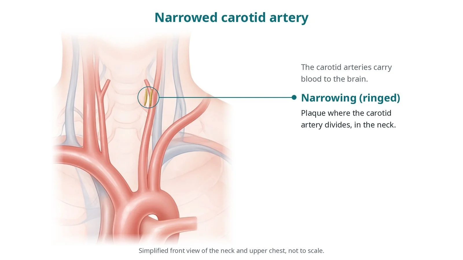
How a narrowed carotid artery in the neck is opened with a stent, usually with a filter to catch debris. Original diagrams; steps, kit, risks and recovery.
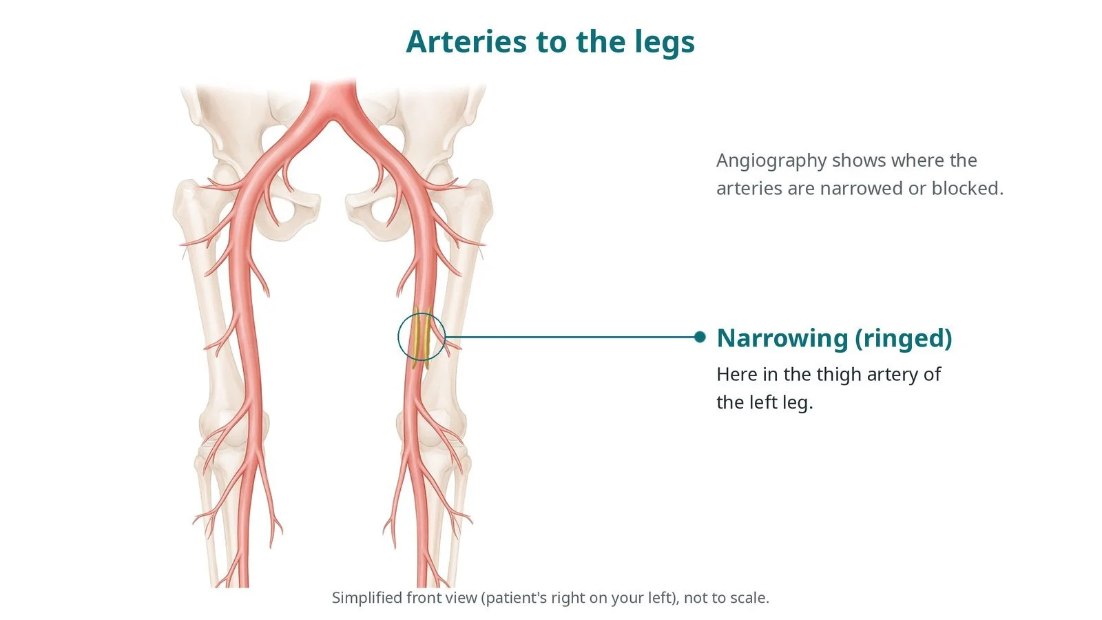
How contrast dye shows narrowings or blockages in arteries outside the heart, such as the legs. Original diagrams; steps, kit, risks and recovery.
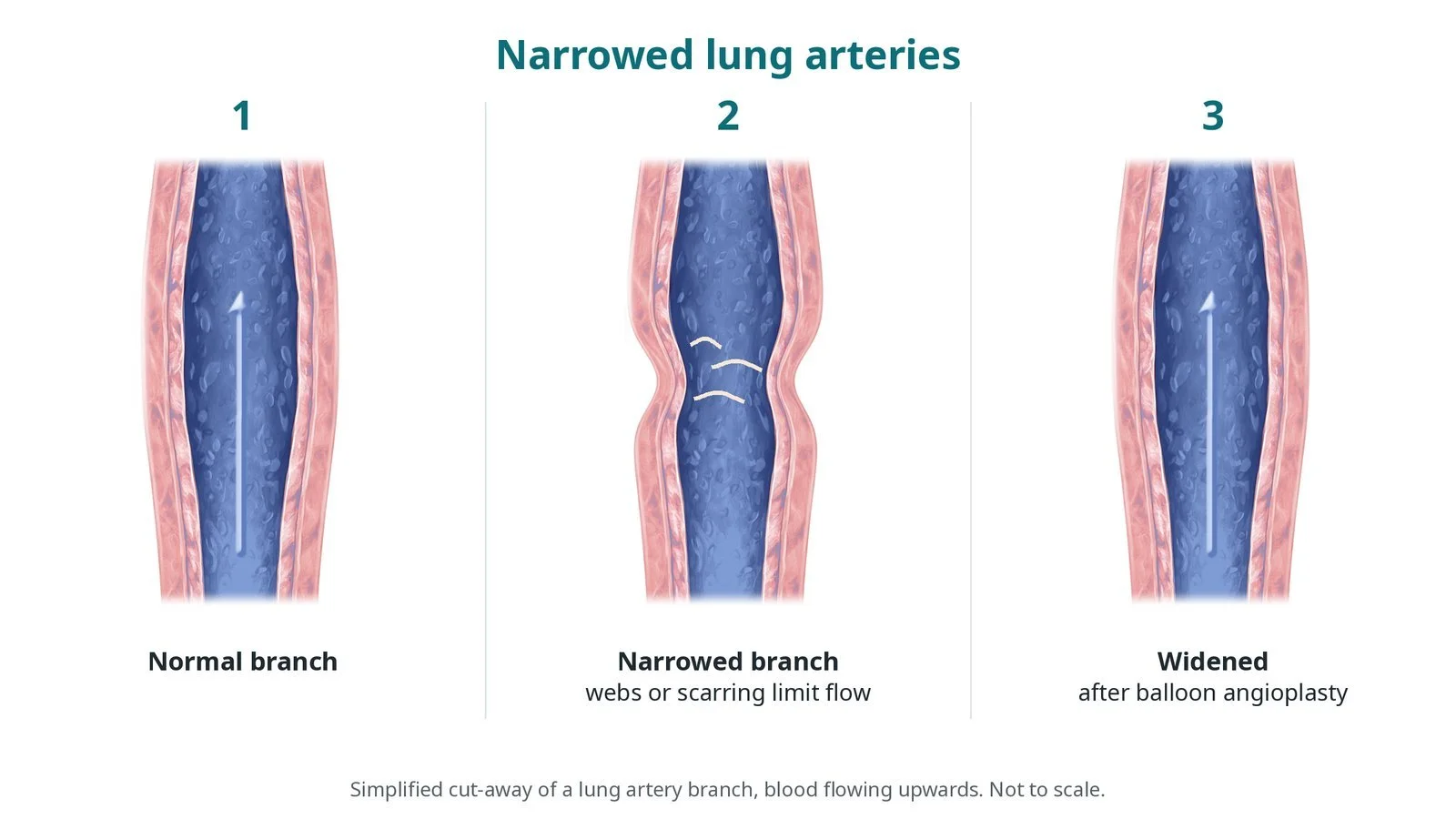
How narrowed arteries in the lungs are widened with a balloon, sometimes with a stent, in CTEPH or congenital heart disease. Original diagrams; steps, kit, risks and recovery.
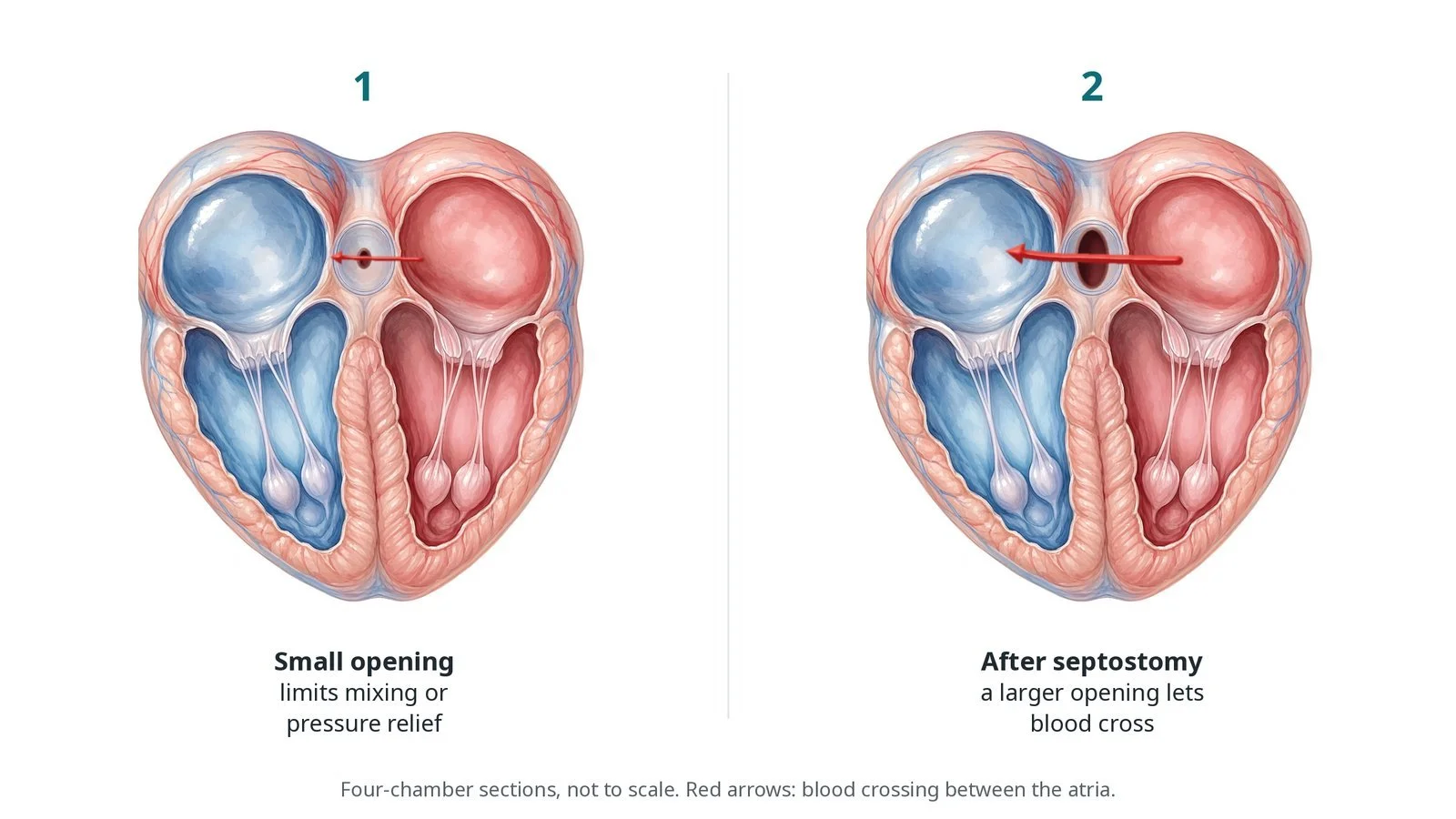
How a balloon catheter makes or enlarges an opening between the upper heart chambers, mainly in newborns with some congenital heart conditions. Original diagrams; steps and risks.
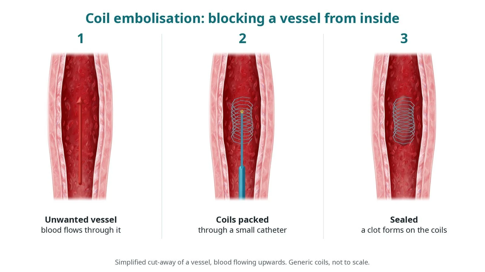
How small metal coils are placed through a catheter to block an unwanted vessel or connection. Original diagrams; steps, kit, risks and recovery.
How a narrowed vein or artery graft from earlier bypass surgery is opened with a balloon and stent. Original diagrams; steps, kit, risks and recovery.
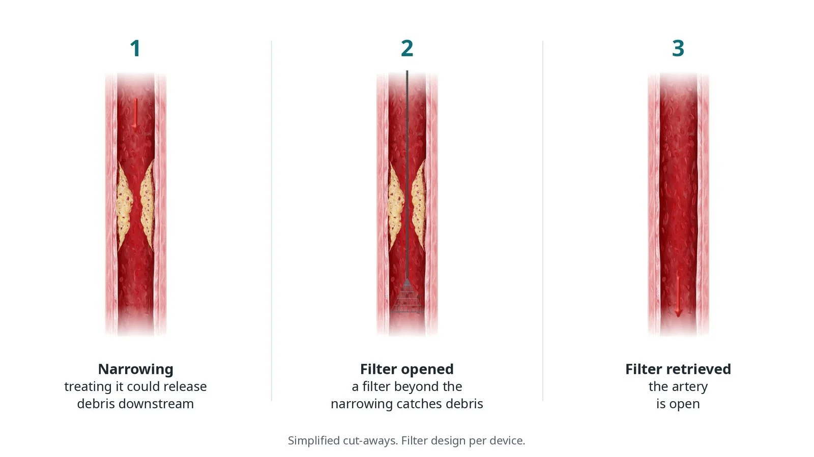
How a small filter or similar device placed beyond a narrowing catches debris released during treatment. Original diagrams; steps, kit, risks and recovery.
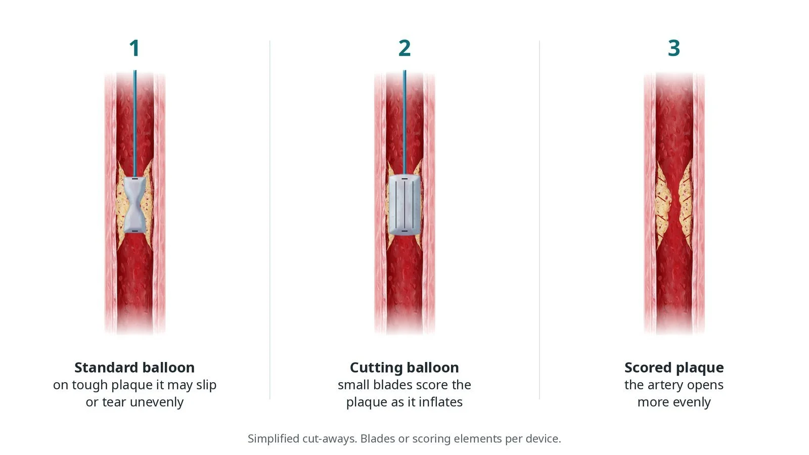
How a balloon with tiny blades or scoring wires makes controlled cuts in tough plaque so the artery opens evenly. Original diagrams; steps, kit, risks and recovery.
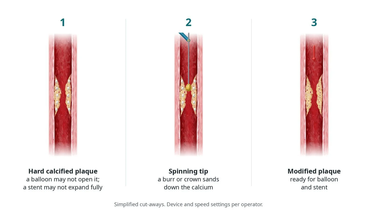
How a spinning burr or crown sands down hard, calcified plaque so a heart artery can be ballooned and stented. Original diagrams; steps, kit, risks and recovery.
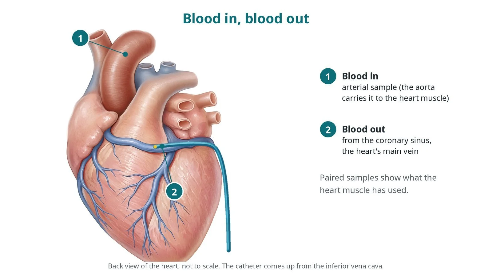
How blood samples from the artery and the heart's own vein (coronary sinus) are compared in research studies of how the heart uses energy. Original diagrams; steps and risks.
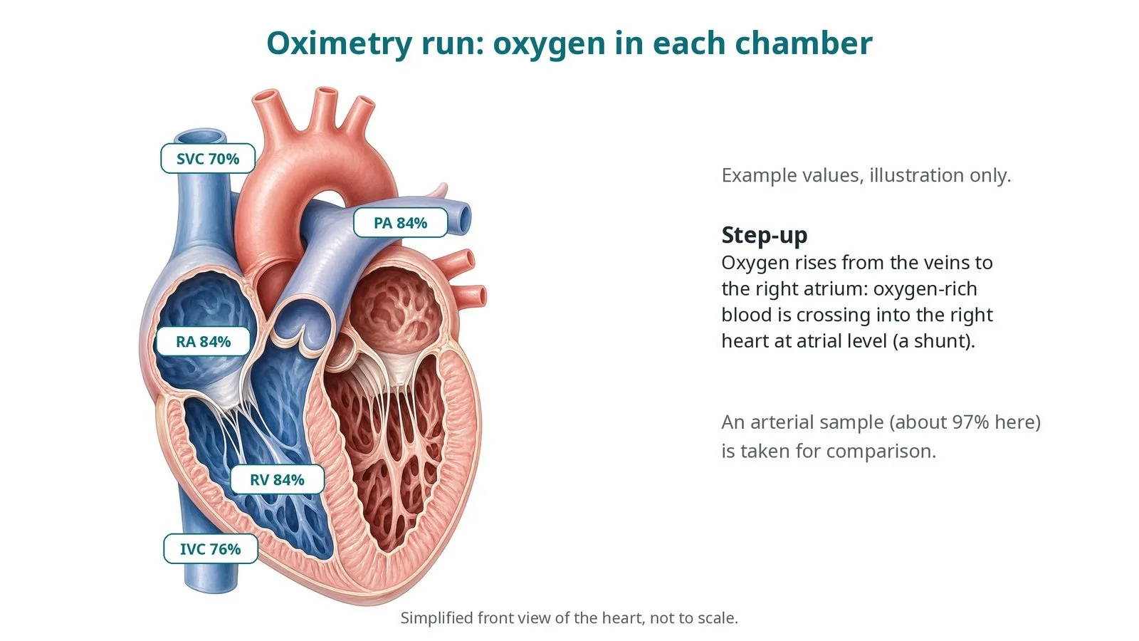
How oxygen samples from each heart chamber show blood flowing between the two sides. Original diagrams; steps, kit, risks and recovery.
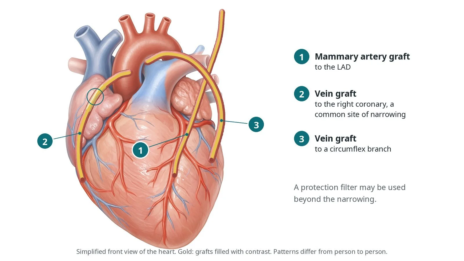
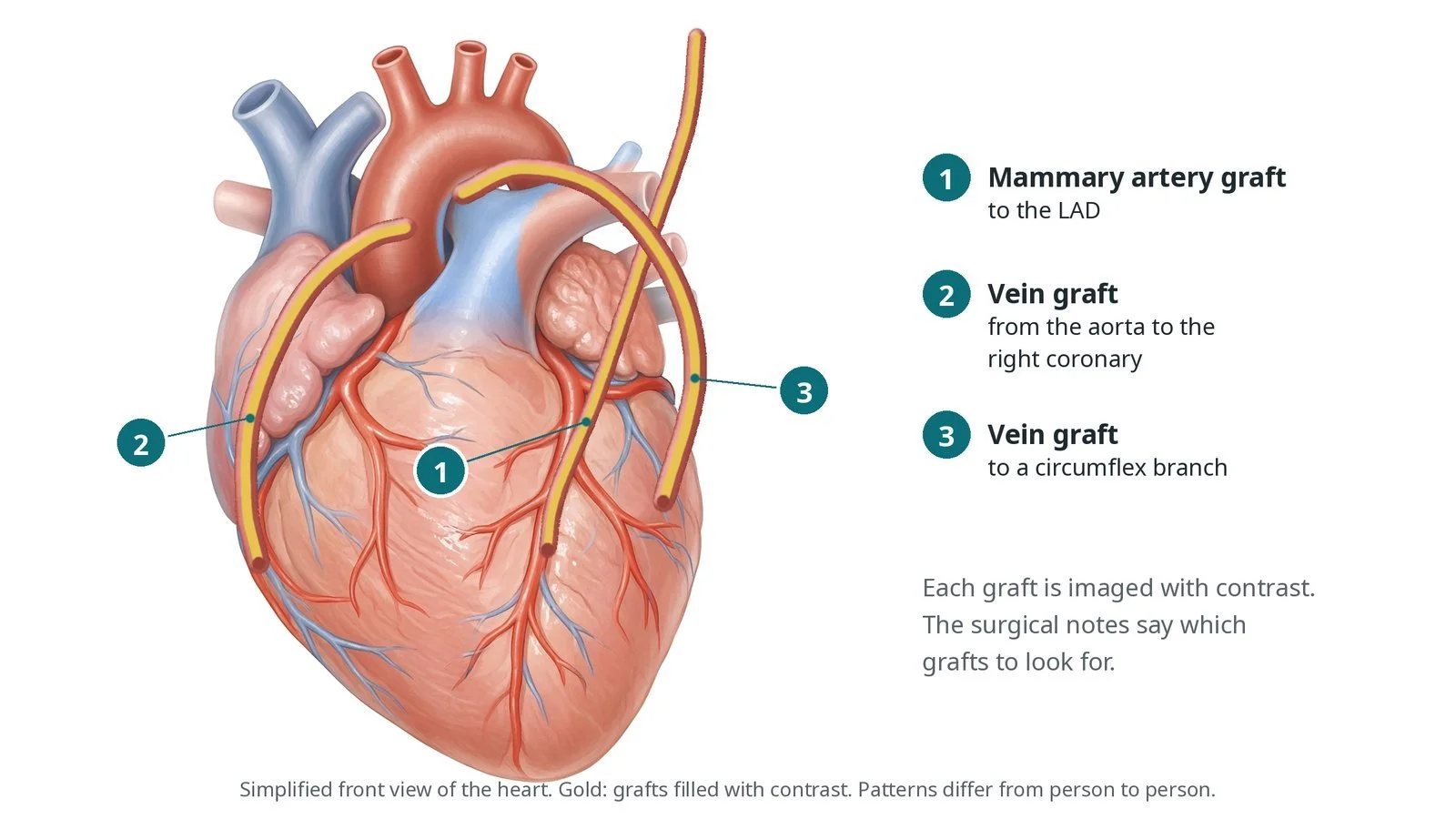
How the grafts from earlier bypass surgery are found and filled with contrast dye to check they are open. Original diagrams; steps, kit, risks and recovery.
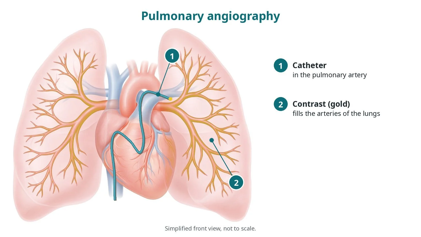
How contrast dye injected through a catheter in the lung artery shows clots or narrowings. Original diagrams; steps, kit, risks and recovery.
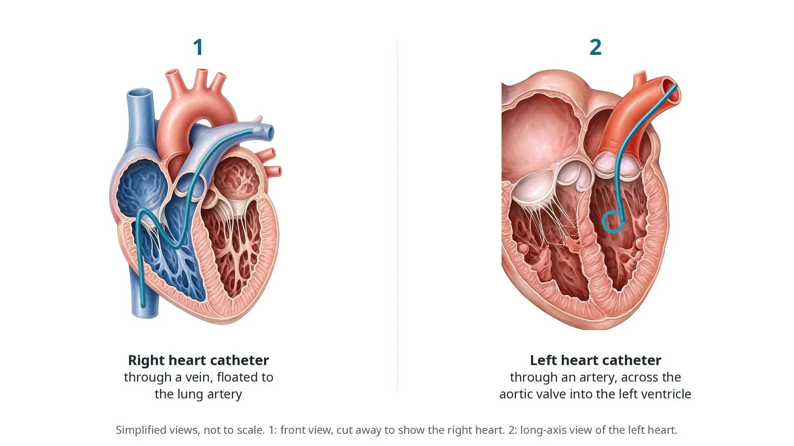
How pressures, oxygen levels and cardiac output are measured on both sides of the heart through thin catheters from a vein and an artery. Original diagrams; steps, kit, risks and recovery.
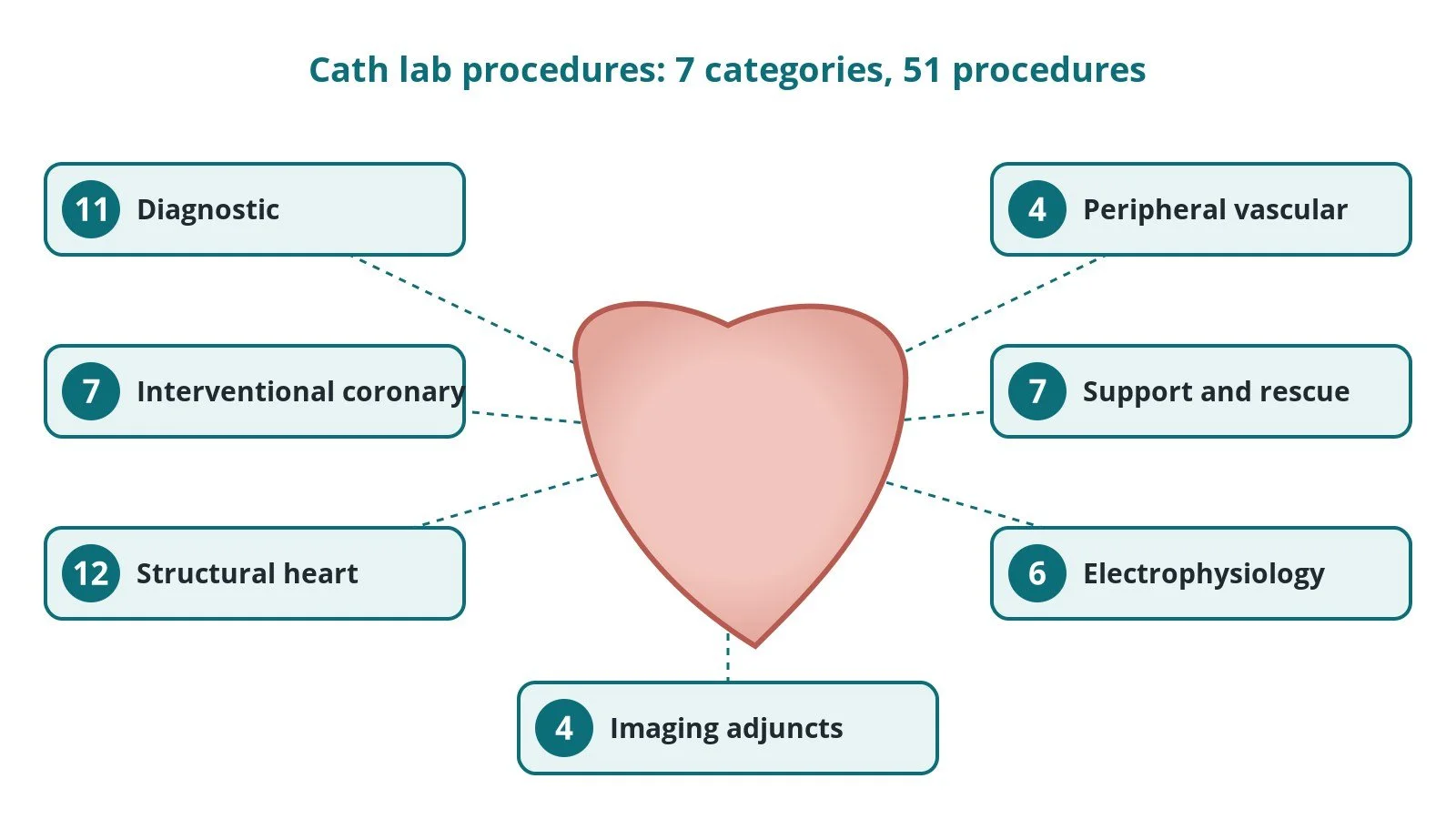
Every procedure done in or alongside a cardiac cath lab, in 7 categories, each with a one-line plain-English description and links to full Cath Lab 101 guides where they exist.
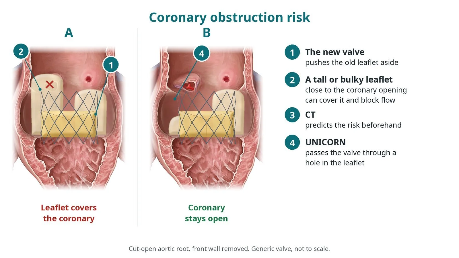
How UNICORN makes a hole at the base of an old valve leaflet and expands the new TAVI valve through it, so the leaflet is pushed away from a coronary artery. Original diagrams; honest about evidence.
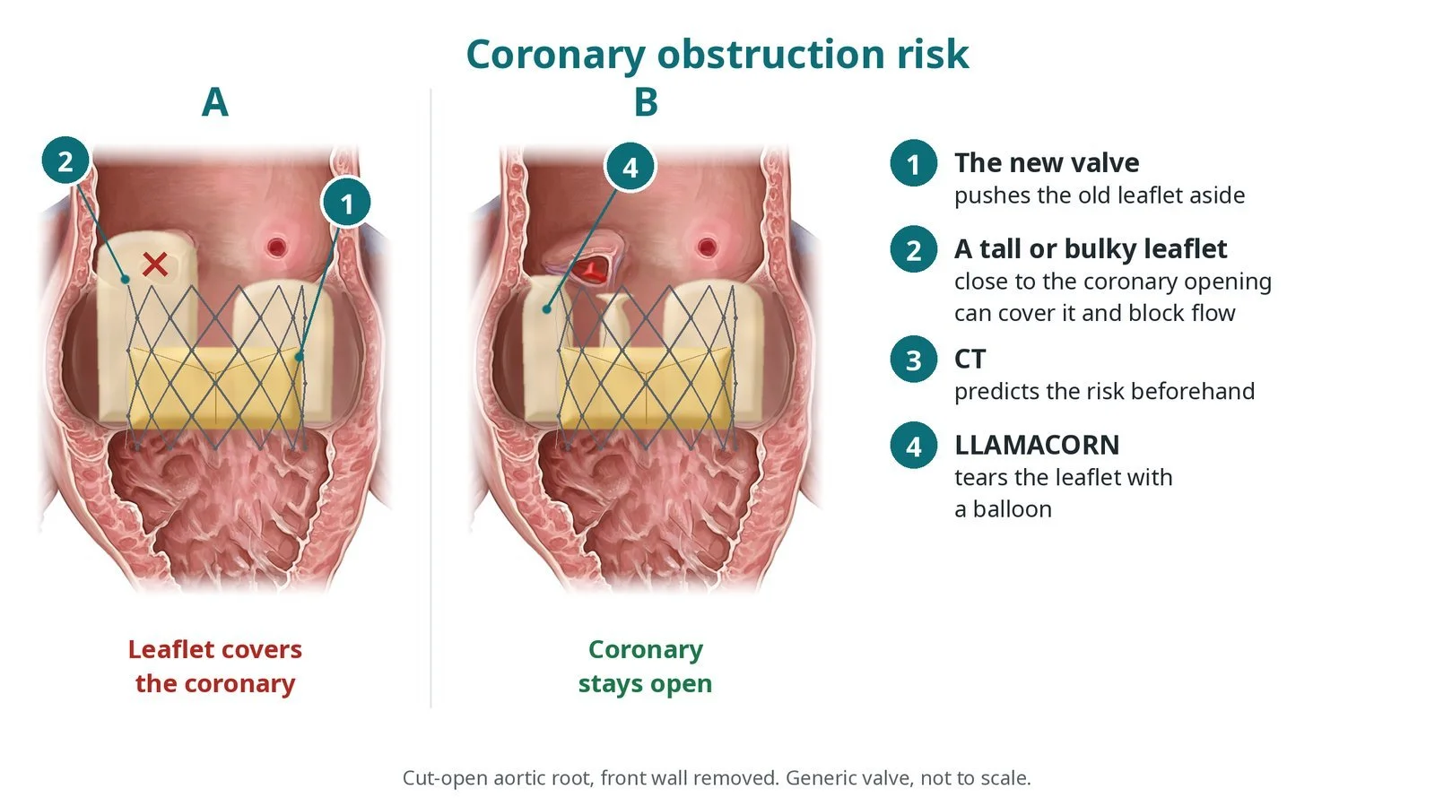
How LLAMACORN uses an electrified wire and then a large balloon to split an old aortic valve leaflet before valve-in-valve TAVI, and how it compares with BASILICA. Original diagrams; honest about evidence.
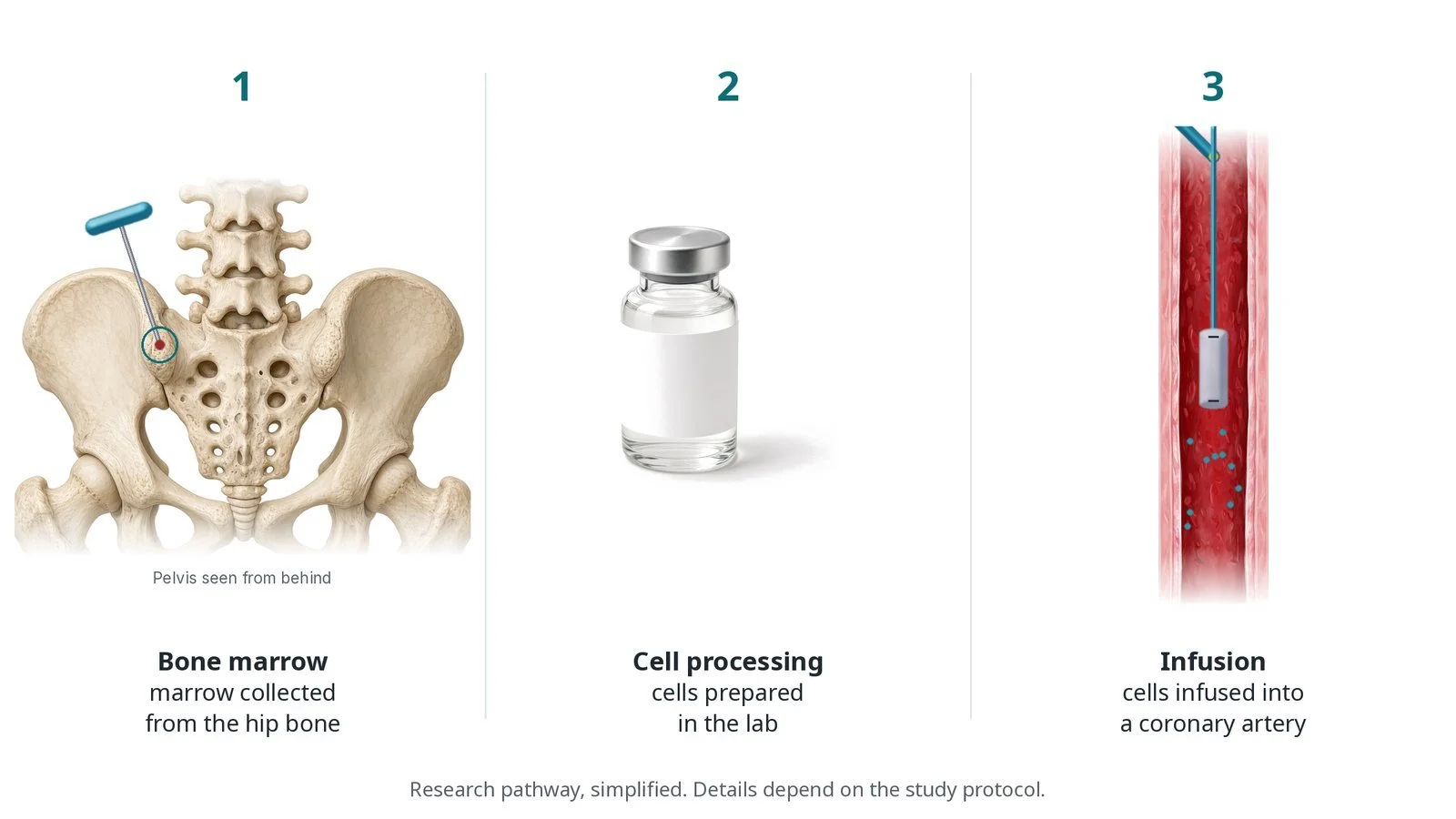
How cells taken from a patient's own bone marrow are delivered into a coronary artery during research studies, using a balloon "stop-flow" technique. Original diagrams; explains risks and the research context.